INTRODUCTION
Three species of Cirratulidae (Polychaeta) are frequent and abundant in areas of intermediate organic contamination affected by the intertidal sewage discharge of Mar del Plata city (38°S 57°W) (Elías et al., Reference Elías, Rivero and Vallarino2003, Reference Elías, Rivero, Palacios, Vallarino, Sarda, San Martin, Lopez, Martin and George2006). The densities of one species of Aphelochaeta Blake, Reference Blake, Blake, Hilbig and Scott1996 and of the new species of Caulleriella Chamberlain, 1919 described here peak in subtidal sand patches affected by sewage outfall (Elías et al., Reference Elías, Vallarino, Scagliola and Isla2004, Reference Elías, Palacios, Rivero and Vallarino2005); other cirratulids are abundant in Mar del Plata harbour (Rivero et al., Reference Rivero, Vallarino and Elías2005). Due to their distribution in areas moderately contaminated by organic matter cirratulids are early indicators of organic enrichment. The taxonomies of southern hemisphere cirratulids and other polychaete species were started by European researchers on their expeditions to Antarctica or the Southern Ocean in the 19th Century cruises or earlier. Consequently, after analysing the material they named the southern species with European names (but most frequently the species was not the European type but a new one). The same circumstances are mentioned by Doner & Blake (Reference Doner, Blake, Sarda, San Martin, Lopez, Martin and George2006) for North American waters. A series of studies around Mar del Plata had revealed several new cirratulid species (Elías & Rivero, Reference Elías and Rivero2008, Reference Elías and Rivero2009a, b), but there are still several more to be described.
Polychaetes have been extensively used in coastal studies for monitoring environmental quality in both soft- and hard-bottom habitats (Pearson & Rosenberg, Reference Pearson and Rosenberg1978; Pocklington & Wells, Reference Pocklington and Wells1992; Giangrande et al., Reference Giangrande, Liccinao and Musco2005; Elías et al., Reference Elías, Rivero, Palacios, Vallarino, Sarda, San Martin, Lopez, Martin and George2006; Dean, Reference Dean2008). They are the best indicators of environmental disturbance because they are both sensitive and tolerant species in a gradient from pristine to heavily disturbed habitats. Opportunistic species that proliferate after increases in organic matter or other disturbances belong mostly to the Capitellidae, Cirratulidae and Spionidae. Some authors have typified polluted areas by the presence of species belonging to these families (Bellan, Reference Bellan1984; Bellan et al., Reference Bellan, Desrosiers and Willsie1988). Due to their distribution in areas moderately contaminated by organic matter, cirratulids are considered early indicators of organic enrichment. It is also well known that the capitellid Capitella capitata is a complex of sibling species (Grassle & Grassle, Reference Grassle and Grassle1974), as well as the Polydora ciliate complex (Manchenko & Radashevsky, Reference Manchenko and Radashevsky1998), the aggregate/complex of the cirratulid Chaetozone setosa (Chambers & Woodham, Reference Chambers and Woodham2003) and probably many of the other small opportunistic polychaetes. Meanwhile, in order to identify other indicator species a quicker taxonomic method is required for monitoring or environmental impact assessments. Here we describe a new cirratulid species that is difficult to place in known genera, and is an important indicator of organically enriched areas in the south-western Atlantic. In order to facilitate its identification we provide an illustrated key to the known cirratulid species from warm-temperate areas of the Argentine coast.
MATERIALS AND METHODS
Material for this study came from subtidal sampling in the sand-bottoms off sewage effluent (ARA Luisito cruises in 1999; Elías et al., Reference Elías, Vallarino, Scagliola and Isla2004, Reference Elías, Palacios, Rivero and Vallarino2005). Replicate van Veen grabs (0.05 m2) were taken and sieved through a 0.5 mm mesh.
Material was examined with optical equipment (microscope and stereomicroscope) and also by scanning electron microscope (SEM) JEOL, JSM–6460 LV. The material for SEM was prepared after fixation for 24 hours with glutaraldehyde 3% in buffer of sodium cacolitate 0.1 mol with a pH between 7.2 and 7.4; followed by dehydration in alcohol (50, 70, 80, 90, 95 and 100%). Samples were dried in HMDS (hexamethyldisilazane), mounted in aluminium discs and metalized with Au–Pd. For small worms, the material was mounted on slides with a Grey and Wess medium (PVA) for optical observation.
Type material was deposited in the Museo de Ciencias Naturales de La Plata (MLP, Argentina), and in the Museu de Zoologia ‘Adão José Cardoso’, São Paulo (ZUEC POL, Brazil).
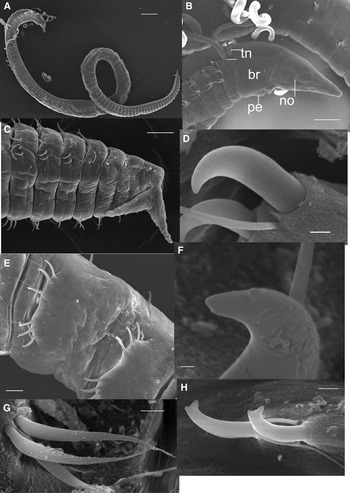
Caulleriella trispina sp. nov. (A) General view; (B) anterior region, lateral view; (C) posterior region and pygidium with two fleshy cirri; (D) unidentate hook (right); (E) posterior region showing tori bearing bidentate hooks; (F) sub-bidentate hook of middle region; (G) anterior serrate capillaries; (H) bidentate hooks of posterior region. Scale bars: A = 500 µm; B, C = 100 µm; F = 1 µm; D, G, H = 5 µm; E = 20 µm.

Caulleriella sp. A: Bolivar, Reference Bolivar1990: p. 54, figure 40a–d.
Caulleriella sp.: Elías et al., Reference Elías, Vallarino, Scagliola and Isla2004: 223, 224, 225, 227, Table 2; Elías et al., Reference Elías, Palacios, Rivero and Vallarino2005 (in part): 240.
MATERIAL EXAMINED
Station 37, 37°55.17′S 57°28.35′W, 13 m depth, medium sand, 6 incomplete specimens; Station 43, 37°57.82′S 57°28.87′W, 15 m, medium sand with shell debris, 2 entire specimens; Station 41, 37°57.16′S 57°29.30′W, 12.5 m, black mud, 2 entire and 5 incomplete specimens; Station 44, 37°57.29′S 57°28.61′W, 16 m, fine sand, 1 entire and 1 incomplete specimens. Another 9 specimens were used for SEM; collected by R. Elías and E.A. Vallarino, August 1999.
TYPE MATERIAL
Holotype: Station 43, 37°57.82′S 57°28.87′W, 15 m depth, medium sand with shell debris, (1 complete specimen MLP No. 6509).
Paratypes: Station 41, 37°57.16′S 57°29.30′W, 12.5 m, black mud, 5 entire and 12 incomplete specimens (MLP No. 6510).
COMPARATIVE MATERIAL EXAMINED FROM SOUTH-EASTERN BRAZIL
Paratypes: Station E1, 22°57′S 42°08′W, GEOCII, 89 m (7 complete specimens, and 16 incomplete specimens, ZUEC POL 5553); Station D1, 22°57′S 42°19′W, GEOCII, 35 m (56 complete and 35 incomplete specimens, ZUEC POL 5554); collected by G. Bolivar, March 1986.
DESCRIPTION
Caulleriella trispina sp. nov. is a moderately-sized species. Holotype is 8 mm long, 0.5 wide for 108 chaetigers. Paratypes vary between 70 and 112 chaetigers. Body elongate, with segments of similar length throughout, weakly curved dorsally, often inflated anteriorly (Figure 1A); chaetigers bearing capillaries, forming a shoulder-like appendage (Figure 1B); ventral groove along length of body; posterior segments sub-moniliform. Body terminates in pygidium with two fleshy cirri, anus ventral (Figure 1C). Colour in alcohol light tan.
Prostomium pointed, elongate (very glandular with SEM), with rounded red spots present laterally just anterior to oral opening, only seen with light microscope. These structures could be pigmented nuchal organs that are visible as vertical slits under SEM (Figure 1B). Peristomium a single enlarged, achaetous segment; paratypes have variable annulations, ventrally or laterally with two or three annuli, only seen under SEM. The peristomium has a slightly dorsal crest extending to chaetiger 1. Dorsal tentacles arise from posterior edge of peristomium. Branchiae present from the posterior edge of the peristomium, arising posterior and laterally to tentacles; subsequent branchiae arising from posterior edge of each chaetiger, just dorsal to notochaete; branchiae long, thin, present only in a few anterior chaetigers in the holotype, but between one- to two-thirds of body in most paratypes.
Parapodia are rudimentary, with low rounded lobes (simple tori) only in chaetigers bearing hooks (Figure 2E); fascicles of capillaries arising directly from the body. Anterior capillaries short, with blade finely denticulate, sawtooth-like (at SEM, Figure 1G). Noto- and neuropodium very close to each other (Figure 1B, E). First 9 chaetigers of holotype have 4–5 capillaries in noto- and neuropodia (7–12 in paratypes), from chaetiger 10 to 34 (8–33 in paratypes) with two hooks in noto- and neuropodia, curved and apparently unidentate under light microscopy. Paratypes examined under SEM reveal that the sub-apical tooth was often greatly reduced yet still present in some of the hooks. Thus, so in the middle section there are both uni- and sub-bidentate sigmoid hooks (Figure 1D, F). From chaetiger 35 (22–34 in paratypes) with three (then four) bidentate hooks vis-à-vis in noto- and neuropodia, with a thin companion smooth capillary for each hook (in some paratypes the capillaries are between hooks; Figure 1E, H); in the posterior region there are 2 hooks and 2 capillaries; in some paratypes the last 2–3 chaetigers are without chaetae (Figure 1C).
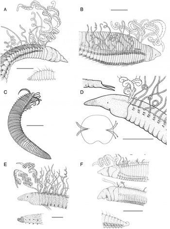
(A) Aphelochaeta malefica, general view and pygidium (from Elías & Rivero, Reference Elías and Rivero2009b); (B) Protocirrineris angelicolliatio, general view (from Elías & Rivero, Reference Elías and Rivero2009b); (C) Dodecaceria meridiana, general view (from Elías & Rivero, 2009a); (D) Caulleriella trispina, sp. nov., pygidium, general view and cross-section (this study); (E) Caulleriella galeanoi, general view and pygidium (from Elías & Rivero, 2008); (F) Caulleriella bremecae, dorsal, general and pygidium view (from Elías & Rivero, 2008). Scale bar A = 400 µm; B = 300 µm; C = 100 µm; D = 150 µm; E = 150 µm; F = 300 µm.

METHYL GREEN STAINING PATTERN
All worms stained dark green, the colour remaining for more than a year.
ETYMOLOGY
The species name refers to the presence of three different kinds of spines.
DISTRIBUTION
Caulleriella trispina sp. nov. is found in subtidal areas between 12 and 15 m depth, in muddy–sand bottoms off Mar del Plata sewage outfall. Bolivar (Reference Bolivar1990) collected it off south-eastern Brazil, between 35 and 89 m depth (no other information given).
REMARKS
Caulleriella trispina sp. nov. is characterized by the presence of three types of spines. The unique feature of this species is the presence of acicular sigmoid spines in the middle region, present simultaneously in noto- and neuropodia. These spines are mostly unidentate, but under SEM some sub-bidentate spines were visible. The posterior region is characterized by the presence of bidentate sigmoid spines, as in the middle region, in noto- and neuropodia. In both regions the spines are directed medially from each ramus. The short spines are markedly curved, while other spines are straighter. Tips (one or two) are always rounded (resulting from abrasion or wear). Specimens from Brazil are quite long compared to Argentina's, and often have more chaetigers (between 100 and 140). According to Bolivar (Reference Bolivar1990) the peristomium is triannulated, but the material examined from Brazil is not annulated under light microscope or SEM. The gap between noto- and neuropodium is almost non-existent. In the Brazilian material bidentate hooks start more anteriorly (20–25) than in Argentinean material (22–34).
Caulleriella trispina sp. nov. is similar to Caulleriella venefica Doner & Blake, Reference Doner, Blake, Sarda, San Martin, Lopez, Martin and George2006 in having a similar body shape, the presence of bidentate hooks that are directed medially from each ramus in posterior chaetigers, and the two fleshy cirri of the pygidium. Other similarities are the non-annulated peristomium with a low crest, and the close proximity of the noto- and neuropodia (these features are not mentioned in the original description, but can be seen in figure 1A, B in Doner & Blake, Reference Doner, Blake, Sarda, San Martin, Lopez, Martin and George2006). However, C. trispina sp. nov. has uni- and sub-bidentate sigmoid spines in addition to bidentate spines, as opposed to only bidentate spines in C. venefica. The staining pattern is similar, except the prostomium, which is more intensely stained in C. venefica. The origin of hooks in C. venefica is different in noto- (13–14) and neuropodia (9–10), while in C. trispina sp. nov. hooks start more anteriorly (chaetigers 7–12) and simultaneously in noto- and neuropodia.
ILLUSTRATED KEY TO KNOWN SPECIES OF CIRRATULIDAE FROM THE MAR DEL PLATA AREA
The key includes images of each species as a guide in order to facilitate the identification for a non-specialist.
1a. Only capillary chaetae in both, noto- and neuropodia………2
1b. Capillaries and modified chaetae in some chaetigers of noto- and neuropodia………3
2a. Only one pair of palps or tentacular filaments arising from posterior part of peristomium
Aphelochaeta malefica (Figure 2A)
2b. Several tentacular filaments arising from three anterior chaetigers
Protocirrineris angelicolliatio (Figure 2B)
3a. Modified chaetae include spoon-shaped hooks present from chaetigers 8–18 in notopodium, and from 7–15 neuropodium
Dodecaceria meridiana (Figure 2C)
3b. Modified chaetae include unidentate spines, sub-bidentate or bidentate………4
4a. Noto- and neuropodium close to each other, spines unidentate (and sub-bidentate) present in middle region, bidentate spines in posterior region
Caulleriella trispina sp. nov. (Figure 2D)
4b. Noto- and neuropodium widely separate, with only bidentate hooks………5
5a. Bidentate hooks from 3rd neuropodia, first two neuropodia with only capillary chaetae
Caulleriella galeanoi (Figure 2E)
5b. Bidentate hooks from 1st neuropodia
Caulleriella bremecae (Figure 2F)
DIAGNOSTIC CHARACTERS OF THE KNOWN SPECIES OF THE MAR DEL PLATA AREA
Aphelochaeta malefica (Figure 2A)
A moderate-size species; prostomium triangular, peristomium triannulated, inflated, with dorsal crest extending over the first chaetiger, two gross palps as long as half of the body, thoracic region not expanded but crowded in first 20–25 chaetigers, dorsum forming a crest and grooved ventrally; anterior chaetigers with shoulders, thereafter with rounded segments but not moniliform, narrowing posterior, then expanding dorsally with a terminal pygidium and anus dorsal. Capillaries are smooth and very long.
Protocirrineris angelicollatio (Figure 2B)
A large size species, anterior region expanded, prostomium rounded, peristomium short with 2–3 annulations, shoulders well developed anteriorly with a dome in the dorsum from chaetigers 3–4 to 25, narrow anteriorly given a tier shape, tentacular cirri from chaetigers 4 to 7, with 2–3 pairs in each segment; annulated branchiae starting on the shoulder of the last chaetiger that bare tentacular cirri, extending to most parts of the body. Chaetigers in the posterior region before pygidium crowded and also expanded, both laterally and dorsally, with a groove in the dorsum. Capillaries are smooth, finely denticulate and last third festooned on one side (only seen at SEM).
Dodecaceria meridiana (Figure 2C)
Moderate-sized specimens, dark brown or black, prostomium spatulated and short, grooved tentacles short arising laterally from peristomium; 7–18 pair of anterior branchiae; first 7 to 17 anterior notopodium with only capillaries, then hooks and capillaries; in neuropodium first 7 to 15 with capillaries only, then alternate hooks + capillaries or only capillaries. Two types of spoon-shaped hooks; pre-pygidium region expanded.
Caulleriella galeanoi (Figure 2D)
Very small species, colour yellowish-tan with brown dots; palps, branchiae and pygidium almost black; the prostomium has a large pair of red spots laterally; peristomium slightly tri-annulated dorsally, bearing two long and slender palps; branchiae present all along the body; pygidium rounded and anus dorsal. Capillary chaetae in notopodium, from chaetiger 8 to 21 with 1–2 hooks accompanied by 1–2 capillaries; neuropodia with first two chaetigers bearing only capillaries, thereafter all bidentate hooks, ranging from 4–6 in anterior and middle part of the body, decreasing to 1–4 in posterior region accompanied by few capillaries.
Caulleriella bremecae (Figure 2E)
A moderate-size species, body fusiform square in cross-section, first 15 to 17 chaetigers crowded. Prostomium triangular and short, with two small lateral red spots; peristomium lacks annulations but extending dorsally over half of the first chaetiger and forming a slight crest; first chaetiger overlapping anterior half of second chaetiger, bearing two long grooved palps. Branchiae first present in chaetiger 1, anterior and lateral to palps, following branchiae dorsal to notopodia, long in anterior region of the body, shorter and less frequent in middle region; pygidium recurved with dorsal anus. Notopodium has short and long capillaries and 3–6 bidentate hooks from chaetigers 14–25, thereafter 6–9 bidentate hooks with 1–2 capillary chaetae, decreasing posteriorly. Neurochaetae all short and bidentate hooks, accompanied by a few capillaries only in first two or three chaetigers.
DISCUSSION
Caulleriella trispina sp. nov. has characters that are considered diagnostic of three different genera. The gap between noto- and neuropodia is quite small, which according to Blake (Reference Blake, Blake, Hilbig and Scott1996) and Dean & Blake (Reference Dean and Blake2007) is a diagnostic character of Chaetozone and Tharyx, while large gaps are characteristic of Caulleriella (Blake, Reference Blake, Blake, Hilbig and Scott1996). In this regard, Chaetozone armata Hartman, 1963 from the southern California continental shelf should be referred to Caulleriella (see Blake Reference Blake, Blake, Hilbig and Scott1996 for a re-description) because it has large gaps between noto- and neuropodia (Dean & Blake, Reference Dean and Blake2007). This species has widely separated noto- and neuropodial chaetal fascicles that contain only a single curved spine. Cinctures are never developed. Although the spines have always been reported as unidentate, a re-examination of the holotype suggests that a few spines have low ridges on the convex side of the curved tip near the apex. Other spines exhibit a closely adhering sheath on the convex side of the curved spine.
Caulleriella trispina sp. nov. has unidentate spines in the middle chaetigers, bidentate hooks in posterior chaetigers, and sub-bidentate spines in the middle region, which according to Blake (Reference Blake1991, Reference Blake, Blake, Hilbig and Scott1996) are characters diagnostic of, respectively, Chaetozone, Caulleriella and Tharyx. Some species of Chaetozone, however, have bidentate spines among unidentate spines in posterior chaetigers, like Chaetozone lunula Blake, Reference Blake, Blake, Hilbig and Scott1996 and C. diodonta Doner & Blake, Reference Doner, Blake, Sarda, San Martin, Lopez, Martin and George2006. In the same way, C. setosa Malmgren, 1867 was reported with bidentate spines among cinctures of unidentate spines (Christie, Reference Christie1985). The presence of clearly bidentate spines is considered diagnostic of Caulleriella (Chamberlain, Reference Chamberlain1919; Hartman, Reference Hartman1961; Fauchald, Reference Fauchald1977; Blake, Reference Blake, Blake, Hilbig and Scott1996). The development of teeth on the bidentate spines is unusual in Caulleriella cucula Dean & Blake, Reference Dean and Blake2007 and C. murilloi Dean & Blake, Reference Dean and Blake2007. In C. cucula many of the spines appear unidentate in posterior chaetigers but, upon closer inspection with oil immersion or SEM, a small subapical tooth is usually present, i.e. a sub-bidentate hook. In cases where no subapical tooth is visible, abrasion of the minute tooth may have occurred. Caulleriella murilloi is difficult to classify because of the unusual structure of the spines. This species has bidentate spines in a few anterior segments of the notopodium, while in middle and posterior segments the spines have a short hood interpreted as derived from a subdistal tooth. Many posterior notopodial spines appear unidentate, but according to the authors this is due to abrasion of the hood (Dean & Blake, Reference Dean and Blake2007). Blake (Reference Blake, Blake, Hilbig and Scott1996) modified the definition of the genus Chaetozone to include species with occasional bidentate spines. According to Dean & Blake (Reference Dean and Blake2007) and based upon the presence of spines which are unidentate in appearance in C. cucula, the generic definition of Caulleriella should also be modified to include those species which may have a small number of spines which appear to be unidentate, probably due to abrasion or wear. Caulleriella zetlandica McIntosh, Reference McIntosh1911 has only unidentate neuropodial spines, but bidentate hooks in juveniles (McIntosh, Reference McIntosh1911).
Nuchal organs of Caulleriella trispina sp. nov. are vertical slits, and according to Blake (Reference Blake, Blake, Hilbig and Scott1996) and Doner & Blake (Reference Doner, Blake, Sarda, San Martin, Lopez, Martin and George2006) correspond to the normal feature described for Caulleriella. Also according to Dean & Blake (Reference Dean and Blake2007) the peristomium in the genus Caulleriella is tri-annulate. The material of Caulleriella trispina sp. nov. examined here could be bi- or triannulate after examination with SEM, but annulation is not visible with light microscopy. There is a lack of peristomial annulation in Caulleriella venefica and C. apicula Blake, Reference Blake, Blake, Hilbig and Scott1996, while in C. pacifica Berkeley, 1929 the peristomium is bi-annulated (Blake, Reference Blake, Blake, Hilbig and Scott1996).
Characters used in cirratulid taxonomy are not well defined. This could be one of the reasons that in this group of polychaetes it is difficult to assign a specimen to a species or even a genus. Monitoring for environmental impact assessment requires identification of many polychaete worms which are abundant and important in impacted sites. Observation of Caulleriella under oil or SEM is not a tool for monitoring and access to SEM is not always possible or quick. Sending the material to a specialist in this family is quite difficult, because they always have a lot to do, and their own agenda. If the diagnostic character is hidden or only seen under oil or SEM then correct classification will always be doubtful. It is clear that the diagnosis of Caulleriella must be revised in order to include the species described here, which has unidentate spines in both noto- and neuropodia of middle segments, starting in the same chaetiger. Otherwise it should be referred to Tharyx, in which case the diagnosis of this genus should be re-defined. A revision of generic diagnoses of these genera is beyond the scope of the present study.
ACKNOWLEDGEMENTS
Dr James Blake has reviewed a manuscript with the first description of this species suggesting that it must be placed in Caulleriella rather than in Chaetozone. The corrections and suggestions were very important for the development of this work; he was very generous with exchange of information and ideas about cirratulids. Paulo Lana (Universidade Federal do Paraná, Brazil) made available the material of Caulleriella sp. A from Brazil. We are very grateful to Dr Susan Chambers (National Museums of Scotland) for giving suggestions that improved the manuscript. Two anonymous referees also greatly improved the manuscript. The Project was supported by PICTO 2004 No. 1-536 (BID 1728/OC-AR), Universidad Nacional de Mar del Plata and Agencia Nacional de Promoción de Ciencia y Técnica (ANPCyT-FONCyT).