Introduction
Over recent decades, it has become common to investigate the mechanisms involved in follicular development among mammalian species. The reserve preantral follicles in the ovary are abundant (≥90%), but a small portion reaches the ovulation route to be fertilized by a sperm (Gomes et al., Reference Gomes, Lisboa, Silva, Max, Marino, Oliveira, González, Santos, Barreiros, Marinho and Sened2015). Understanding these mechanisms in vivo and in vitro makes it possible to improve the in vitro culture system for ovarian follicles, as the regulation of early follicular development is unknown (Gougeon, 1996; Farin et al., Reference Farin, Rodriguez, Alexander, Hockney, Herrick and Kennedy-Stoskopf2007; Araújo et al., Reference Araújo, Gastal, Figueiredo and Gastal2014). The in situ culture mimics the in vivo physiological events by featuring specific medium supplemented with substances that enable the activation, development, and maintenance of follicular integrity and adjacent ovarian stromal cells. Therefore, the in situ in vitro culture of follicles is one of the most effective tools available to unlock folliculogenesis and enable assisted animal reproduction (Gomes et al., Reference Gomes, Lisboa, Silva, Max, Marino, Oliveira, González, Santos, Barreiros, Marinho and Sened2015; Silva et al., Reference Silva, van den Hurk and Figueiredo2016).
The follicular growth in the ovaries is established by the presence of gonadotropins and growth factors such as intraovarian regulators (Fortune, Reference Fortune2003). The molecular mechanisms and factors produced by the ovaries have been studied regarding performance in preantral follicles. Recently, findings have identified substances that allow survivability, maturation and follicular growth, and reduced follicular death. Although this technique has satisfactory results, many studies have addressed attempts to use combinations of different substances, such as growth factors, hormones, and antioxidants (Araújo et al., Reference Araújo, Gastal, Figueiredo and Gastal2014; Silva et al., Reference Silva, van den Hurk and Figueiredo2016). These associated components act to stimulate follicular growth and survival, and inhibit atresia processes.
Gonadotropic hormones are responsible for regulating the development of follicles in the ovaries. So far, it is believed that the early stages of ovarian folliculogenesis usually progress independent of gonadotrophins, while the late stages regulate its development by the presence of gonadotrophins (Salomon et al., Reference Salomon, Leon, Campbell and Young2018). Therefore, cases are depicting the stimulating effect of follicle-stimulating hormone (FSH) on follicular activation. The respective receptors are arranged in a specific manner, acting in synchrony and promoting stimulation and/or inhibitory effects (Almeida et al., 2011). FSH is involved in stimulating steroidogenesis, acting in the in vitro proliferation and differentiation of granulosa cells, and FSH is apparently involved in early folliculogenesis (Wandji et al., Reference Wandji, Eppig and Fortune1996; Kreeger et al., Reference Kreeger, Fernandes, Woodruff and Shea2005). FSH is also related to the development and viability of follicles isolated in different concentrations and species (goats: Magalhães et al., Reference Magalhães, Duarte, Araújo, Brito, Soares, Lima, Lopes, Campello, Rodrigues and Figueiredo2011; sheep: Rodrigues et al., Reference Rodrigues, Silva, Faustino, Bruno, Pinto, Lopes, Campello and Figueiredo2010; bovine: McLaughlin et al., Reference McLaughlin, Bromfield, Albertini and Telfer2010).
FSH is one of the important substances in the process of folliculogenesis. The hypothesis is that the specific concentration of FSH may affect the activation, growth, and integrity of preantral cattle follicles. Therefore, the objective of this study was to assess the concentration curve of FSH; 10, 50, 100 and 200 ng/ml) in the in vitro cultivation of the development, survival and integrity of preantral follicles bovine.
Materials and methods
All chemicals used in this study were purchased from Sigma-Aldrich (St. Louis, MO, USA) unless otherwise noted during the study.
Collection and transport of ovaries
Ovaries (n = 10) were collected from a local slaughterhouse (latitude 23°17′34″ and longitude 51°10′24″) from five adult Bos indicus females that were cyclical, judging by the corpus luteum presence, and in body condition (body score of 2.5 to 3.5 on a scale of 0 to 5). After recovery, the ovaries were washed in 70% ethanol and buffer solution (phosphate-buffered saline (PBS); Embriolife®, Vitrocell, Campinas, São Paulo, Brasil), processed, and transported to a temperature-controlled laboratory (20–24°C; approximately 30 km). The transport of bovine ovaries used minimal essential medium (MEM; Gibco BRL, Rockville, MD, USA; osmolarity 300 mOsm/l, pH 7.2) supplemented with 200 mg/ml penicillin and 200 mg/ml streptomycin.
Cultivation of preantral follicles and experimental protocol
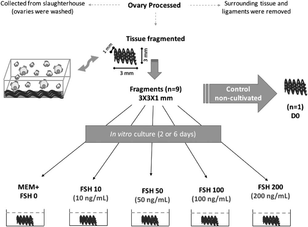
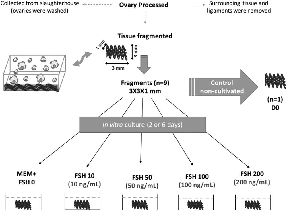
The pair of ovaries from each animal was carefully processed; surrounding tissue and ligaments were removed, and then the ovary was sectioned longitudinally. The medulla, large antral follicles and corpora lutea were removed. Subsequently, the cortex of the ovary was divided into fragments of approximately 3 × 3 × 1 mm. (Fig. 1) For each animal, a fragment was randomly selected and immediately fixed in Bouin’s solution (treatment control non-cultivated, D0). The remaining fragments of the ovarian cortex were cultured individually in 1 ml aliquots of culture medium in 24-well culture plates in an incubator at 38.5°C in an atmosphere of 5% CO2 in air and saturated humidity. The control culture comprised MEM, Gibco BRL, Rockville, MD, USA; osmolarity 300 mOsm/l, pH 7.2) supplemented (MEM+) with insulin–transferrin–selenium (ITS) (insulin 6.25 mg/ml, transferrin 6.25 mg/ml and selenium 6.25 ng/ml), 0.23 mM pyruvate, 2 mM glutamine, 2 mM hypoxanthine, 1.25 mg/ml bovine serum albumin (BSA Gibco BRL, Rockville, MD, USA), 20 UI/ml penicillin and 200 mg/ml streptomycin. For treatments, the MEM+ was supplemented with porcine pituitary FSH (Folltropin®, Bioniche Canada Inc., Ontario, Canada) at concentrations of 10, 50, 100 or 200 ng/ml (FSH 10, FSH 50, FSH 100, FSH 200). Ten fragments of the ovarian cortex of each animal were cultured in medium and tested at 2 (D2) or 6 (D6) days. At each interval of 2 days, the culture medium was replaced by fresh medium. The cultivation period used was based on published studies on the activation time course of primordial follicles in other species (Braw-Tal & Yossefi, Reference Braw-Tal and Yossefi1997; Fortune et al., Reference Fortune, Kito, Wandji and Srsen1998).
Experimental protocol used for in vitro culture of bovine preantral follicles in control medium (MEM+) or medium containing follicle-stimulating hormone (FSH).
Histological processing
For the analysis of ovarian morphology and ovarian follicles, control ovarian cortex fragments (non-cultivated) were cultured for 2 or 6 days and fixed by immersion in Bouin’s solution for 24 h. After fixation the fragments, the tissues were kept in 70% ethanol for 24 h and dehydrated in a graded series of solutions of increasing ethanol, subjected to clarified and cleared in xylene, and embedded in paraffin for preparation of blocks for histological analysis. Subsequently, each block was sectioned to a thickness of 5 µm with an interval of five sections of tissue with a rotary microtome (Leica®, Wetzlar, Germany) for mounting microscope slides (Andrade et al., Reference Andrade, van den Hurk, Lisboa, Hertel, Melo-Sterza, Moreno, APFRL, Landim-Alvarenga, Seneda and Alfieri2012; Max et al. , Reference Max, Silva, González, Lindquist, Búfalo, Gomes, Morotti, Costa, Barreiros, Lisboa and Seneda2017). The slides were stained with periodic acid–Schiff (PAS) and haematoxylin (Gomes et al., Reference Gomes, Silva, González, Oliveira, Max, Lisboa, Barreiros, Santos, Sarapião, Gastal and Seneda2018) stains. For PAS staining, the tissue slides were first deparaffinized with xylene and then rehydrated using 70% ethanol prior to staining with periodic acid for 5 min and the Schiff reaction for 10 min.
Morphologic analysis and assessment of in vitro follicular growth
All sections were examined using optical microscopy ( × 10 and × 40; Nikon, Tokyo, Japan). Follicles were ranked according to the following detailed specifications (Hulshof et al., Reference Hulshof, Figueiredo, Bekers, Bevers and van den Hurk1994). Preantral follicles were classified as: (1) primordial (one layer containing somatic cells, known as granulosa cells, flat or flattened around the oocyte) or in development; (2) primary (a single layer of cuboidal granulosa cells around the oocyte); or (3) secondary (two or more layers of granulosa cells cubic). To evaluate the activation and follicle growth, the follicles were quantified at different stages of development (primordial, primary and secondary) in control and after in vitro cultivation in different treatments.
Follicles also were classified according to morphology integrity according to the structure, in intact or degenerated. The follicle present was called morphologically intact when it had a regular nucleus and was surrounded by granulosa cells arranged in discrete layers; the follicle was called degenerated when it was huddled with a pyknotic nucleus and surrounded by granulosa cells isolated from disorganized basement membrane (Búfalo et al., Reference Búfalo, González, Silva, Lindquist, Bergamo, Costa, Marinho and Seneda2016). Then, approximately 250 follicles per treatment were studied for the duration of cultivation. To prevent recounts, preantral follicles were counted only in the section in which the nucleus of the oocyte was observed. To evaluate the growth rates and follicular activation, only intact follicles were considered, and the percentages of primordial, primary and secondary follicles were calculated after 0 (control), 2 or 6 days of culture at varying concentrations of the culture medium.
Morphometry analysis of preantral follicles cultured in vitro
For the analysis of follicles and cultured oocyte diameters, five morphologically intact preantral follicles were observed per treatment (MEM+, FSH 10, FSH 50, FSH 100, FSH 200) among the five replicates, and images were captured. In total, 250 preantral follicles were evaluated in the cultured treatments at 2 days (n = 125) and 6 days (n = 125). The measurements of follicles and oocytes were performed according to Silva-Buttkus et al. (Reference Silva-Buttkus, Jayasooriya, Mora, Mobberley, Ryder, Baithun, Stark, Franks and Hardy2008), and the oocyte and follicular diameters were calculated from the arithmetic mean of two perpendicular measurements using the Motic Plus 2.0 software program.
Statistical analysis
The data were initially submitted to normality tests (Shapiro–Wilk) and homogeneity of variance (Bartlett). The mean number of morphologically intact preantral follicles, primordial follicles and developing follicles obtained in the control treatments and different concentrations of FSH cultured for 2 or 6 days were subjected to analysis of variance (ANOVA) and Tukey’s test (P≤0.05). All analyses were performed with the Action 3.1 software, version of R 3.0.2 (Campinas, SP, Brazil), and the values were considered statistically significant when P≤0.05.
Results
Morphologic analysis and assessment of in vitro follicular growth
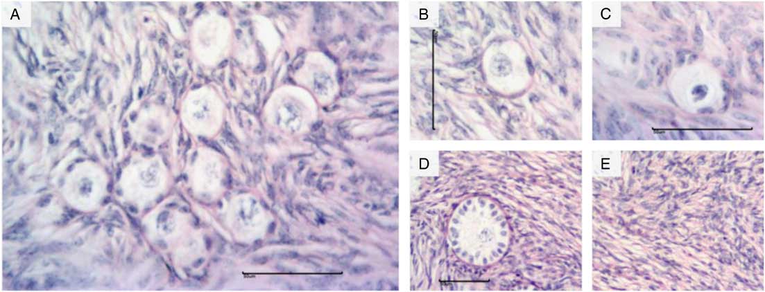
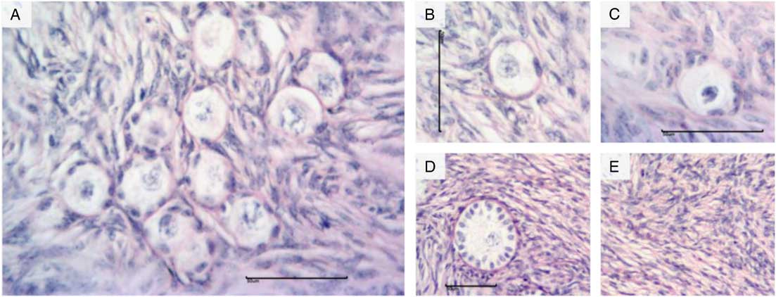
In total, 2250 preantral follicles were evaluated in the experiment, with 250 follicles assessed per treatment (Fig. 2 A). Considering the stage of development, 772 were classified as primordial follicles and 1478 as developing follicles (Fig. 2 B, D). For the histological analysis, the integrity and degeneration of follicles were observed during the in vitro culture of ovarian cortex (Fig. 2 B, C). Normal stromal tissue was observed between follicles (Fig. 2 E).
Histological illustration of bovine preantral follicle morphology. (A) Preantral follicles. (B) Intact primordial follicle. (C) Degenerated primordial follicle. (D) Intact development follicle. (E) Normal stroma. The fragments were stained using periodic acid–Schiff (PAS) and haematoxylin stains. Scale bar represents 50 μm.
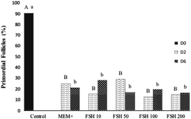
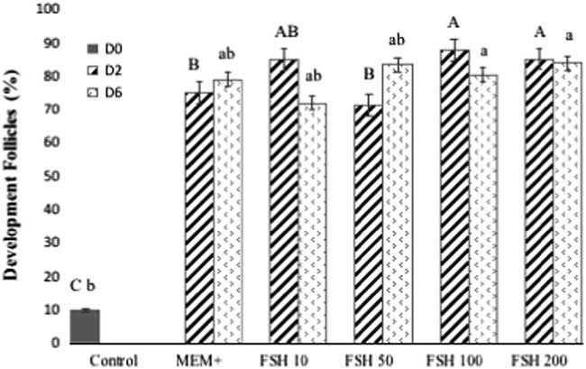
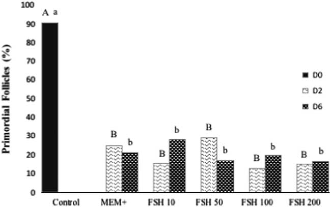
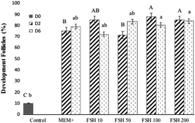
The non-cultivated control (D0) contained largely intact primordial follicles (90.4%; Fig. 3) and a small portion of developing follicles (9.6%; Fig. 4). The distribution of intact primordial follicles and intact developing follicles present in the in vitro cortical tissue at 2 or 6 days cultivation in the presence or absence of FSH is shown in Fig. 3 and Fig. 4, respectively.
Percentage of intact follicles from female Bos indicus at early stage (primordial) and non-cultured ovarian tissue (control, day 0) and after in vitro culture for 2 or 6 days in MEM+ or MEM+ supplemented with different concentrations of FSH (10, 50, 100 or 200 ng/ml). A,BValues within a treatment (culture day – D2) with different superscripts differ significantly at P<0.05 (between treatments – Control, MEM+, FSH 10, FSH 50, FSH 100 and FSH 200). a,bValues within a treatment (culture day – D6) with different superscripts differ significantly at P<0.05 (between treatments – Control, MEM+, FSH 10, FSH 50, FSH 100 and FSH 200).
Percentage of intact follicles from female Bos indicus at development (primary and secondary) in non-cultured ovarian tissue (control, day 0) and after in vitro culture for 2 or 6 days in MEM+ or MEM+ supplemented with different concentrations of FSH (10, 50, 100 or 200 ng/ml). A,BValues within a treatment (culture day – D2) with different superscripts differ significantly at P<0.05 (between treatments – Control, MEM+, FSH 10, FSH 50, FSH 100 and FSH 200). a,bValues within a treatment (culture day – D6) with different superscripts differ significantly at P<0.05 (between treatments – Control, MEM+, FSH 10, FSH 50, FSH 100 and FSH 200).
In general, after the start of cultivation, the proportion of primordial follicles was reduced between the different treatments. Therefore, there was an increased percentage of developing follicles. In all treatments for 2 or 6 days, the mean percentage of primordial follicles decreased compared with the non-cultured control, however there was no significant difference between the groups (P>0.05; Fig. 3).
Regarding the development of primordial follicles, the ovarian tissue supplemented with 10, 100 or 200 ng/ml FSH for 2 days efficiently led to the development of preantral follicles. However, regarding initiation, the concentrations of 100 and 200 ng/ml FSH were better than 10 ng/ml and MEM+ (P<0.05; Fig. 4). The percentage of follicles in developing D6 did not obtain significant differences between treatments (MEM+, 10, 50, 100 or 200 ng/ml FSH; P<0.05; Fig. 4).
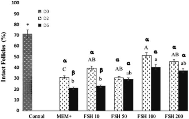
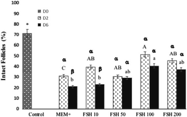
Survival rates and follicular integrity in the two-dimensional culture are shown in Fig. 5. The percentages of intact follicles were significantly higher in the 100 ng/ml FSH group cultured for 2 or 6 days than those in the other treatments (P≤0.05).
Percentage of intact follicles from Bos indicus females in early development stage (primordial, primary + secondary) in non-cultured ovarian tissue (control, day 0) and after in vitro culture for 2 or 6 days in MEM+ or MEM+ supplemented with different FSH concentrations (10, 50, 100 or 200 ng/ml). A,BValues within a treatment (culture day – D2) with different superscripts differ significantly at P<0.05 (between treatments – Control, MEM,+ FSH 10, FSH 50, FSH 100 and FSH 200). a,bValues within a treatment (culture day – D6) with different superscripts differ significantly at P<0.05 (between treatments – Control, MEM+, FSH 10, FSH 50, FSH 100 and FSH 200). α,βValues within the same treatment (D2 vs. D6) with different superscripts differ significantly at P<0.05 (treatments – Control, MEM+, FSH 10, FSH 50, FSH 100 and FSH 200).
Morphological changes identified in the atretic preantral follicles were mainly characterized as retracted oocyte, pyknotic nucleus disorganized granulosa cells and low cell density.
Morphometry analysis of preantral follicles cultured in vitro
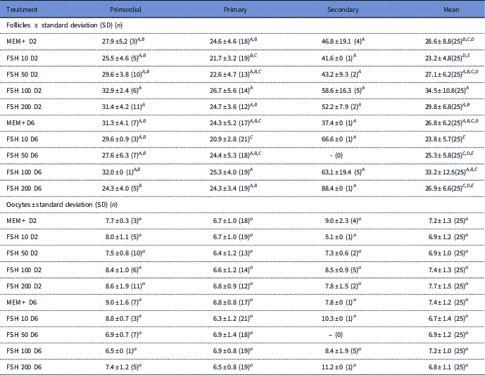
For the morphometric analysis, the follicle and oocyte dimensions were measured in ovarian tissue cultured for 2 or 6 days. The mean follicular and oocyte diameters at days 2 and 6 of culture can be found in Table 1.
Mean (± standard deviation, SD) of follicle/oocyte diameter in micrometres (μm) of bovine preantral follicles cultured for 2 and 6 days with MEM + or MEM + supplemented with different concentrations of FSH (10, 50, 100 and 200 ng/ml)

A,B Values within a column with different superscripts differ significantly at P<0.05 (between follicular diameter and treatments – MEM+, FSH 10, FSH 50, FSH 100 and FSH 200).
a,b Values within a column with different superscripts differ significantly at P<0.05 (between oocyte diameter and treatments – MEM+, FSH 10, FSH 50, FSH 100 and FSH 200).
After culture, the mean diameter of the preantral follicles (primordial, primary and secondary) was higher at the concentration of 100 ng/ml FSH than that with the other treatments at 2 days and 6 days of culture (34.5 ± 10.8 μm and 33.2±12.5, respectively; P<0.05). The mean follicular diameter of primordial follicles was higher at concentrations of 100 and 200 ng/ml at 2 days of culture (P<0.05), whereas at 6 days all treatments had similar alterations in morphology (P>0.05).
The mean follicle diameters showed significant differences, as the concentration of 100 ng/ml FSH at 2 and 6 days led to mean diameters of 26.7 ± 5.6 and 25.3±4.0 μm for primary follicles, respectively (P<0.05). The mean diameters for secondary follicles did not show differences between measurements at 2 or 6 days of culture (P>0.05). Regarding the diameter of the oocytes present in the follicles, at all stages of development, no significant difference was observed (P>0.05; Table 1).
Discussion
This is the first study to evaluate the effect of different concentrations of FSH on the survival and development of bovine preantral follicles enclosed in ovarian tissue cultured in vitro in situ. We identified positive effects of the addition of FSH on survival and growth of follicles in the initial stage of development.
The addition of 100 ng/ml FSH exhibited better conditions for follicular growth and development and maintained the viability and integrity of preantral follicles in cultivation. The effect of adding FSH observed in this study showed that the follicles present in ovarian tissue (in situ) of cattle for 2 and 6 days cultivation responded to FSH in a dose-dependent manner. Similarly, 100 ng/ml FSH also efficiently maintained the survival of follicle cultures isolated from goats and cross-bred cows (6 days: Barros et al., Reference Barros, Cavalcante, Macedo, Barberino, Lins, Gouveia, Menezes, Queiroz, Araújo, Palheta, Leite and Matos2013; 12 days: Rossetto et al., Reference Rossetto, Saraiva, Bernuci, Silva, Brito, Alves, Magalhães-Padilha, Báo, Campello, Rodrigues and Figueiredo2016; respectively). However, other studies have described a lower concentration requirement (1 μm/ml and 50 ng/ml) of FSH for supplementation of the culture medium (isolated follicles: bovine – Gutierrez et al., Reference Gutierrez, Ralph, Telfer, Wilmut and Webb2000 and in situ follicles: goats – Matos et al., Reference Matos, Lima-Verde, Luque, Maia, Silva, Celestino, Martins, Báo, Lucci and Figueiredo2007; Magalhães et al., Reference Magalhães, Fernandes, Araújo, Almeida, Matos and Figueiredo2009; sheep – Lima et al., Reference Lima, Rocha, Alves, Saraiva, Araújo, Lima, Lopes, Báo, Campello, Rodrigues and Figueiredo2013; equine – Aguiar et al., Reference Aguiar, Lunardi, Lima, Rocha, Bruno, Magalhães-Padilha, Cibin, Nunes-Pinheiro, Gastal, Rodrigues, Apgar, Gastal and Figueiredo2016). Those differences between the FSH concentration and also culture time for follicular development may be related to the differences between the species and also the methods for culture of follicles (isolated or in situ).
At 6 days of culture, the development of cultured follicles seemed to be non-dose dependent (10, 50, 100 and 200 ng/ml FSH). We believe that a small amount of FSH was sufficient to allow the development of the preantral follicles. Our results demonstrated that FSH is a possible substance needed in the base medium of bovine preantral follicle cultures, as it maintains cellular organization and it is responsive to the stimulatory effects of this gonadotrophin. This corroborates results described by Roy & Albee (Reference Roy and Albee2000), in which the presence of FSH is essential for the differentiation of somatic cells of granulosa cells during early development of primordial follicles.
The morphometric responses at 2 days of culture showed that the mean follicle growth was higher with the 100 ng/ml FSH treatment than those of the other treatments. Different results were found for follicular growth in other species, including goats (Matos et al., Reference Matos, Lima-Verde, Luque, Maia, Silva, Celestino, Martins, Báo, Lucci and Figueiredo2007), dog (Serafim et al., Reference Serafim, Duarte, Silva, Souza, Magalhães-Padilha, Moura, Silva, Campello and Figueiredo2015) and equine (Aguiar et al., Reference Aguiar, Lunardi, Lima, Rocha, Bruno, Magalhães-Padilha, Cibin, Nunes-Pinheiro, Gastal, Rodrigues, Apgar, Gastal and Figueiredo2016), in which treatment with 50 ng/ml FSH allowed a larger follicle diameter. However, at 6 days of culture, there was no significant difference in follicular diameter for any concentration of FSH.
FSH is essential for normal development of the gonads, control of steroidogenesis (Souza et al., Reference Souza, Salles, Camargo, Gulart, Costa e Silva, Lima, FAG and Silva2017) and to complete the folliculogenesis. The importance of FSH for the growth of antral follicles in vivo is well known. Conversely, the action of FSH in preantral follicles (primordial, primary and secondary) remains to be better understood (Adriaens et al., Reference Adriaens, Cortvrindt and Smitz2004). In vitro studies have identified FSH as responsible for stimulation of oocyte growth and the transition from primary to secondary stage of follicles (Hulshof et al., Reference Hulshof, Figueiredo, Beckers, Bevers, Donk and Van Den Hurk1995). In addition, FSH also contributes to formation of the antrum, estradiol production, follicle survival and meiotic resumption (Silva et al., Reference Silva, van den Hurk and Figueiredo2016).
Moreover, the FSH concentration curve provided evidence for the role of this gonadotropin in the proliferation and differentiation of granulosa cells in primordial follicles as described by Thomas et al. (Reference Thomas, Ethier, Shimasaki and Vanderhyden2005) and Duarte et al. (Reference Duarte, Araújo, Chaves, Silva, Luz, Haag, Magalhães-Padilha, Almeida, Lobo, Campello and Figueiredo2013). Other studies have also shown the importance of FSH in the early stages of follicular development (primordial, primary and secondary), with or without growth factors (Derrar et al., Reference Derrar, Price and Sirard2000; Cushman et al., Reference Cushman, Wahl and Fortune2002; Vasconcelos et al., Reference Vasconcelos, Saraiva, Costa, Passos, Silva, Rossi, Portela, Duarte, Magalhães-Padilha, Campelo, Figueiredo, van den Hurk and Silva2013).
In this study, follicles were cultured in the ovarian tissue from cattle, which favours the interaction of follicular cells with the adjacent cells. The information about in situ cultivation in bovine ovary is scarce. However, research has shown the cultivation of isolated preantral follicles with FSH supplementation in various species (humans: Wright et al., Reference Wright, Hovatta, Margara, Trew, Winston, Franks and Hardy1999; goats: Matos et al., Reference Matos, Lima-Verde, Luque, Maia, Silva, Celestino, Martins, Báo, Lucci and Figueiredo2007; sheep: Barros et al., Reference Barros, Cavalcante, Macedo, Barberino, Lins, Gouveia, Menezes, Queiroz, Araújo, Palheta, Leite and Matos2013; swine: Wu et al., Reference Wu, Xu and Wang2007; canine: Serafim et al., Reference Serafim, Duarte, Silva, Souza, Magalhães-Padilha, Moura, Silva, Campello and Figueiredo2015 and bovine: Rossetto et al., Reference Rossetto, Saraiva, Bernuci, Silva, Brito, Alves, Magalhães-Padilha, Báo, Campello, Rodrigues and Figueiredo2016).
Considering these perspectives, our study demonstrated advantages for including FSH in the in vitro culture of bovine follicles. The interaction of FSH with granulosa cells was observed by Saraiva et al. (Reference Saraiva, Rossetto, Brito, Celestino, Silva, Faustino, Almeida, Bruno, Magalhães, Matos, Campello and Figueiredo2010) and Rossetto et al. (2016). In addition, this hormone exerts influence during steroidogenesis, which in turn provides the interaction of granulosa cells with theca cells (Wandji et al., Reference Wandji, Eppig and Fortune1996; Thomas et al., Reference Thomas, Armstrong and Telfer2003). In addition to the direct participation of FSH on granulosa cells, the role of FSH in other steps of folliculogenesis has also been studied (Hsueh et al., Reference Hsueh, Kawamura, Cheng and Fauser2015).
FSH receptor expression was also performed for the transition from primary to secondary follicles and subsequently follicle (Oktay et al., Reference Oktay, Briggs and Gosden1997). In the same context, the presence of FSH receptors was described in primordial follicles (Médure et al., Reference Médure, Charnaux, Driancourt, Combettes, Granet, Vannier, Lossfelt and Milgrom2002). More recently, it has been shown that FSH may influence the in vitro differentiation and proliferation of granulosa cells (Vasconcelos et al., Reference Vasconcelos, Saraiva, Costa, Passos, Silva, Rossi, Portela, Duarte, Magalhães-Padilha, Campelo, Figueiredo, van den Hurk and Silva2013), which corroborates the results found in the present study.
Therefore, it is possible to note that for 2 days of growth, a high concentration of FSH was required (100 ng/ml) to initiate follicular activation, which is measured by the differentiation capacity of granulosa cells. This finding may suggest that in early follicular development, FSH acts at high concentrations. For 6 days of culture, lower concentrations were also effective (10, 50, 100 or 200 ng/ml), demonstrating that demand cannot exceed gonadotropin dose dependence in more advanced stages of follicular development.
The exact mechanism by which FSH acts on follicular cells is still unknown, considering viable stimulation of cell growth by indirect influence on local interactions of mesenchymal cells present in the ovarian epithelium (Nilsson & Skinner, Reference Nilsson and Skinner2001). In this context, our in situ culture model provides more interesting conditions than the isolated growing follicles, in which no such interaction exists between the follicular cells and ovarian stroma.
Despite consistent advances in the beneficial effects of FSH, it is important to consider the action of the other constituents in the medium that can exert synergistic action with this gonadotropin. Several substrates are used for the supplementation of culture medium, such as pyruvate, hypoxanthine, glutamine, ITS, and BSA. The medium used for the in vitro culture are rich in oxygen, nutrients and other elements present in the ovarian cortex (Rossetto et al., Reference Rossetto, Santos, Silva, Duarte, Silva, Campello and Figueiredo2013), and all of these components need to be considered for a correct understanding of follicular physiology.
In conclusion, the addition of FSH demonstrated better conditions for follicular growth and development and for maintaining the viability and integrity of preantral follicles in cultivation for a short period. The optimal concentration of FSH for growing preantral follicles of bovine was 100 ng/ml, which allowed for the development and maintenance of integrity during ovarian tissue in in vitro culture for a short period.