Introduction
Cervical metastasis from an unknown primary site accounts for less than 5 per cent of head and neck cancers annually.Reference Strojan, Ferlito, Medina, Woolgar, Rinaldo and Robbins1 The majority of primary tumours are identified within the palatine tonsils (44.7 per cent) and tongue base (43.9 per cent).Reference Strojan, Ferlito, Medina, Woolgar, Rinaldo and Robbins1 Small tongue base tumours can be easily overlooked on imaging and endoscopy.Reference Cianchetti, Mancuso, Amdur, Werning, Kirwan and Morris2 Furthermore, tissue diagnosis ranges from 53 per cent, when biopsy is targeted, to 9 per cent when ‘blind’.Reference Pattani, Goodier, Lilien, Kupferman, Caldito and Nathan3 If diagnostic modalities are unsuccessful, treatment comprises primary (chemo)radiation therapy to all mucosal subsites, or neck dissection followed by radiation.Reference Cianchetti, Mancuso, Amdur, Werning, Kirwan and Morris2
The five-year survival rates remain disheartening, at 69, 58 and 30 per cent for nodal N1, N2 and N3 disease, respectively.Reference Cianchetti, Mancuso, Amdur, Werning, Kirwan and Morris2 Contrariwise, identification of the primary tumour can increase survival rates to 100 per cent and obviate the need for pan-mucosal irradiation.Reference Cianchetti, Mancuso, Amdur, Werning, Kirwan and Morris2, Reference Pattani, Goodier, Lilien, Kupferman, Caldito and Nathan3 Intensity-modulated radiotherapy can significantly ameliorate associated side effects, including xerostomia, dysphagia and dysphonia.Reference Bhide, Ahmed, Newbold, Harrington and Nutting4–Reference Haas, Hoffmann, Engers and Ganzer6
Tongue base mucosectomy via transoral robotic surgery has purported mucosal primary tumour detection rates of 90 per cent, with minimal additional morbidity.Reference Mehta, Johnson, Tassler, Kim, Ferris and Nance7 Though recommended in UK national guidelines, the technique is currently restricted to centres with such facilities.Reference Mackenzie, Watson, Jankowska, Bhide and Simo8 This paper aims to report the author's experience in the translation of transoral robotic surgery techniques to resect mucosectomy specimens using electrocautery, in the context of wider implementation of this technique.
Materials and methods
Ethical considerations
The author is trained in transoral robotic surgery, and the use of electrocautery represents a modification of an established technique. Patients were operated upon according to national guidelines, and within the outcome framework of the head and neck oncology multidisciplinary team. Operations were conducted in the regional tertiary head and neck centres of both The Royal Sussex County Hospital, Brighton, and The Queen Victoria Hospital, East Grinstead.
Subjects
Subjects represented consecutive patients prospectively recruited between May 2017 and June 2018 through Brighton and Sussex University Hospitals multidisciplinary meeting. All patients had biopsy-proven metastatic cervical squamous cell carcinoma, and were assessed by a consultant surgeon with flexible laryngoscopy to refute primary disease of the upper aerodigestive tract.
Magnetic resonance imaging of the neck, with concurrent positron emission tomography/computed tomography (PET/CT), was performed in all patients; only those patients with no identified primary malignancy or pathological fludeoxyglucose avidity, respectively, were included. Imaging was reviewed by a dedicated head and neck radiologist.
Further inclusion criteria stipulated prior examination under anaesthetic, tonsillectomy and ‘blind’ tongue base biopsies without consequent tumour identification, prior to mucosectomy. Patients were excluded if a primary tumour was identified.
All patients underwent ultrasound-guided core biopsies whereby p16 status was elucidated. Patients were scheduled for mucosectomy irrespective of access considerations, so as not to introduce selection bias.
Technique
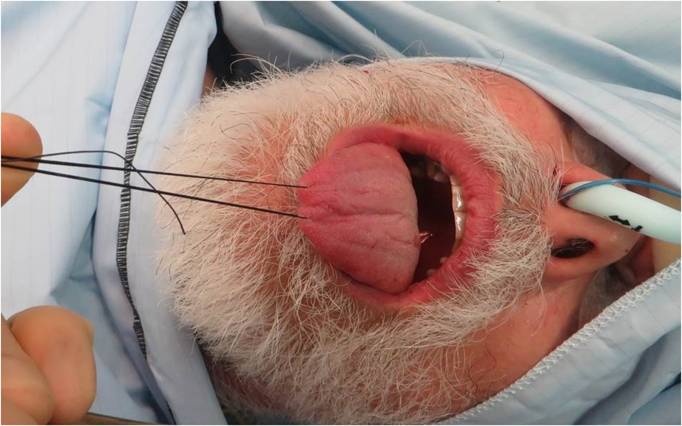
The procedure is performed under general anaesthetic. The patient is placed supine and with their neck extended. A size 2-0 silk suture is double-passed through the tongue, and traction is applied anteriorly (Figure 1).
Double-passed, size 2-0, silk suture, with anterior traction of the tongue before placement of the Boyle–Davis gag and tongue blade; a damp tonsil swab is inserted between the ventral tongue and the lower incisors to avoid traumatising the mucosa. The patient is intubated with a nasal endotracheal tube.

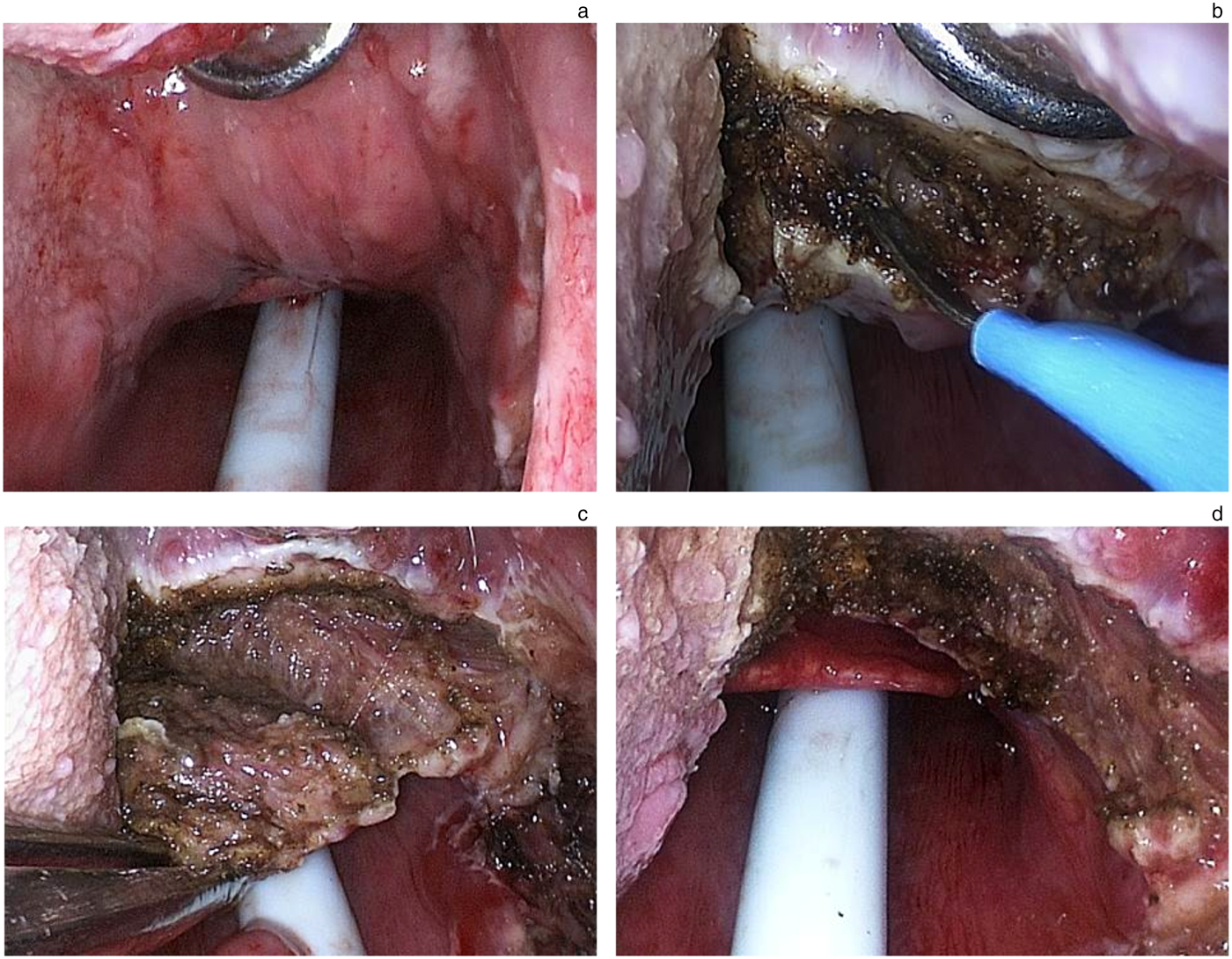
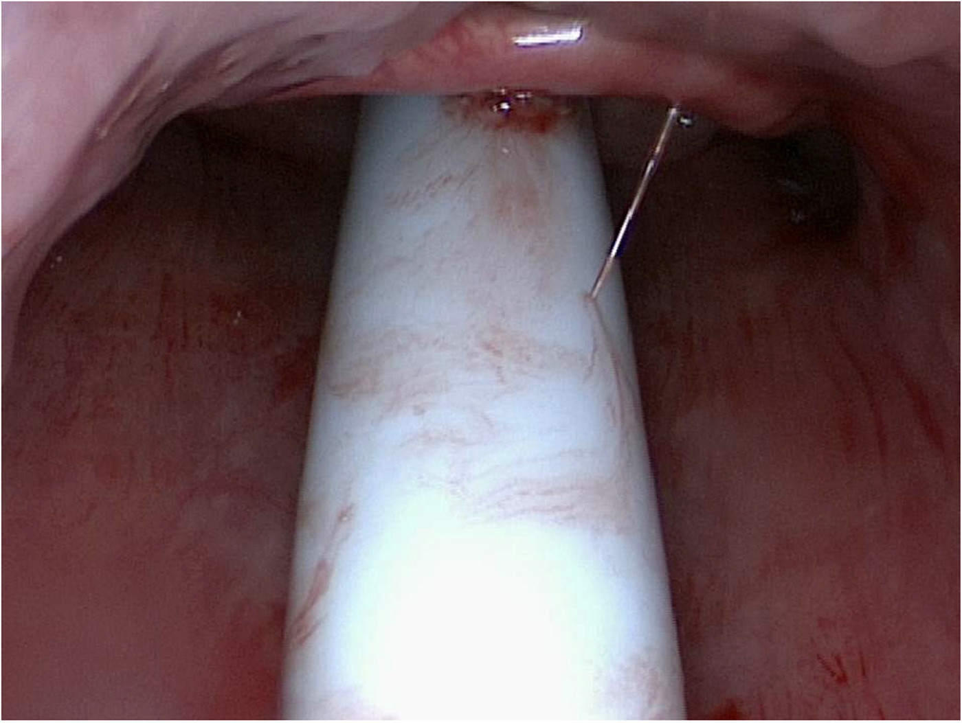
A Boyle–Davis gag is inserted and stabilised with Draffin rods. A 0-degree or 30-degree endoscope is inserted to inspect the tongue base, epiglottis and vallecula (Figure 2a). With an assistant holding the endoscope within the oropharynx, the midline of the tongue is incised with monopolar cautery using a 100 mm extension tip with smoke evacuation. The incision proceeds to the intrinsic musculature. Dissection extends laterally to the glossopharyngeal sulcus along the demarcation of the circumvallate papillae (Figure 2b). Simultaneous traction of the tongue anteriorly and tongue base posteriorly facilitates dissection to the vallecula, using the intrinsic musculature as a pseudo-plane (Figure 2c). The length of the Boyle–Davis blade can be changed to encourage anterior pressure in the dissection bed, conveniently delivering the tongue base into the field. Visualisation of the epiglottis confirms the line of dissection (Figure 3).
(a) Right tongue base exposure after suspension utilising a Boyle–Davis retractor. Previous palatine tonsillectomy is evident (right). (b) Initial vertical midline and subsequent lateral cut demonstrated using monopolar electrocautery. (c) Allis forceps used to provide counter-traction during right tongue base mucosectomy (akin to Maryland retractor during transoral robotic surgery); intrinsic tongue musculature can be visualised as a deep limit of dissection. (d) Completion of right tongue base mucosectomy, with epiglottis and vallecula mucosa clearly visible.

Views of the epiglottis using a 0-degree, rigid endoscope to establish a projected line of dissection.

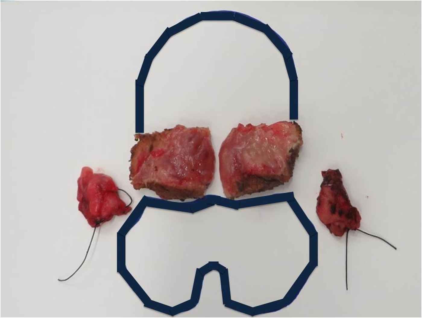
The lateral half of the tongue base is excised once the pharyngoepiglottic folds are visualised (Figure 2d). The tongue and gag are repositioned, and the contralateral tongue base mucosa is resected in the same fashion. Specimens are orientated for histology (Figure 4). Haemostasis is achieved with bipolar cautery, and is confirmed following: the release of tongue traction, the removal of the Boyle–Davis gag and endoscopic re-assessment. Fibrin sealant can be discretionally applied.
Example of orientation method for tonsillectomy (seen laterally) and mucosectomy specimens following procedure completion (if resected concurrently). Tongue base specimen dimensions (width × length × depth): 37 × 35 × 11 mm (left); 32 × 28 × 10 mm (right).

Results
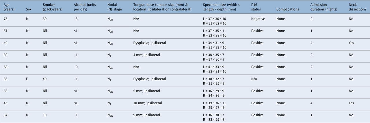
Over 13 months, 9 patients fulfilled criteria for inclusion (Table 1). The majority were male, with a mean age of 60.2 years. Two patients smoked heavily, correlating with p16 status (i.e. negative).
Patient demographics, nodal stage, final pathology*, complications and admission duration

*Determined after transoral electrocautery tongue base mucosectomy. M = male; N/A = not applicable; L = left; R = right; F = female
A mucosal primary was identified in four patients (44.4 per cent). Intensity-modulated radiotherapy was advocated in three of these patients and completion ipsilateral tongue base resection was undertaken in the fourth. All patients were pathologically staged as having tumour T1 disease and were p16 positive. The female patient was diagnosed with primary lung cancer and incidental dysplasia of the ipsilateral tongue base. Patients without tumour identification underwent concurrent pan-mucosal chemoradiation; exceptions included a 75-year-old male patient who underwent radiotherapy alone and the female patient for whom surveillance was recommended. Of the two patients who consented to undergo neck dissection, one was upstaged from recurrent N2a to pathologically confirmed N2b disease, without extracapsular spread. In this patient, mucosectomy revealed ipsilateral dysplasia; the patient received intensity-modulated radiotherapy to the ipsilateral neck and tongue base. The other patient who underwent neck dissection, after informed discussion, elected for completion tongue base resection. This revealed a 4 mm residual tumour of the ipsilateral tongue base, which was excised with clear margins (greater than 10 mm); therefore, no adjuvant radiotherapy was advocated.
No significant surgical complications were identified in the cohort. No procedures were abandoned because of access limitations. All patients experienced post-operative pain and transient dysphagia.
Normal diet was resumed within 48 hours in all patients. The mean duration of admission was 1.4 nights, excluding the two patients who underwent concurrent neck dissection, for whom their surgical drains restricted discharge. The mean specimen size was 36.4 × 33.0 × 9.1 mm for the left side and 32.3 × 30.7 × 9.2 mm for the right.
Discussion
Selected case series have demonstrated improvement in the detection rates of cervical metastasis from an unknown primary site with the use of transoral robotic surgery or transoral laser surgery.Reference Nagel, Hinni, Hayden and Lott9 These methods require specific training, and have inherent complications, and cost and resource implications. Cervical metastasis from an unknown primary site represents a rare scenario and, unsurprisingly, expert opinion remains divided regarding appropriate management.Reference Mehta, Johnson, Tassler, Kim, Ferris and Nance7–Reference Winter, Ofo, Meikle, Silva, Fraser and O'Hara11
We present a series of patients in whom mucosectomy was performed simply, cost-effectively and safely with electrocautery and tonsillectomy equipment. The skillset required represents a hybrid of tonsillectomy and transoral laser surgery. There is the advantage of both tactile feedback and direct access if dorsal lingual artery ligation is required. We believe that performing mucosectomy with electrocautery expands the potential breadth of use among surgeons and prevents prolonged diagnostic pathways.
In nine patients who underwent PET/CT, panendoscopy, palatine tonsillectomy and ‘blind’ tongue base biopsies, electrocautery mucosectomy was subsequently able to identify primary malignancy in four patients. Dysplasia was demonstrated in two further patients, which collectively provided information relevant to radiotherapy fields and post-treatment surveillance.
Electrocautery mucosectomy did not identify primary malignancy with the same frequency as transoral robotic surgery (i.e. 44.4 per cent vs 53–90 per cent).Reference Mehta, Johnson, Tassler, Kim, Ferris and Nance7, Reference Winter, Ofo, Meikle, Silva, Fraser and O'Hara11 Firstly, the three-dimensional, high-definition views obtained from transoral robotic surgery are exceptional. Both Paleri's and Weinstein's groups have reported the benefit this approach confers.Reference Winter, Ofo, Meikle, Silva, Fraser and O'Hara11, Reference Hatten, O'Malley, Bur, Patel, Rassekh and Newman12 The images obtained from rigid endoscopy are comparatively inferior, though the microscope may also be implemented, which could improve detection rates further.Reference Nagel, Hinni, Hayden and Lott9, Reference Hatten, O'Malley, Bur, Patel, Rassekh and Newman12 Secondly, our patient group had entirely negative findings on imaging. Conversely, while Mehta et al. reported a 90 per cent detection rate, the patients included had PET/CT imaging findings indicative of lateralised disease: one particular tumour excised was 20 mm in maximal diameter.Reference Mehta, Johnson, Tassler, Kim, Ferris and Nance7 One would be inclined to hypothesise that this would be either palpable or visible on cross-sectional imaging, respectively. Inclusion of such data invariably skews the results towards a favourable correlation. Thirdly, the technique we describe is new and will have an inherent learning curve. Accordingly, only through continual use may we establish whether the technique remains valid in obtaining sufficient pathological specimens to allow primary tumour identification.
Maintaining a standardised approach ensured adequate size and consistency of the resected specimens (Table 1). Surgical borders should comprise the vallecula posteriorly, the circumvallate papillae anteriorly, the glossotonsillar sulcus laterally (as it becomes contiguous with lateral pharyngeal mucosa) and the intrinsic musculature of the tongue. Striated muscle cells were identified in the deep aspect of all specimens, confirming adequate deep extent of resection.
While it is fortuitous that none of the procedures in our series were aborted because of access limitations, it is readily appreciated that there will inexorably be patients with unfavourable anatomy, including prominent dentition, restricted neck extension or mouth opening, in whom the endoscopic approach may not be successful. Such patients should be considered for referral to a centre with robotic facilities, as stipulated within UK guidelines.Reference Mackenzie, Watson, Jankowska, Bhide and Simo8 It is prudent, however, to simultaneously conceptualise this recommendation: multidisciplinary teams may not instinctively explore this referral pathway in the context of a diagnostic procedure, particularly if treatment is to be commenced expeditiously and chemoradiation remains institutionally habitual. Therefore, if intra-oral access to the tongue base is indeed possible with conventional equipment (which can be ascertained at the time of initial rigid pharyngoscopy), we recommend that surgeons endeavour to do so, when considering the possible sequelae for the patient. If a straightforwardly executed technique as mucosectomy can ameliorate the necessity for pan-mucosal irradiation and reduce the long-term morbidity this treatment confers, its role should not be understated.
Competing interests
None declared