Introduction
Members of the genus Paratylenchus Micoletzky, 1922 are ectoparasitic plant nematodes, widely distributed worldwide and associated with a large variety of plants (Raski, Reference Raski and Nickle1991; Siddiqi, Reference Siddiqi2000; Van den Berg et al., Reference Van den Berg, Tiedt and Subbotin2014). The nematodes of this genus always feed on epidermal cells of roots or the root cortical tissues and may hinder the growth of their hosts (Siddiqi, Reference Siddiqi2000). For example, Paratylenchus neoamblycephalus damages roots of myrobalan plum and causes poor growth of this plant (Braun & Lownsbery, Reference Braun and Lownsbery1975). To date, more than 120 species of Paratylenchus have been described and almost all species of this genus are extremely small in body size (0.2–0.6 mm) (Siddiqi, Reference Siddiqi2000; Van Den Berg et al., Reference Van den Berg, Tiedt and Subbotin2014). The morphological identification of this genus remains difficult due to the small number and overlapping of diagnostic characters (Geraert, Reference Geraert1965; Brzeski & Háněl, Reference Brzeski and Háněl2000). Presently, only a few characters can be used for species identification, such as stylet length, body length, vulva position, lateral lines, head shape and vulval lateral flaps, etc. Exploration of the usefulness of molecular data for species identification and phylogenetic reconstruction within the genus Paratylenchus is just beginning (Van den Berg et al., Reference Van den Berg, Tiedt and Subbotin2014). In China, approximately 29 known species of Paratylenchus have been reported (Liu, 2004; Chen et al., Reference Chen, Ni, Tsay and Yen2008; Wang et al., Reference Wang, Xie, Li, Xu, Yu and Wang2013). In July 2012, a Paratylenchus species was found from the rhizosphere of Pinus massoniana in Nanjing, a city in Jiangsu Province of China. Morphological and morphometric studies of the nematode revealed that it differed from all other species of this genus, and it is described here as Paratylenchus nanjingensis n. sp. The internal transcribed spacer ribosomal RNA (ITS rRNA) gene from P. nanjingensis n. sp. was amplified and sequenced and the phylogenetic relationships of the new species with other Paratylenchus species were studied based on the ITS rRNA gene sequences.
Materials and methods
Collection and examination of nematodes
Soil samples were collected from the rhizosphere of P. massoniana in the Sun Yat-sen Mausoleum, Nanjing, Jiangsu Province. Nematodes were extracted from soil by the modified Baermann funnel method (Xie, Reference Xie2005).
Nematodes were killed by heat, and fixed in FG solution (10:1:89, formalin:glycerin:water) (Xie, Reference Xie2005). The fixed nematodes were transferred to anhydrous glycerin as described by Seinhorst (Reference Seinhorst1959) and mounted on permanent slides. Measurements were made using a Nikon Eclipse 90i microscope with a specialized software (NIS-Elements microscope imaging software) (Nikon, Tokyo, Japan). Photographs were taken using a Zeiss Axio Scope A1 microscope equipped with an AxioCam MRm camera (Zeiss, Jena, Germany). Drawings were obtained using an Olympus CX40 microscope with a drawing tube (Olympus, Tokyo, Japan).
Molecular analysis
For molecular study, genomic DNA was extracted from one individual nematode using proteinase K according to Wang et al. (Reference Wang, Zhang and Gu2011). The ITS rRNA gene was amplified using one pair of primers: rDNA1 (5’-TTGATTACGTCCCTGCCCTTT-3’) and rDNA2 (5’-TTTCACTCGCCGTTACTAAGG-3’) (Vrain et al., Reference Vrain, Wakarchuk, Levesque and Hamilton1992). Polymerase chain reaction (PCR) amplifications were performed in 25 μl volumes with the components as follows: 1 × PCR buffer for KOD FX; 0.4 mm of deoxy-ribonucleoside triphosphates (dNTPs); 0.3 μm of each primer; and 0.5 units of KOD FX DNA polymerase (Toyobo, Osaka, Japan). The PCR cycling conditions were as follows: 94°C for 2 min, followed by 35 cycles (98°C for 10 s, 58.2°C for 30 s, 68°C for 90 s) and final extension at 72°C for 10 min. PCR products were purified using the Gel Extraction Kit (Omega, Norcross, Georgia, USA), and sequenced by Sangon Biotech Co. Ltd (Shanghai, PR China) after cloning into pJET 1.2/blunt cloning vectors (Thermo Scientific, Waltham, Connecticut, USA). The newly obtained sequences were submitted to the GenBank database (National Center for Biotechnology Information; NCBI).
The sequences of P. nanjingensis n. sp. were compared with sequences of other Paratylenchus species in the GenBank database using the blastn program implemented in NCBI (http://blast.ncbi.nlm.nih.gov/Blast.cgi_). The sequence (GU253921) of Sphaeronema alni was chosen for the outgroup taxon, according to Van den Berg et al. (Reference Van den Berg, Tiedt and Subbotin2014).The ITS rRNA gene sequences were aligned by ClustalW implemented in MEGA 5.05 (Tamura et al., Reference Tamura, Peterson, Peterson, Stecher, Nei and Kumar2011). Phylogenetic relationships of P. nanjingensis n. sp. with other Paratylenchus species were established by Bayesian inference (BI) using MrBayes3.1.1 (Huelsenbeck & Ronquist, Reference Huelsenbeck and Ronquist2001). The best-fit model (GTR+I+G) was selected by MrModeltest 2.3 based on Akaike Information Criterion (AIC) (Pereira et al., Reference Pereira, Santos, Schaefer, Souza, Paiva, Malabarba, Schmidt and Dergam2013). Bayesian analysis was initiated with a random starting tree. Four Markov chains (one cold chain and three heated chains) were run for 1,000,000 generations and sampled every 100 generations. After discarding 2500 samples, the remaining samples were retained to generate a 50% majority rule consensus tree. Posterior probabilities (PP) were given on appropriate clades.
Results
Description of Paratylenchus nanjingensis n. sp.
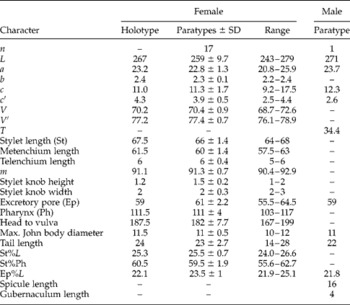
Measurements. See table 1.
Morphometics (μm) of Paratylenchus nanjingensis n. sp. from the rhizosphere of Pinus massoniana from Nanjing, Jiangsu Province, China; n=number of specimens observed, L=body length, a=L/max. width, b=L/pharyngeal length, c=L/tail length, c′=tail length/anal body width, V=distance of vulva from anterior end×100/L, V′=distance from anterior end to vulva×100/distance from anterior end to anus, T=distance between cloaca and anterior-most part of testis×100/L, m=metenchium length×100/stylet length.

Type material. Holotype female, 17 paratype females and 1 paratype male are deposited in the Laboratory of Plant Nematology, South China Agricultural University, Guangzhou, Guangdong Province, PR China.
Type habitat and locality. Rhizosphere soil of pine (P. massoniana) was from the Sun Yat-sen Mausoleum, Nanjing, Jiangsu Province, China. The material was collected in July 2012.
Morphological description
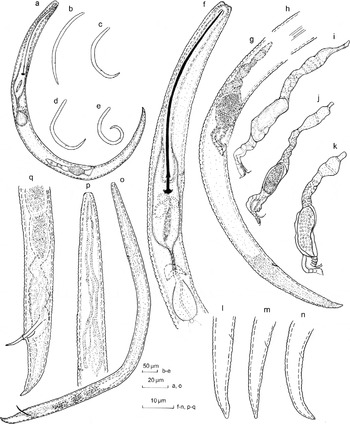
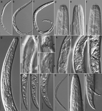
Female (fig. 1a–n, fig. 2a–p). Body slender, vermiform, posture ranging from an open C-shape to a figure of 6 when relaxed. Body annuli 1 ± 0.1 (0.8–1.1) μm wide at mid-body. Lateral field 1.8 ± 0.2 (1.4–2) μm wide, with four lines. Head not set off, submedian lobes protruding slightly upward; lateral lobes at the same level as submedian lobes or at a slightly lower level, forming a flattened anterior end or a small depression at the oral area in lateral view. Cephalic framework weak. Stylet slender, curved or straight with small, anchor-shaped knobs. Cone occupying 91.1 (89.3–92.9)% of stylet length. Median pharyngeal bulb elongate, valve 8 ± 0.8 (6.5–10) μm long. Isthmus slender, 11 ± 1.4 (8–15) μm long and 2 ± 0.2 (1.5–2.2) μm wide, surrounded by nerve ring; oesophago-intestinal valve small, rounded. Excretory pore situated anterior to the level of stylet knob. Hemizonid situated directly posterior to excretory pore, 2–3 annuli wide. Hemizonion situated 8–18 annuli posterior to hemizonid, one annulus wide. Lateral vulval flaps distinct, small, crenate or smooth. Spermatheca oblong to elongate, 11 ± 2.6 (6–16) μm long and 4.5 ± 0.8 (3–6) μm wide, with or without spermatozoa. Post-vulval uterine sac absent or depauperate. Anus indistinct. Tail slender and long, conoid, ending in a finely rounded or subacute terminus.
Morphology of Paratylenchus nanjingensis n. sp. Female: (a–e) entire body, (f) anterior end, (g) posterior end, (h) lateral lines, (i–k) reproductive system, (l–n) tail. Male: (o) entire body, (p) anterior end, (q) posterior end.

Light micrographs of Paratylenchus nanjingensis n. sp. Female: (a–c) entire body, (d–f) anterior end, (g) anterior part, (h) lateral lines, (i–j) lateral vulval flap (arrowed), (k–l) spermatheca, (m) post-uterine sac, (n–p) tail. Male: (q) entire body, (r) anterior end, (s) tail and spicule. Scale bars: 20 μm (a–c, q); 10 μm (g); 5 μm (d–f, h–p, r, s).

Male (fig. 1o–q, fig. 2q–s). Only one male was found. Body posture assuming open C-shape after heat relaxation. Body annuli more distinct than those of females, approximately 1.2 μm wide at mid-body. Lateral field with four lines. Head conical–truncate, anterior end flattened. Cephalic framework very weak. Stylet lacking. Pharynx degenerate. Testis outstretched, with small spermatozoa. Spicules distinct, slightly curved. Penile sheath short, with spicule protruding from the cloaca. Bursa not observed. Tail slightly curved ventrad, with distinct annuli, tapering gradually to a subacute tip.
Diagnosis and relationships
Paratylenchus nanjingensis n. sp. is characterized by having females with a slender, vermiform body 243–279 μm long, stylet 64–68 μm long, knobs anchor-shaped, lateral field with four lines, head not offset, with distinct submedian lobes, cephalic framework weak, excretory pore situated anterior to the level of the stylet knob, spermatheca oblong to elongate, more anterior vulva and small lateral vulval flaps; and by males with more distinct body annuli, stylet lacking and pharynx degenerate.
When using the diagnostic compendium proposed by Esser (Reference Esser1992) and using the stylet length, lateral lines as a guide, P. nanjingensis n. sp. is close to P. aonli Misra & Edward, 1971, P. capitatus (Adams & Eichenmuller, 1962) Siddiqi & Goodey, Reference Siddiqi and Goodey1964, P. crenatus Corbett, 1966, P. longilabiatus (Huang & Raski, Reference Huang and Raski1986) Brzeski, 1998, P. marylandicus Jenkins, Reference Jenkins1960, P. pandatus (Raski, Reference Raski1976) Siddiqi, 1986, P. robustus Wu, 1974, P. steineri Golden, Reference Golden1961, P. verus (Brzeski, Reference Brzeski1995) Brzeski, 1998, and P. vitecus (Pramodini, Mohilal & Dhanachand, Reference Pramodini, Mohilal and Dhanachand2006) Ghaderi, Kashi & Karegar, Reference Ghaderi, Kashi and Karegar2014. When following the key of Ghaderi et al. (Reference Ghaderi, Kashi and Karegar2014), the new species appears to be close to P. pandatus (Raski, Reference Raski1976) Siddiqi, 1986. However, the new species differs from P. aonli by having a more anterior vulva (V= 68.7–72.6 vs. 74–84), a shorter distance from excretory pore to anterior end (55.5–64.5 μm vs. 75–90 μm) in the female, and a shorter body length (L= 271 μm vs. 280–343 μm) in the male (Raski, Reference Raski1976, Reference Raski and Nickle1991; Van den Berg & Cadet, Reference Van den Berg and Cadet1991; Esser, Reference Esser1992). It differs from P. capitatus by the head being unexpanded and not offset vs. expanded and offset, having a shorter body length (L= 243–279 μm vs. 410–510 μm) in the female and stylet absent vs. present in the male (Huang & Raski, Reference Huang and Raski1986; Esser, Reference Esser1992); and from P. crenatus mainly by having a shorter body length (L= 243–279 μm vs. 300–400 μm), a more anterior vulva position (V= 68.7–72.6 vs. 76–81) and body annuli smooth vs. crenate in the female (Raski, Reference Raski and Nickle1991; Esser, Reference Esser1992; Kashi, et al., Reference Kashi, Karegar and Kheiri2009). It differs from P. longilabiatus mainly by the head being unexpanded and not offset vs. expanded and markedly offset and having a lower V value (68.7–72.6 vs. 76.4–80.0) in the female (Huang & Raski, Reference Huang and Raski1986); from P. marylandicus mainly by having a shorter body length (L= 243–279 μm vs. 340–420 μm) and a more anterior excretory pore position (anterior to the level of stylet knob vs. in the region of the nerve ring) in the female (Jenkins, Reference Jenkins1960); and from P. pandatus mainly by the female having a shorter body length (L= 243–279 μm vs. 330–420 μm), a lower b value (2.2–2.4 vs. 2.8–3.2), a more anterior excretory pore position (anterior to the level of the stylet knob vs. at the level of the isthmus) and by the male having a shorter spicule length (16 μm vs. 19–22 μm) (Raski, Reference Raski1976). It differs from P. robustus by having a shorter body length (L= 243–279 μm vs. 327–432 μm), a more anterior vulva (V= 68.7–72.6 vs. 81–85), the excretory pore being anterior to the level of the stylet knob vs. opposite the isthmus, and spermatheca present vs. absent in the female (Raski, Reference Raski1976, Reference Raski and Nickle1991; Esser, Reference Esser1992); and from P. verus mainly by having a more anterior vulva (V= 68.7–72.6 vs. 76–79), a shorter distance from the excretory pore to the anterior end (55.5–64.5 μm vs. 68–92 μm) in the female and a shorter body length (L= 271 μm vs. 284–340 μm) in the male (Brzeski, Reference Brzeski1995). The new species differs from P. steineri and P. vitecus mainly by having lateral vulval flaps present vs. absent in females (Golden, Reference Golden1961; Pramodini et al., Reference Pramodini, Mohilal and Dhanachand2006). It also differs from P. steineri by having a more anterior vulva (V= 68.7–72.6 vs. 76–79) in the female (Brzeski, Reference Brzeski1995) and also from P. vitecus by having a longer pharynx (b= 2.2–2.4 vs. 2.7–2.9) and different excretory pore position (anterior to the level of the stylet knob vs. at the base of median bulb or at the level of the isthmus) in the female (Pramodini et al., Reference Pramodini, Mohilal and Dhanachand2006).
Molecular characterization and phylogenetic relationships
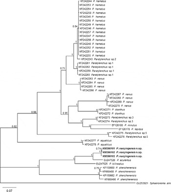
Three 1032-bp ITS rRNA gene sequences of P. nanjingensis n. sp. were obtained and submitted to the GenBank database (NCBI) under accession numbers KM366101–KM366103. Variation of the three ITS sequences was 0–2 bp. The results of BLAST search showed the sequences from the new species were closest to the sequence from P. aculentus (EU247526). The identity of the ITS sequences from P. nanjingensis n. sp. and P. aculentus was 92%. The phylogenetic relationships of P. nanjingensis n. sp. with other Paratylenchus species are shown in fig. 3. Relationships among species were well resolved. Three ITS sequences of P. nanjingensis n. sp. clustered together, and P. nanjingensis n. sp. is in a 100% supported clade with P. aculentus.
The 50% majority rule consensus tree inferred from Bayesian analysis using the ITS rRNA gene sequence dataset under the GTR+I+G model; posterior probabilities are given for appropriate clades and newly obtained sequences are shown in bold.

Discussion
Identification of Paratylenchus species is always an acute problem. There is only small number of useful diagnostic characters in the genus Paratylenchus and, morphologically, many of the described populations resemble each other closely (Geraert, Reference Geraert1965; Brzeski & Háněl, Reference Brzeski and Háněl2000). Raski (Reference Raski1962) proposed that the genus Gracilacus should include those Paratylenchus species having a stylet longer than 48 μm. Siddiqi & Goodey (Reference Siddiqi and Goodey1964) synonymized Gracilacus with Paratylenchus and this opinion was accepted by Brzeski (Reference Brzeski1963), Geraert (Reference Geraert1965) and Allen & Sher (Reference Allen and Sher1967), but was not agreed with by Thorne & Malek (Reference Thorne and Malek1968), Golden (Reference Golden, Zukerman, Mai and Rohde1971) and Andrássy (Reference Andrássy2007). Siddiqi (Reference Siddiqi2000) regarded Gracilacus as a subgenus of Paratylenchus. Geraert (Reference Geraert1965) divided this genus into ten groups, depending on the morphological characters such as stylet length, vulva position, lateral field, head shape and tail shape.
Van den Berg et al. (Reference Van den Berg, Tiedt and Subbotin2014) found that the ITS rRNA gene sequences were more useful in revealing the phylogenetic relationships within the Paratylenchus species than other rRNA gene sequences, and that four major clades distinguished in their phylogenetic trees generally corresponded to several groups given by Geraert (Reference Geraert1965). In this study, the Bayesian tree (fig. 3) showed that P. nanjingensis n. sp. and P. aculentus occupied the same clade with high posterior probability (100%). The two species shared the same range of stylet length and V value and fitted into Geraert's group VI (stylet = 54–85 μm; V= 68–79). However, in the ITS rRNA gene tree, P. shenzhenensis (stylet = 17–21 μm) was more closely related to the species with a stylet longer than 48 μm (P. aculentus, P. bilineatus and P. nanjingensis n. sp.) than to the species with a stylet less than 48 μm. When using molecular criteria, Van den Berg et al. (Reference Van den Berg, Tiedt and Subbotin2014) noticed that the morphospecies P. aquaticus and P. nanus might contain two sibling species. The existence of species complexes in this genus (Van den Berg et al., Reference Van den Berg, Tiedt and Subbotin2014) may make the identification of this genus more difficult. The use of molecular traits has been demonstrated to be helpful for diagnosis in many other genera (Hu & Gasser, Reference Hu and Gasser2006; Powers et al., Reference Powers, Harris, Higgins, Sutton and Powers2010; De Luca et al., Reference De Luca, Reyes, Troccoli and Castillo2011). However, the majority of Paratylenchus species have yet to be sequenced. As more molecular data become available, the use of molecular characterization may help to re-evaluate the classification of Paratylenchus.
Acknowledgement
The authors express their thanks to Rui Hao and Xin Huang for collecting soil samples.
Financial support
This work was supported by a Special Project of Scientific and Technological Basis of the Ministry of Science and Technology of the People's Republic of China to H.X. (grant no. 2006FY120100).
Conflict of interest
None.