Zhang et al. (Reference Zhang, Proenca, Maffei, Barone, Leopold and Friedman1994) discovered leptin, a 16-KDa polypeptide hormone as the product of the obese (ob) gene. Leptin is mainly but not exclusively produced by white adipose tissue (Zhang et al. Reference Zhang, Proenca, Maffei, Barone, Leopold and Friedman1994; Ahima et al. Reference Ahima, Saper, Flier and Elmquist2000). Recent studies have shown that leptin plays a critical role in the regulation of the development of ducts (Hu et al. Reference Hu, Juneja, Maihle and Cleary2002), formation of gland alveolus, gene expression of milk protein (Feuermann et al. Reference Feuermann, Shamay and Mabjeesh2008) and the onset involution of mammary gland (Baratta et al. Reference Baratta, Grolli and Tamanini2003). Importantly, OB-Rb is the only leptin receptor isoform that contains intracellular tyrosine residues (Tartaglia, Reference Tartaglia1997). Ligand binding to the OB-Rb results in activation of JAK2 by transphosphorylation and the subsequent tyrosine phosphorylation of tyrosine residues on the intracellular OB-Rb (Ihle, Reference Ihle1995; Ghilardi & Skoda, Reference Ghilardi and Skoda1997) then stimulates the activation of signal transducer and activator of transcription (STAT) protein (Bole-Feysot et al. Reference Bole-Feysot, Goffin, Edery, Binart and Kelly1998). The wide pattern of leptin and leptin receptor expression suggests very different functions of the hormone, including control of mammary gland development processes such as mammogenesis, lactogenesis, galactopoiesis and involution. However, these studies only reported the quantification of leptin mRNA level by real-time quantitative RT-PCR (Aoki et al. Reference Aoki, Kawamura and Matsuda1999; Bonnet et al. Reference Bonnet, Gourdou, Leroux, Chilliard and Djiane2002; Ren et al. Reference Ren, Wegner, Bellmann, Brockmann, Schneider, Teuscher and Ender2002; Sayed-Ahmed et al. Reference Sayed-Ahmed, Elmmorsy, Rudas and Bartha2003) and there are few data available for mammary leptin regulation in dairy goat. The purpose of this study was to investigate its presence and regulatory effect in Chinese Guan Zhong goat mammary gland and to define the cell signalling pathway by which leptin affects mammary gland development. This research may provide some new techniques for improving the production and quality of milk.
Materials and Methods
Animals, tissue sampling and preparation
All animal experimentation protocols were approved by the local ethics committee of the Institute. Chinese Guanzhong dairy goats used in this study were healthy and their mammary glands were palpated and found to be free of abnormality. All the categories of animals were fed with natural pastures. Lactating goats reamained with their lambs throughout lactation.
Mammary glands from dairy goats (n=42) were removed within 20 min after slaughter during defined stages. Small pieces (1–2 g) of mammary tissue were frozen in liquid nitrogen and stored at −80°C. Slices of mammary tissue for immunohistochemistry were prepared as described below. Classification of the animals was established as follows (n=3, each group): (i) ductal growth, day 30 of virgin, day 90 of virgin, day 150 of virgin; (ii) lobuloalveolar development, day 30 of pregnancy, day 90 of pregnancy and day 150 of pregnancy; (iii) lactogenesis, onset of secretion, days 2 and 10 post partum; peak lactation, days 35 and 60 post partum; late lactation, day 120 post partum; (iv) involution, days 3, 7 and 21 after dry-off and non-pregnant.
Other materials
Rabbit anti-goat leptin and leptin receptor (long form) polyclonal antibody were purchased from BIS (Beijing, China). Dulbecco's modified Eagle's medium-F12 (DMEM-F12) was from Invitrogen (Carlsbad CA, USA). Penicillin and streptomycin were obtained from Wantai (Harbin, China). Chicken serum confining liquid, FITC-conjugated chicken anti-rabbit IgG, horseradish peroxidase–conjugated chicken anti-rabbit IgG were purchased from Immunology Consultants Laboratory (Newberg OR, USA). Propidium Iodide, bovine insulin, cortisol and ovine prolactin were purchased from Sigma-Aldrich (St. Louis MO, USA). Ovine recombinant leptin was from PeproTech EC (London, UK). DeadEndTM Fluorometric TUNEL System kit was from Promega Biotech (Madison WI, USA). PhosphoPlus Stat3(Tyr705) Antibody Kit, phosphoPlus p44/42 MAPKinase(Thr202/Tyr204) Antibody Kit, phosphoPlus Stat5(Tyr694) Antibody Kit were purchased from Cell Signaling Technology (Danvers MA, USA).
Leptin and OB-Rb localization by indirect immunofluorescence
Mammary gland tissues were frozen in liquid nitrogen vapour, samples were taken and cut into 8-μm sections at −25°C with a Reichert Cryocut (Leica), then the sections were fixed with 2% paraformaldehyde in PBS buffer (pH 7·2) for 10 min. Tissue sections were successively incubated with chicken serum (1:10, 1 h) and rabbit anti-goat leptin and leptin receptor polyclonal antibody (1:50, overnight at 4°C) then incubated with FITC-conjugated chicken anti-rabbit IgG (1:100 for 45 min). Sections were stained with Propidium Iodide for 5 min, mounted on a drop of DABCO [1,4-diazobicyclo(2,2,2)octane] and observed with a confocal laser scanning microscope (Leica). Negative control sections were treated similarly with omission of polyclonal antibody. Three goats at each development stage were used, seven sections were made for each goat, one was used as negative control, the other six were used to detect the location of leptin and OB-Rb; five different visual fields were observed for each section.
Leptin and OB-Rb quantification by western blotting
Mammary gland tissues from virgin, pregnancy, lactation and involution stages were rinsed respectively in cold PBS. To enrich for epithelial cells, tissue was minced and digested with 5 mg of collagenase III in 1·5 ml of PBS for 1·5 h at 37°C with rotation. Digested tissues were then centrifuged at 8000 rpm at 4°C for 5 min and the floating adipocytes and supernatant were discarded. The remaining epithelial-enriched cell pellet was washed thoroughly with PBS to remove collagenase and homogenized on ice in 100–200 μl of lysis buffer. Tissue lysates were sonicated on ice and clarified by centrifugation at 8000 rpm at 4°C for 10 min. Protein concentrations of cell lysates were determined using a u.v. spectrophotometer; 5 μl sample was taken to determine OD590nm. Samples were boiled for 5 min and 15 μl of lysate from each development stage sample was used for western blot analysis (Stull et al. Reference Stull, Richert, Aimee, Loladze and Teresa2002).
Protein lysates were separated on 12% SDS-PAGE for leptin and 8% SDS-PAGE for leptin receptor under reducing conditions and transferred to nitrocellulose membranes. The nitrocellulose membranes were blocked with 5% instant skimmed milk powder for 1 h at room temperature, washed with TBS-T (1×PBS, 0·1% Tween-20) and then incubated at 4°C overnight with antibodies: rabbit anti-goat leptin polyclonal antibody (1:500, overnight at 4°C), rabbit anti-goat leptin receptor (long form) polyclonal antibody (1:500, overnight at 4°C) and β-actin antibody (1:5000, overnight at 4°C). After washing 3 times with TBS-T, the membranes were incubated at room temperature with horseradish peroxidase–conjugated chicken anti-rabbit IgG at a 1:2000 dilution in TBS-T for 1 h. After washing, specifically bound antibody was detected using the enhanced chemiluminescence kit after exposure in the dark room. Results were analysed by Glyko BandScan 4.3 software.
Explant culture
Mammary tissues obtained from dairy goats in mid-pregnancy (pregnancy 90 d), mid-lactation (lactation 60 d) and mid-involution (involution 7 d) were removed within 20 min after slaughter and transferred into medium DMEM-F12 containing 100 U/ml of penicillin and 100 μg/ml of streptomycin, then put on a super-clean bench and washed with PBS to obtain the inner part of the tissue for culture. Twenty explants (50–80 mg total weight) were placed on an impregnated lens paper floating in 3 ml of DMEM-F12, and were cultured at 37°C in medium supplemented with insulin at 5 μg/ml, cortisol at 5 μg/ml and prolactin at 5 μg/ml. The tissues were incubated in duplicate in the absence or presence of ovine leptin (3 μg/ml) for 5 d with the medium being changed every 24 h (Robinson,Reference Robinson, Roberts and Daniel1993; Lin et al. Reference Lin and Li2007). For leptin signal transduction pathway research, mammary glands tissues were cultured in serum- and insulin-free basal DMEM-F12 for 48 h with a media change after 24 h; the tissues were incubated in duplicate in the absence or presence of ovine leptin at 3 μg/ml.
After tissue culture was completed, the mid-pregnancy and mid-involution mammary gland explants were used to make paraffin section (HE stain) to detect the effect of leptin on the structural changes of mammary gland. The mid-lactation mammary gland explants were used to perform capillary electrophoresis (CE); the peak area of CE is positively correlated with β-casein concentration, so the changes of peak area represented β-casein concentration.
Expression of β-casein protein
Culture medium from cultured mid-lactation mammary gland at 24 h, 48 h, 72 h, 96 h and 120 h was filtered with 0·22-μm microporous membrane, then prepared for CE. Bovine milk β-casein was used as a standard, the peak appearing at 5·59 min, so the peak area in the sample at 5·59 min represented β-casein peak area. For in-capillary complexation, the capillary was filled with a running electrolyte consisting of Na2B4O7⋅10H2O and boric acid buffer (20 mm, pH 9·0). Samples were injected at the cathodic end of the capillary by applying a pressure of 0·5 psi for 5 s. Throughout the work, direct u.v. detection was made at 200 nm, and the capillary temperature was held at 25°C.
Apoptosis analysis by TUNEL
Explants from cultured mid-involution mammary gland were used to make the frozen section to detect the effect of leptin on mammary epithelial cell apoptosis. The slide was immersed in 4% methanol-free formaldehyde in PBS and then treated according to the instructions of DeadEnd™ Fluorometric TUNEL System kit.
Western blot analysis of signal transduction pathway
Protein lysates were separated on 8% SDS-PAGE under reducing conditions and transferred to nitrocellulose membranes. Nitrocellulose membranes were blocked with 5% instant skimmed milk powder for 1 h at room temperature, washed with TBS-T and then incubated at 4°C overnight with antibodies: anti-phospho ERK1/2(1:1000), anti-phospho Stat3 (1:1000), anti-phospho Stat5 (1:1000); after washing, the membranes were incubated at room temperature with horseradish peroxidase–conjugated anti-rabbit IgG at a 1:2000 dilution in TBS-T for 1 h. After washing, specifically bound antibody was detected using the enhanced chemiluminescence kit, after exposure in the dark room.
Statistical analysis
The fluorescence intensity of mammary gland apoptosis was analysed by Image Pro 5.0 Plus software. All data analyses including the correlations of the amount of leptin and OB-Rb were carried out using the SPSS 11.0 software package. Differences between means were analysed using student's t test
Results
Immunofluorescent location of leptin and OB-Rb
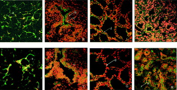
During virgin and early pregnancy stages, leptin was found in adipocytes, ductal epithelial cells and basal lamina around the duct (Fig. 1A). In late pregnancy and lactation, leptin was detected in alveolar epithelial cells near to the basal lamina and extracellular matrix (Fig. 1B,C); in involution, mammary fat pad was restored and leptin existed in adipocytes and ductal epithelial cells again (Fig. 1D). For leptin receptor, in virgin and early pregnancy stages, OB-Rb was found in adipocytes and ductal epithelial cells (Fig. 1E); in late pregnancy and lactation, OB-Rb was detected on the membrane of alveolar epithelial cells near to the basal lamina (Fig. 1F,G), and in involution, OB-Rb was localized in adipocytes and ductal epithelial cells (Fig. 1H). At all stages, leptin immunostaining was eliminated when rabbit anti-goat leptin and leptin receptor (long form) polyclonal antibody were omitted.
Location of leptin and OB-Rb in mammary gland of goat (400×). Representative photographs of immunofluorescent location of leptin and OB-Rb in mammary tissue from virgin to involution (400×). Pictures A–D represent leptin location: A, virgin; B, pregnancy; C, lactation; D, involution. Pictures E–H represent OB-Rb location: E, virgin; F, pregnancy; G, lactation; H, involution. Red colour represent nucleus, and green colour represent positive signal in the picture. a. adipocytes; b. basal lamina; c. ductal epithelial cell; d. alveolus epithelial cells. Leptin was mainly located in adipocytes, ductal epithelial cells, basal lamina around the duct, alveolus epithelial cells near to the basal lamina and extracellular matrix; OB-Rb was found in adipocytes, ductal epithelial cells, membrane of alveolus epithelial cells.

Leptin and OB-Rb expression
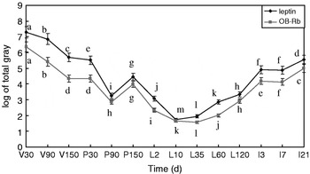
Amounts of leptin protein varied significantly (P<0·01) among virgin, pregnancy, lactation and involution stages (Fig. 2). During the virgin stage and at the beginning of pregnancy, the amount of leptin protein was high. During pregnancy, the leptin protein amount decreased strongly between days 30 and 90 (P<0·05) before increasing at day 150 (P<0·05 for day 150 v. day 90 of pregnancy) to levels similar to those assayed at day 30. Throughout lactation, leptin protein amounts did not vary significantly but were lower (P<0·05) than the level assayed at day 90 of pregnancy. Moreover, at days 10 and 35 of lactation, the amount of leptin protein was significantly (P<0·05) lower than those assayed at days 2, 60 or 120 of lactation. In involution, the amount of leptin increased and recovered to the original virgin level. The amount of OB-Rb was similar to leptin and showed significant positive correlation (r=0·98382, P<0·01).
Expression of leptin and OB-Rb in mammary gland of goat. V, virgin; P, pregnancy; L, lactation; I, involution. β-actin was used as standard. Expression of leptin and OB-Rb in the mammary gland was measured by western blot. Results were analysed by Glyko BandScan 4.3 software. Values are means±sd (n=3); means with different superscripts differ significantly (P<0·05).

Expression of β-casein protein
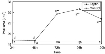
Leptin induced gene expression of β-casein within 120 h, the expression of β-casein protein was elevated from 48 h to 120 h, the maximum value being achieved at 96 h. The amount of β-casein protein in culture medium with leptin was significantly different from control at 72 h, 96 h and 120 h (P<0·01); the concentration of β-casein in control medium showed no significant change (P>0·05) (Fig. 3).
Effects of leptin on β-casein expression in mammary gland explants of lactating goats. Expression of β-casein was measured by CE after incubation with or without leptin (24 h, 48 h, 72 h, 96 h and 120 h). Leptin induced the gene expression of β-casein within 120 h and the amount of β-casein protein was elevated from 48 h to 120 h. The β-casein protein amount in the culture medium with leptin was significantly different from the control at 72 h, 96 h and 120 h (P<0·01); the β-casein protein amounts in the control medium were not significantly different from one another (P>0·05). Values are means±sd (n=9); means with different superscripts differ significantly (P<0·05): *P<0·05, **P<0·01.

Haematoxylin and eosin (HE) stain
In pregnancy the quantity of branches of mammary gland cultured with leptin were more than for the control cultured without leptin (Table 1). In involution, leptin made the alveolar disappear and only epithelial cell cords remained, while in control the ducts were still evident (Table 2).
The effects of leptin on development of mammary gland epithelium in pregnancy
Values are mean optical density readings with sd for n=6

† means with different superscripts differ significantly (P<0·05)
The effects of leptin on development of mammary gland epithelium in involution
Values are mean optical density readings with sd for n=6

† means with different superscripts differ significantly (P<0·05)
TUNEL analysis
TUNEL results also showed that the structure of mammary gland cultured without leptin remained normal and there was little apoptosis, while in the explants cultured with leptin, the ducts and gland alveolus collapsed and the apoptotic cells increased (Table 3).
The effects of leptin on apoptosis of mammary gland epithelium in involution
Values are mean optical density readings with sd for n=6

† means with different superscripts differ significantly (P<0·05)
Leptin-activated signalling in goat mammary gland
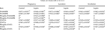
Throughout the mammary gland development stage, there was non-phosphorylation of MAPK, STAT3 and STAT5. In pregnancy, leptin activated p42MAPK/p44MAPK specifically. In lactation, leptin induced the phosphorylation of STAT5 mainly. In involution, leptin induced the phosphorylation of STAT3 specifically (Table 4).
Tyrosine phosphorylation of MAPK, STAT3 and STAT5 by leptin in goat mammary gland
Values are means with sd for n=3

For the items detected in each period (cultured with or without leptin), means with different lowercase superscripts differ significantly (P<0·05); means with different uppercase superscripts differ significantly (P<0·01)
Discussion
Our results suggested a strong synthesis of leptin by mammary adipocytes at the virgin stage and at the beginning of pregnancy. In contrast, after full cell differentiation, just before parturition, adipose tissue had completely regressed. Leptin protein amount decreased to a lower level during the second part of pregnancy; at 150 d of pregnancy, the mammary duct completely developed to a functional gland alveolus. We found that the gland alveolar epithelial cell itself could secrete leptin in late pregnancy and lactation, so at 150 d of pregnancy leptin protein amount increased slightly, and leptin was present in mammary epithelial cells, whereas during lactation when the mammary gland was full of functioning gland alveolus, leptin protein amount remained lower because of the effect of prolactin (PRL) which has an inhibitory effect on leptin expression in lactation (Aoki et al. Reference Aoki, Kawamura and Matsuda1999). In involution, the mammary fat pad regenerated, and leptin protein amount recovered to the virgin level. In the whole development cycle of the mammary gland, the expression of leptin and OB-Rb was positively correlated. These sequential changes of leptin and OB-Rb cellular location indicate that leptin could induce OB-Rb expression, and leptin together with OB-Rb acts as a paracrine and autocrine factor to regulate mammary gland growth, development and function.
OB-Rb is a member of the interleukin-6-receptor family of class I cytokine receptor. In mammary gland, ligand binding to OB-Rb results in the activation of JAK2 by transphosphorylation and the subsequent tyrosine phosphorylation of tyrosine residues on the intracellular OB-Rb, which then initiates the transmission of downstream phosphotyrosine-dependent signals (Bjorbaek et al. Reference Bjorbaek, Uotani, da Silva and Flier1997). Two tyrosine residues in the intracellular domain of OB-Rb become phosphorylated to mediate OB-Rb signalling; Tyr985 controls the tyrosine phosphorylation of SHP-2, and Tyr1138 controls STAT3 activation.
Leptin, like other cytokines, is effective in influencing the growth of various cell types (Silva et al. Reference Silva, Etchebarne, Nielsen, Liesman, Kiupel and VandeHaar2008; Akther et al. Reference Akther, Khan, Begum, Parveen, Kaiser and Chowdhury2009). Our results showed that the quantity of branches of mammary gland cultured with leptin was more than in the control cultured without leptin; we also found in this experiment that leptin activated p42MAPK/p44MAPK specifically in pregnancy, indicating that leptin induced the proliferation and differentiation of ductal epithelial cells by JAK-MAPK. Tyr985-mediated recruitment of SHP-2 resulted in GRB-2 binding to tyrosine-phosphorylated SHP-2 and subsequently activated the ERK Ser/Thr kinases transmission (Banks et al. Reference Banks, Davis, Bates and Myers2000). ERKs translocated to the nucleus following activation, whereupon they phosphorylated a set of nuclear transcription factors to mediate cell proliferation and differentiation. Physiological concentrations of leptin can activate ERK/AP1 signalling pathway (Hu et al. Reference Hu, Juneja, Maihle and Cleary2002); ERK is the main signalling pathway of leptin to induce cell proliferation.
We report here that leptin elevated amounts of β-casein protein, in agreement with the results of Baratta et al. (Reference Baratta, Grolli and Tamanini2003) who found that leptin seemed to exert a positive influence on milk protein induction in the mammary epithelium HC11 cell line (Baratta et al. Reference Baratta, Grolli and Tamanini2003). We suggest that leptin affects milk protein expression in Chinese Guan Zhong goat. This result, together with the finding that leptin-deficient mice induced to become pregnant delivered normally, but failed to lactate (Mounzih et al. Reference Mounzih, Qiu, Ewart-Toland and Chehab1998) supports the hypothesis that leptin plays an important role in regulating mammary function. In lactation, leptin stimulated phosphorylation of STAT5, which accords with the function of STAT5. STAT5 can regulate milk protein synthesis, milk protein genes being target genes of STAT5. At least one STAT5-binding site is found in the promoter of β-casein (Liu et al. Reference Liu, Robinson, Wagner, Garrett, Wynshaw Boris and Hennighausen1997). The JAK-STAT5 signalling pathway is activated in lactation to regulate milk protein expression.
Leptin also activated STAT3 in lactation. JAK-STAT3 cell signalling plays an inhibitory role in mammary gland development (Motta et al. Reference Motta, Accornero, Taulli, Bernabei, Desrivières and Baratta2007). But this experiment showed that during lactation in the mammary gland of the goat, leptin protein amount was maintained at a lower level, indicating that though there was activation of STAT3 in lactation, apoptosis was not obvious. Moreover there were a lot of other cytokines and hormones which could promote cell proliferation leading to a large expression of SHP-2, overexpression of SHP-2 decreased tyrosyl phosphorylation of STAT3, and thus inhibiting the apoptotic effect of STAT3 and SOCS3.
In conditional knock-out mice lacking the STAT3 gene in epithelial cells, the involution process was strongly delayed (Kritikou et al. Reference Kritikou, Sharkey, Abell, Came, Anderson, Clarkson and Watson2003). We suggested that in involution leptin induced apoptosis and mammary gland restitution by JAK-STAT3. Tyr1138 could bind to the SH-2 domain-containing transcription factor STAT3 (Li et al. Reference Li and Friedman1999), activated STAT3 translocated to the nucleus to mediate the inhibitory SOCS3 pathway; SOCS3 is an SH-2 domain containing JAK2 inhibitor that may block JAK-MAPK signalling (Bjorbaek et al. Reference Bjorbaek, Lavery, Bates, Olson, Davis, Flier and Myers2000). Moreover STAT3 can combine to the promoter of C/EBPδ to induce mammary epithelial cell apoptosis (Hutt et al. Reference Hutt, O'Rourke and DeWille2000).
This research was supported by National Natural Science Foundation of China (30671538) and Northeast Agricultural University Foundation (2009RC26).