Implications
In multiparous sows, embryonic mortality at day 21 of gestation is already 24%, and is independent of space. This means that either a percentage of embryos is intrinsically unfit to survive, or there are interactions other than competition for space between embryos in a litter, direct or through influences on the uterine environment, that limit the survival of some embryos.
Introduction
Prenatal losses in the pig range between 30% to 50% in commercial genetic lines (Pope et al., 1972; Geisert and Schmitt, 2002). The majority of these losses occur during the embryonic phase (before day 35), with 20% to 30% of embryos lost by the 3rd week and another 10% to 15% being lost by the end of the embryonic phase (Ford et al., Reference Ford, Vonnahme and Wilson2002). Experimental alteration of the available uterine space per embryo, by using a range of techniques (e.g. Dziuk, 1968; Père et al., Reference Père, Dourmad and Etienne1997; Town et al., Reference Town, Putman, Turchinsky, Dixon and Foxcroft2004) has shown that by day 30 to 35 of gestation more embryos are lost when uterine space is limiting, hence the term uterine capacity. Even though uterine length is plastic and increases with the number of occupying embryos, Chen and Dziuk (Reference Chen and Dziuk1993) established that a minimum length of around 25 cm per embryo is required at fertilisation to maximise survival.
It is not clear when exactly uterine space becomes limiting to the survival of porcine embryos, since most of the experiments mentioned above made observations between day 30 and 40 of gestation. Most embryos survive until day 12 (93% to 96%; Anderson et al., Reference Anderson, Christenson, Christenson and Ford1993). After that, elongation (day 11 to 13), spacing, and implantation (day 15 to 17) occur, however there are few reports of losses during these specific periods. The few studies available before day 30, report losses to be 18% to 35%, and mostly independent of space. However, these reports are all based on gilts, and in some studies there is no clear description of stage of the embryos (Dziuk, Reference Dziuk1968), controls were super-ovulated (Pope et al., Reference Pope, Christenson, Zimmerman-Pope and Day1972; Webel and Dziuk, Reference Webel and Dziuk1974), or number of observations were limited (Knight et al., Reference Knight, Bazer, Thatcher, Franke and Wallace1977). Studies in multiparous sows have only been reported by King and Williams (Reference King and Williams1984) and Town et al. (Reference Town, Putman, Turchinsky, Dixon and Foxcroft2004), for day 30 gestational age. Multiparous sows have a much higher ovulation rate than gilts and the available area for each embryo is smaller than in gilts, despite the greater uterine length. Therefore, the current study was undertaken to provide data on embryonic survival in multiparous sows rather than gilts, and to determine whether space limits embryonic survival before day 30 in multiparous sows specifically, using a unilateral oviduct ligation model.
Material and methods
Multiparous sows (having had 2 to 11 litters) of a Large White×Landrace cross were weaned and then either left intact (CTR, n=42) or subjected to unilateral oviduct ligation (LIG, n=23) after the first post weaning oestrus.
Oviduct ligated sows
Oviduct ligation was performed in LIG sows within 10 days following the first oestrus and sows were mated and ovulation was assessed as described below, at the second oestrus.
Sows were fasted for 12 h before surgery. The animals were anaesthetised by thiopentone sodium at a dose rate of 10 mg/kg of BW administered by injection via an ear vein. Anaesthesia was maintained using a combination of isoflurane and oxygen. Unilateral oviduct ligation was performed by mid-ventral laparotomy. Randomly, either the right or the left oviduct was looped and then tied off at the base of the loop with one absorbable suture. The strictured part of the loop was then excised. The operation wound was closed using vicryl absorbable sutures. Animals were given 250 mg IM (intramuscular) of Flunixil (Flunixin-Meglumine, Norbrook Laboratories, Northern Ireland) as an analgesic and 1050 mg IM of Moxylan (amoxicillin; Jurox, Rutherford, NSW, Australia) as an antibiotic. Sows then received 1050 mg of IM per day of Moxylan for 2 days post-surgery. The rationale for the unilateral oviduct ligation was to reduce the number of embryos entering the uterus after fertilisation by 50% on average, and as a result doubling the available uterine space per embryo, compared with intact sows.
Heat detection and ultrasound
All sows received boar contact twice a day (morning and afternoon) from 5 days before expected (second) oestrus, in a detection-mating-area, until oestrus had ceased. Sows were mated with 3 billion sperm cells from pooled semen at first standing and then every 24 h until ovulation was detected. Ovulation was assessed using trans-rectal ultrasound (3.5 MHz sector probe; Aquila Vet, Pie Medical, the Netherlands), once per day. Once the pre-ovulatory follicles had disappeared, time of ovulation was estimated as having occurred in the 24 h prior.
Reproductive tracts
Intact sows were slaughtered at day 9, day 21 or day 35 of gestation (day 0 is day of ovulation), and LIG sows at day 21 and 35. The day 9 observations were included to demonstrate that the heat detection and insemination strategy resulted in >90% fertilisation in order to prove that losses at later stages occurred after day 9. We assumed that there would be no crowding before day 9, and more importantly, that at this stage there is negligible mortality, based on the observations by Anderson et al. (Reference Anderson, Christenson, Christenson and Ford1993) and earlier work from our lab, and that ovulations that are not presented by an embryo probably reflect flushing inefficiency or unfertilised oocytes. In LIG sows, the ligated oviduct was double-checked for (non) patency by infusing saline into the infidibulum. Reproductive tracts were collected to assess ovulation rate, weight of individual corpora lutea after excision, number of embryos, and gross morphological characteristics of uterine horns, placentae and implantations (day 21 and day 35). Length of implantations was measured and embryos were weighed. Placentae were removed from the implantation sites and then spread on wax paper and traced. After drying the size of the traced area was calculated by relating its weight to a standard size piece of wax paper. Arbitrarely, the 10% of embryos lowest in weight were considered as unviable. This included embryos that were obviously unviable based on haemorrhagic appearance or showing signs of disintegrating and a brownish colour.
For the day 9 gestational stage, uterine horns were flushed using phosphate buffered saline, and subsequently embryos were counted and dimensions were measured under a dissecting microscope. Presence of unfertilised oocytes was also recorded.
Embryonic survival was calculated as the percentage of ovulations that were represented by an embryo. For LIG sows, only the corpora lutea on the ovary with the intact oviduct were counted.
Statistical analysis
Statistical analyses were performed using the SAS statistical package (9.3 edition; SAS Institute Inc, Cary, NC, USA). Differences between treatments (LIG v. controls) and between stages of gestation were tested using the GLM procedure. Regression analysis, for example between the number of embryos and the number of ovulations, was also performed using the GLM procedure.
Results
Overall, ovulation rate was 22±0.9. Ovulation rate was 18.7±0.7 (n=18) for second litter sows, 20.5±0.9 (n=12) for third litter sows, 19.5±2 (n=7 for fourth litter sows, and 22.8±0.8 (n=28) for older sows. Luteal tissue mass was 14.6±0.9 g per sow at day 9, 8.5±0.3 g at day 21 and 9.3±0.4 g at day 35. At day 21 and day 35, the distribution of embryos across horns in ligated sows was even, with 52% of the embryos in the horn ipsilateral to the patent oviduct (range 33% to 73%).
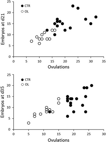
At day 9 of pregnancy almost every (92%) ovulation was represented by an embryo, and, every extra ovulation resulted in 0.9 extra embryos (r=0.6; P<0.01). At later stages too, the number of embryos was correlated to the number of ovulations (r=0.78; P<0.05), and each extra ovulation resulted in 0.6 extra embryos at day 21 and 0.5 embryos at day 35 of gestation (P<0.05). Embryonic survival decreased with ovulation rate (r=−0.47; P<0.01), by 0.6% for every ovulation at day 21, and by 1.3% for every ovulation at day 35 (P<0.01). The relationship between embryonic survival and ovulation rate is reported in Figure 1.
Number of embryos surviving at day 21 (top panel) and at day 35 (bottom panel) of gestation in relation to ovulation rate for intact sows (closed circles) and oviduct ligated sows (open circles). At day 21, each extra ovulation resulted in 0.73 extra embryo (R 2=0.68). At day 35, each extra ovulation resulted in 0.8 extra embryo in ligated sows, and 0.6 extra embryo in intact sows (R 2=0.52).

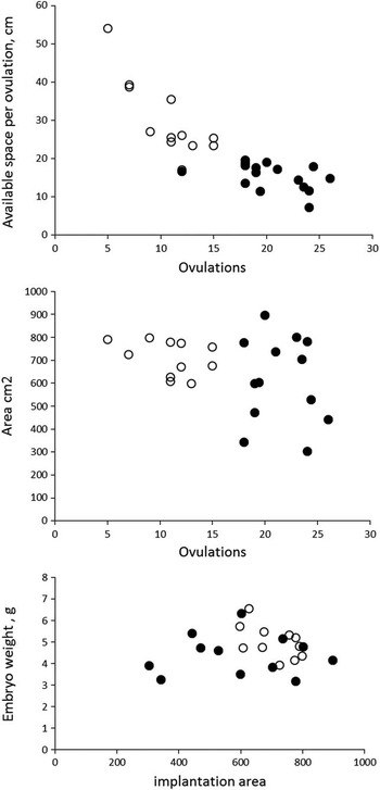
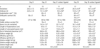
In intact animals, the available space per embryo was strongly negatively correlated (r=−0.85) to the number of ovulations. At day 21, the length of the uterine horns was unaffected, however, at day 35 the length of the uterine horn increased by 6 cm with each extra embryo (r=0.41; P<0.05). Nevertheless, at day 35 the available space per ovulation in intact sows was half that in ligated sows, with all intact sows having an average available space per ovulation smaller than 25 cm (Figure 2). At day 21, the extra space available due to ligation did not affect the area of the placentas (67±9 v. 84±22 cm2, ns), however, at day 35 placentas in ligated animals spanned an 18% greater area (600±54 v. 709±23 cm2). The crowding effect at day 35 was also evident from the length of the implantation sites in intact animals (15.5±1.3 cm) being almost similar to the available space (16.1±0.8 cm), whereas in ligated sows the space taken up by implantation sites (19±1 cm) was longer than for intact sows but far smaller than the available space (30±2.9 cm) (Table 1; Figure 2).
Top two panels: Available length (cm) of uterine horn per ovulation (calculated) and actual mean area of placentation (cm2), in relation to the number of available ovulations for intact sows (closed circles) and sows with unilateral oviduct ligation (open circles). Bottom panel: Embryo weight in relation to area of placentation. All data from day 35.

Embryonic survival and characteristics of placentation and embryonic development at day 9, day 21 and day 35 of gestation

a,b P<0.05; x,yP<0.10 within rows.
* 70% survival when excluding outliers.
** Percentage of ovulations represented by an implantation mark, including ‘unoccupied’ implantations where there are visible marks of implantation but no embryos present.
The frequency distribution for area of individual placentas followed a similar pattern for intact and ligated sows at day 21, which is reflected by the mean placenta area in Table 1. At day 35 however, in ligated sows there was a shift towards larger placentas (area), which is reflected in both mean placenta area and mean implantation length. For example, placenta area was smaller than 500 cm2 in 45% of placentas in intact animals, but only in 12% of ligated animals (P<0.01). The size of unoccupied areas of the uterine horns showed a similar (normal) distribution across treatments, although the mean length of unoccupied spaces was larger for ligated sows at day 21 (Table 1). The size of unoccupied spaces did not increase from day 21 to day 35, whereas that of occupied spaces did.
By day 21 most embryonic mortality (~24%) had already occurred and was not influenced by available space. In ligated animals there was no more embryonic mortality after day 21. In intact animals, however, another 8% to 14% mortality occurred, depending on whether outliers were included, bringing the viable embryonic survival by day 35 down to 59% in intact animals as opposed to 77% in ligated sows. Although the reduced available space in intact animals clearly reduced the placenta area, there was no clear relationship between mean placenta area and embryonic survival rate, at sow level. There was also no clear relationship between mean placenta area and embryo weight (Figure 2), however, in the ligated group the extra available space tended to increase embryo weight. Moreover, at the embryo level embryo weight increased by 0.16 g per 100 cm2 placenta at day 21 (r=0.6; P<0.01), and with 0.28 g per 100 cm2 placenta at day 35 (r=0.4; P<0.01).
Discussion
In this study, two-thirds (24% out of 41%) of embryonic mortality in multiparous sows occurred before day 21. The mortality before day 21 was not due to space limitation. This has not been reported before for multiparous sows. Père et al. (Reference Père, Dourmad and Etienne1997) and Town et al. (Reference Town, Putman, Turchinsky, Dixon and Foxcroft2004) also used unilateral oviduct ligation as a means to increase the available space for embryos, but the earliest stage of gestation they reported on was day 30 to 35. In gilts, oviduct ligation models have been used to show that early embryonic mortality is not related to space (Dziuk, Reference Dziuk1968; Webel and Dziuk, Reference Webel and Dziuk1974), and Freking et al. (Reference Freking, Leymaster, Vallet and Christenson2007) showed that selection for uterine capacity did not change early embryonic losses. However, these studies did not investigate embryonic survival before day 25 of gestation.
Assuming fertilisation rate is 90% to 95%, there was minimal embryonic loss before day 9 and recovery rates lower than 100% at this stage were presumably due to efficiency of the flushing procedure. Since implantation rate was 82% of the potential number of embryos, 18% did not even implant, indicating that some of the early mortality occurred during the period of spacing (after day 9 but before implantation) and some around implantation. Implantation rate includes those placentations that do not have an embryo attached, and is therefore higher than embryonic survival rate, which only includes ovulation represented by an embryo. From our observations in this study and numerous other studies we have performed, it appears that at postmortem some implantation sites show an implantation mark and remnants of the chorio-allantois, without the presence of an embryo. This is evidence of embryos that have commenced implantation but failed to survive, leaving an ‘unoccupied’ implantation site. These areas are generally small. Embryos that would have implanted but later been resorbed, would have left implantation marks on the endometrium.
By doubling the available space in the LIG sows, embryonic mortality from day 21 to day 35 was eliminated. In intact sows, embryo mortality between day 21 and day 35 was 14% when including all sows, and 8% when excluding two sows that had embryo mortality lower than 40%. This threshold was based on the distribution of embryonic mortality which clearly showed that observations below 40% were well outside the normal distribution. It is debatable whether the outliers should be included in the analysis of mortality. On one hand, they may be a consequence of crowding as they only occurred in the intact animals. On the other hand, the outliers were animals with ovulation rate below the mean, which would suggest that factors other than crowding were a cause of the increased embryonic mortality. Either way, embryonic mortality after day 21 was only observed in intact animals with limited space per embryo, and not in the ligated sows. Similarly, studies by Town et al. (Reference Town, Putman, Turchinsky, Dixon and Foxcroft2004) and Père et al. (Reference Père, Dourmad and Etienne1997) show that doubling the uterine space per embryo by ligation reduces embryonic mortality at day 30 to 35. Clearly, at this stage the area of the placenta becomes a limiting factor in facilitating nutrient uptake, which was reflected by the smaller placentas in intact sows, and by the fact that nearly all the available space was occupied by implantations, leaving hardly any space to increase physical area of the implantations. The element of crowding was also reflected in the positive relationship between implantation area and embryo weight, and it may be speculated that the higher percentage of smaller placentas in intact sows may cause additional mortality of foetuses later in gestation. As a consequence, sows with a high ovulation rate may not only suffer a higher embryonic mortality post day 21, but also in the foetal stages because of insufficient placental interface.
If embryonic mortality before day 21 is independent of space, the question is what then causes embryonic mortality around spacing and implantation. From day 8 to day 10, embryos migrate through the uterine horns and some will cross the bifurcation between the left and right horn. Between day 10 and day 13, embryos elongate and space along the uterine horns. Around day 12, embryos start to secrete oestrogens, and at the same time they start the process of spacing, in which they position themselves throughout the uterine horns aligning themselves in preparation for implantation at around day 15 (Dziuk, Reference Dziuk1985). The process of spacing is a co-ordinated mechanism, during which the available uterine space is distributed fairly evenly among the embryos. Studies with inert, oestrogen-soaked beads have shown that this process may be facilitated by mild contractile activity of the uterus, induced by oestrogens originating from the embryos, with presence of embryos in localised areas of the uterus inducing more or less uterine contractility (Pope et al., Reference Pope, Lawyer and First1986). Spacing is independent of the number of embryos, and small and large litters both migrate and space evenly (Dziuk, Reference Dziuk1968). Whether there is direct communication between embryos is not clear, however, from our own observations implantation sites hardly ever overlap, which would suggest that there is. In the process of spacing and implantation, developmental stage of the embryos and variation between embryos play a role in determining which embryos survive (Geisert and Schmitt, Reference Geisert and Schmitt2002).
Before implantation, variation in embryonic development may be such that advanced embryos influence the uterine environment in a way that is detrimental to more delayed embryos. Geisert et al. (Reference Geisert, Ross, Ashworth, White, Johnson and Da Silva2006) showed that treatment of sows with oestrogens before day 12 had a negative impact on embryonic survival. Embryos will still elongate, but not survive to day 16 (Morgan et al., Reference Morgan, Geisert, Zavy and Fazleabas1987). This suggests that there is a window, before elongation, in which oestrogens are detrimental to embryonic development through their effect on the uterine environment relative to the developmental stage of the embryos. Treatment with oestrogens at a later stage, when embryos start to secrete oestrogens themselves, is not harmful to embryonic survival (Pope et al., Reference Pope, Lawyer and First1986). Advanced embryos may start to secrete oestrogens at a stage when retarded embryos are compromised by the same oestrogens, and this may be another mechanism through which variation in embryonic development causes embryonic mortality. In this respect, it is noteworthy that Meishan embryos develop slower, are smaller in general and secrete less oestrogens (Anderson et al., Reference Anderson, Christenson, Christenson and Ford1993). This would reduce the variation between embryos once a litter reaches the critical stage when the first embryos start elongating and secreting oestrogens. The amount of oestrogen secreted in white breeds is probably well above what is necessary for embryonic development, since reduction of secretion with aromatase inhibitors by 57% did not impede embryonic development (O’Neill et al., Reference O’Neill, Geisert, Zavy, Morgan and Wettemann1991).
In conclusion, in multiparous sows around 24% of embryos are lost during spacing and implantation, and this loss is not related to uterine space, but probably due to intrinsic embryonic quality or variation, interaction between embryos and/or the uterine environment, or a combination of these factors. After implantation, uterine space is limiting, and causes another 10% mortality between day 21 and day 35.
Acknowledgements
The authors gratefully acknowledge the CRC for High Integrity Australian Pork for their funding in this project.