Introduction
The p53 gene codes for a small protein (53 kDa) called ‘the guardian of the genome’ (Lane, Reference Lane1992). Different factors, such as DNA damage, hypoxia, nitric oxide or heat shock, may activate the p53 gene (Oren, Reference Oren1999) and lead to apoptosis or cell cycle arrest. These processes are especially important for reproductive cell development. The first physiological wave of germ cell apoptosis regulates the ratio between Sertoli cells and germ cells in the testes of 2–4-week-old male rodents. This process affects mainly spermatogonia, but also affects spermatocytes, and seems to be p53-dependent as an elevated p53 protein level was demonstrated in young male mice and in rat gonads (Rodriguez et al., Reference Rodriguez, Ody, Araki, Garcia and Vassalli1997; Yan et al., Reference Yan, Suominen, Samson, Jegou and Toppari2000). In adult mice gonads p53 mRNA and p53 protein were shown to be present in spermatocytes, but no, or only a weak, p53 protein level was observed in spermatogonia (Schwartz et al., Reference Schwartz, Goldfinger and Rotter1993; Rodriguez et al., Reference Rodriguez, Ody, Araki, Garcia and Vassalli1997; Beumer et al., Reference Beumer, Roepers-Gajadien, Gademan, van Buul, Gil-Gomez, Rutgers and de Rooij1998; Yan et al., Reference Yan, Suominen, Samson, Jegou and Toppari2000), while apoptosis was shown to eliminate spermatogonia rather than spermatocytes (Allan et al., Reference Allan, Harmon and Roberts1992; Rodriguez et al., Reference Rodriguez, Ody, Araki, Garcia and Vassalli1997). Overall, it seems that during normal spermatogenesis the role of p53 in pachytene spermatocyte DNA quality control is more important than its role in apoptosis (Rotter et al., Reference Rotter, Schwartz, Almon, Goldfinger, Kapon, Meshorer, Donehower and Levine1993).
It is possible to activate 53-dependent apoptosis in the differentiating spermatogonia and early spermatocytes of adult animals with X-ray irradiation (Beumer et al., Reference Beumer, Roepers-Gajadien, Gademan, van Buul, Gil-Gomez, Rutgers and de Rooij1998; Hasegawa et al., Reference Hasegawa, Zhang, Niibe, Terry and Meistrich1998). Also cryptorchid testes are characterized by germ cell loss. In such testes, cell death is regulated initially by p53, but followed by a delayed, p53-independent apoptosis (Ohta et al., Reference Ohta, Aizawa and Nishimune2003; Yin et al., Reference Yin, Stahl, DeWolf and Morgentaler2002). Trp53 function was also examined in the knock-out mice, but contradictory results were obtained. For example, the C57BL/6 × 129 males that lack p53 protein were fertile and possessed normal structure of seminiferous tubules, while males from the 129/Sv inbred strain with the mutated p53 gene were reported to be infertile with characteristic multinucleated giant cells in the tubules (Rotter et al., Reference Rotter, Schwartz, Almon, Goldfinger, Kapon, Meshorer, Donehower and Levine1993). Therefore, both the genetic background, and internal and external factors play an important role in p53-dependent spermatogenesis control.
The genetic background of mice plays a role in p53 gene functionality. Not only autosomal genes (Mop1 and Ovca1) can modifying p53 function significantly, but sex chromosomes have an effect as different levels of mortality have been reported in mutant male and female embryos (Evans et al., Reference Evans, Liang, Amos, Gu and Lozano2004; Liang et al., Reference Liang, Ayanga, Du, Godwin and Hartsock2008). Male mice from the B10.BR-Ydel strain possess a Y chromosome with a partial deletion of the long arm. The deletion of gene that control spermatogenesis can reduce semen quality. Ydel males have a high level of sperm head abnormality (Styrna et al., Reference Styrna, Imai and Moriwaki1991) and decreased sperm motility parameters have been observed (Grzmil et al., Reference Grzmil, Golas, Müller and Styrna2007). In addition, abnormal seminiferous tubules in the testes of B10.BR-Ydel males are more apparent than in control B10.BR males (Styrna et al., Reference Styrna, Bilinska and Krzanowska2002). Several authors have demonstrated the role of p53 in germ cell DNA quality control (Beumer et al., Reference Beumer, Roepers-Gajadien, Gademan, van Buul, Gil-Gomez, Rutgers and de Rooij1998; Hasegawa et al., Reference Hasegawa, Zhang, Niibe, Terry and Meistrich1998; Schwartz et al., Reference Schwartz, Goldfinger, Kam and Rotter1999). It was, therefore, interesting to investigate the combined effects of the Ydel and the p53 mutation on spermatogenesis. We designed experiments to analyse the role of p53 in gamete quality in mice that have a large chromosome aberration, i.e. Y chromosome deletion (Ydel). Our experiments were conducted using four groups of animals: B10.BR-p53 +/+, B10.BR-p53 −/−, B10.BR-Ydel-p53 +/+ and B10.BR-Ydel-p53 −/−. If the elimination of sperm depends on the p53 status, a decrease in sperm quality will be observed in B10.BR-Ydel-p53 −/− mice, but not in B10.BR-p53 −/− males, when compared with the appropriate control animals.
Materials and methods
Animals
Trp53 knock-out (C57BL/6 × 129-p53 −/−) and control mice (C57BL/6 × 129-p53 +/+) on the mixed (heterozygous) C57BL/6 × 129 genetic background were obtained from Swiss Federal Institute of Technology Zurich. We backcrossed the C57BL/6 × 129-p53 −/− females to B10.BR males to derive control B10.BR and mutant B10.BR-p53 −/− mice in B8–B9 backcross generation. Then, C57BL/6 × 129-p53 −/− females were crossed with B10.BR-Ydel males to develop control B10.BR-Ydel and mutant B10.BR-Ydel-p53 −/− mice. Six to nine adult male mice from B8–B9 backcross generation of B10.BR, B10.BR-p53 −/−, B10.BR-Ydel and B10.BR-Ydel-p53 −/− genotype were used to address the significance of p53 in animals with poor sperm quality caused by the deletion on Y chromosome.
The animals were bred in the Department of Genetics and Evolution in a 12 h:12 h light:dark conditions with pelleted food and water ad libitum. Mice were killed by cervical dislocation and weighed. The experiments were performed under license of the Commission of Bioethics at the Jagiellonian University, Poland.
Sperm quality parameters
Testes were dissected and weighed. Both vas deferens were dissected and their contents were squeezed gently out into 100 μl of M2 medium (Sigma-Aldrich, USA). Sperm was allowed to disperse (5 min) and sperm suspension was analysed under a light microscope (×400 magnification, unless indicated otherwise).
Fresh sperm suspension was mixed with 0.2% eosin Y (1:1, v/v; Sigma-Aldrich, USA) and incubated for 10 min at 37°C. Red and green sperm heads observed on smears were classified as either dead or viable sperm, respectively (eosin test). A hypo-osmotic (HOS) test was applied to analyse sperm tail membrane integrity. In hypotonic conditions (water; Sigma-Aldrich, USA) sperm with proper integrity in the tail membrane possess swollen tails (for detailed protocols see Golas et al. (Reference Golas, Dzieza, Kuzniarz and Styrna2008)).
The percentage of motile spermatozoa in the sperm suspension was calculated. For motile gametes, the number of sperm without droplets on the tails (mature sperm) and the number of sperm that possessed distal and proximal droplets (immature sperm) were also calculated. Ten μl of sperm suspension were diluted 10 times with phosphate-buffered saline (PBS) (Biomed, Poland) using a Potain pipette for white blood cells. Sperm count per 250 small squares was assessed in Bürker's counting chamber. Total amount of sperm ×106/ml in the ejaculate was calculated.
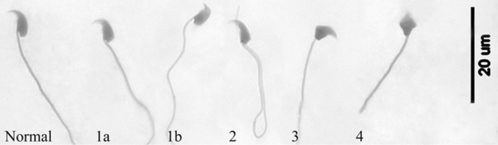
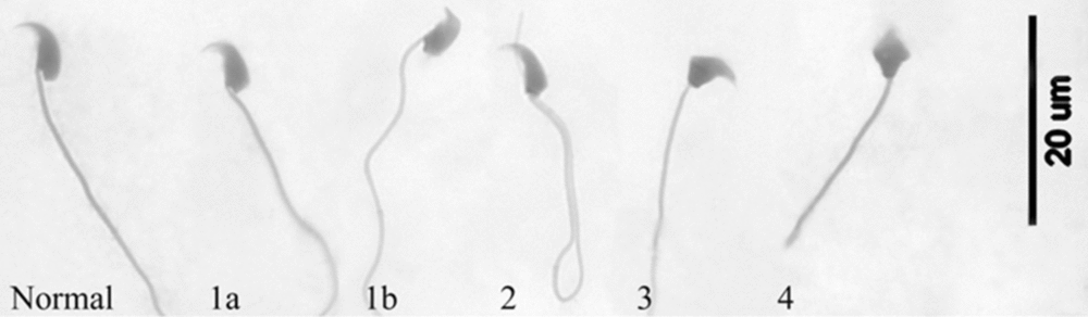
Smears were prepared from 10 μl of sperm suspension. Smears were fixed in a mixture of ethanol and acetic acid (3:1, v/v), and then were stained with eosin (Sigma-Aldrich, USA). The morphology of 200 sperm heads (Fig. 1) for each smear was evaluated under a ×1000 magnification.
Classes of sperm head abnormalities that were discriminated according to Styrna et al. (Reference Styrna, Imai and Moriwaki1991). Class 1a – slightly affected sperm, with bent acrosome or abnormal curvature of the distal head part; class 1b – sperm with flattened acrosomes, observed in mice with the deletion on chromosome Y. Class 2 – sperm with elongated and narrow head, sometimes not stained canals are also present. Class 3 – sperm with abnormalities in the apical and the distal part of head. Class 4 – sperm with severely misshapen head, unstained vacuoles can be also present.
Statistical analysis
Data for specific sperm quality parameters were pooled for each strain. All data are presented as percentages and expressed as means ± standard deviation (SD). Data were arcsin transformed before statistical analysis. The parameters examined for B10.BR-p53, B10.BR-p53 −/−, B10.BR-Ydel and B10.BR-Ydel-p53 −/− males were compared by one-way analysis of variance (ANOVA) followed by Tukey's post-hoc test (p < 0.05).
Results
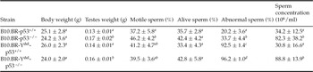
There was no statistical difference in the body weight of males from the four experimental groups. Testes weight was not related to Y chromosome status, but was controlled by p53, as we found that testes from p53 −/− mutants were heavier than testes from control males (Table 1). The percentage of motile sperm was not dependent upon Y chromosome structure, but was statistically higher in B10.BR-p53 −/− mutants than in control males (Table 1). An increased percentage of viable sperm was observed in B10.BR-p53 −/− and B10.BR-Ydel-p53 −/− males when compared with control mice, but these differences were not statistically significant (Table 1).
Body weight, testes weight and the four sperm quality parameters (mean ± SD) in B10.BR-p53+/+, B10.BR-p53−/−, B10.BR-Ydel-p53+/+ and B10.BR-Ydel-p53−/− mice

a–dDifferent superscripts indicate significant differences within a column (p < 0.05).
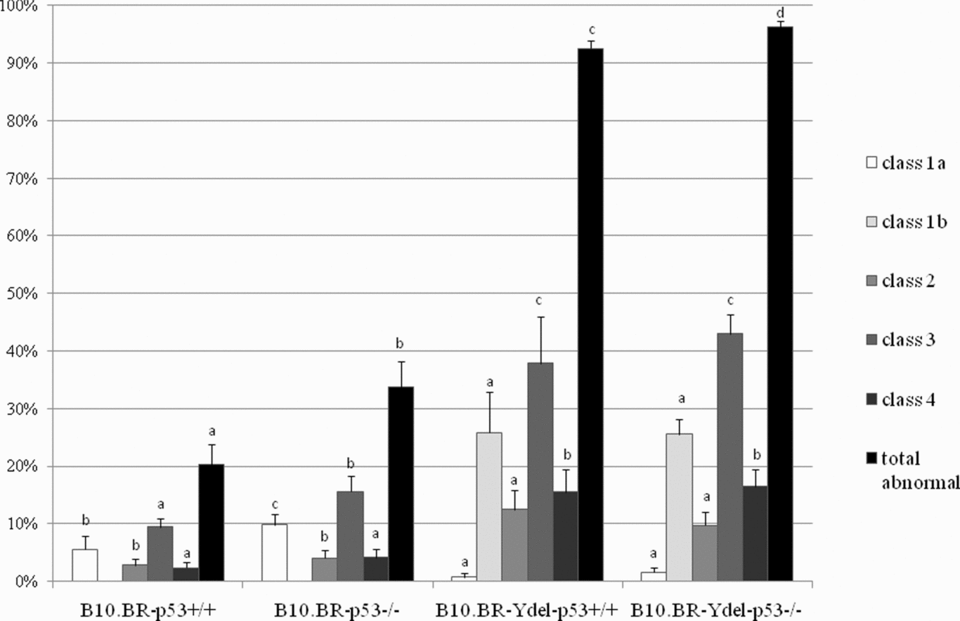
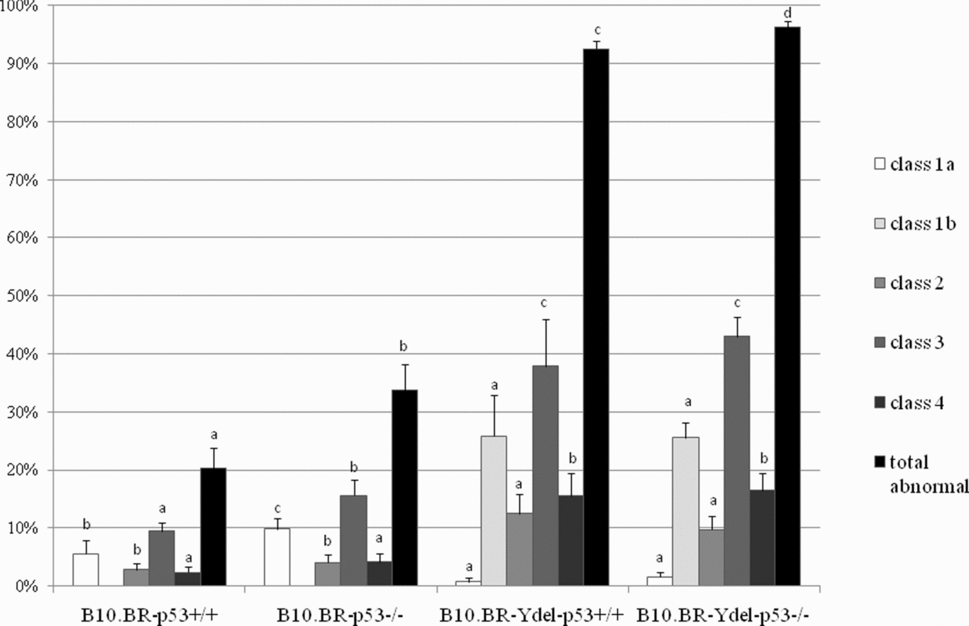
Sperm head morphology was strongly affected by Y chromosome deletion, with head abnormality class 1b characteristic for mice that had the Ydel chromosome (Fig. 1). Mutant B10.BR-p53 −/− and B10.BR-Ydel-p53 −/− males were characterized by significantly higher number of abnormal sperm than did control animals (Table 1, Fig. 2). Sperm count in the mutants was about 2.5 times higher than that in control animals, but was not affected by absence of the chromosome Y deleted genes (Table 1).
Percentage of particular sperm head abnormality classes in B10.BR-p53+/+, B10.BR-p53−/−, B10.BR-Ydel-p53+/+ and B10.BR-Ydel-p53−/− mice. Note the presence of a high percentage of class 1b abnormality in mice with deletion on Y chromosome. Different superscripts indicate significant differences within an abnormality class as well as within total percentage of normal sperm morphology (p < 0.05).
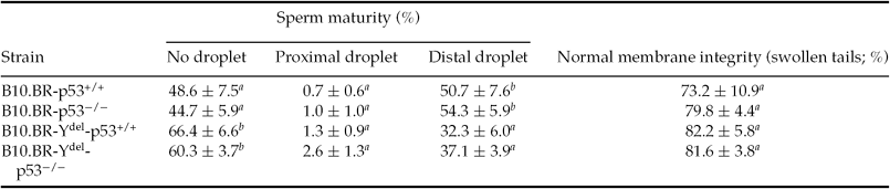
Maturation of sperm in B10.BR and B10.BR-Ydel males was not correlated with p53 genotype. This parameter was affected by the genes within the deleted region of Y chromosome, so that more sperm without droplets were found in control and mutant B10.BR-Ydel male mice than in B10.BR and B10BR-p53 −/− animals (Table 2). Neither Y chromosome genes nor the p53 gene controlled HOS test values in control and mutant B10.BR and B10.BR-Ydel males (Table 2).
Sperm maturity and sperm tail membrane integrity (mean ± SD) in B10.BR-p53+/+, B10.BR-p53−/−, B10.BR-Ydel-p53+/+ and B10.BR-Ydel-p53−/− mice
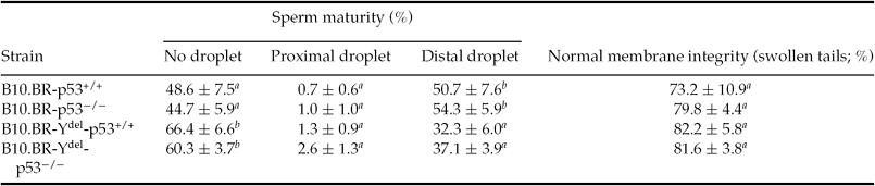
Sperm maturity was determined by location of a cytoplasmic droplet on the sperm tail. Tail membrane integrity was evaluated by hypoosmotic test. a, bDifferent superscripts depict significant differences within a column (p < 0.05).
Discussion
The loss of functional p53 protein leads to a disruption in the apoptosis process. Germ cells use two types of cell division and retain pluripotency, and therefore they need efficient control of DNA integrity and cell cycle regulation. Thus, both p53-dependent and p53-independent apoptosis is described in male germ cells dependent upon the germ cell type, inducing factors and genotype (Odorisio et al., Reference Odorisio, Rodriguez, Evans, Clarke and Burgoyne1998; Yin et al., Reference Yin, Stahl, DeWolf and Morgentaler2002; Salazar et al., Reference Salazar, Joshi, Liu, Wei, Persson and Wolgemuth2005; Paul et al., Reference Paul, Povey, Lawrence, Selfridge, Melton and Saunders2007). Also, the chromosome Y structure plays an important role in male mice spermatogenesis, and has been found to affect mainly sperm head morphology (Krzanowska Reference Krzanowska1976; Burgoyne Reference Burgoyne1998; Styrna et al., Reference Styrna, Bilinska and Krzanowska2002). The four experimental groups of animals used in our study allowed us to show the effect of Trp53 genotype alone and of chromosome Y deleted genes alone on sperm quality parameters and testis histology. We also wished to test if there was an interplay between the Trp53 gene and Y chromosome genes in the control of measured parameters.
Our study showed that testes weight was controlled by p53 status, as p53-deficient males had significantly heavier testes when compared with control males (Table 1). It was demonstrated that testes of C57BL/6 p53 −/− and FvB p53 −/− mutant mice contained higher numbers of A1 spermatogonia than did testes from control males (Beumer et al., Reference Beumer, Roepers-Gajadien, Gademan, van Buul, Gil-Gomez, Rutgers and de Rooij1998), which could be the factor responsible for heavier testes in the mutants. Also, detailed examination of C57BL/6 p53 −/− males revealed that, prior to delayed weight loss, cryptorchid testes were even heavier that scrotal ones (Yin et al., Reference Yin, DeWolf and Morgentaler1998a). Thus, testes weight depended not only on the p53 status, but also on the time of analysis and treatment. Higher testes weight in B10.BR-p53 −/− and B10.BR-Ydel-p53 −/− mutants may be a result of disrupted apoptosis and cell accumulation. We observed a significantly increased sperm concentration in germ cells in the mutants. Previous results by Yin et al. (Reference Yin, Stahl, DeWolf and Morgentaler1998b) have also described elevated number of haploid cells in the testes of p53 mutant males.
In adult males’ testes, a major function of Trp53 in primary spermatocytes is to control DNA integrity, however apoptosis affects mainly spermatogonia and is p53-independent. Interestingly, our mutants had a significantly elevated sperm count. This finding could be the prolonged effect of the disrupted first apoptotic wave, which is known to be mediated by p53. In addition, the process of germ cell elimination had comparable intensity in both B10.BR and B10.BR-Ydel males. We speculate that in adult males p53-mediated apoptosis is activated and, according to Trp53 gene expression pattern, affects spermatocytes. The p53-dependent apoptotic pathway, if present in young or adult males, is the most effective.
It is interesting that the percentage of viable sperm was not affected in B10.BR-Ydel males when compared with B10.BR males. It is possible that large chromosome Y aberrations cannot activate germ cell apoptosis at the primary spermatocyte stage. Our experiments showed that lack of the genes within deleted region on the Y chromosome affects spermiogenesis, and accelerates cytoplasmic droplet retention (Table 2). Also, as it was shown before by Styrna et al. (Reference Styrna, Imai and Moriwaki1991, Reference Styrna, Bilinska and Krzanowska2002), the percentage of abnormal sperm was strongly increased in males from the B10.BR-Ydel strain (Table 1). Thus, among the genes located on the Y chromosome are those that code for transcription factors that playing regulatory functions during spermiogenesis. Some of these factors affect acrosome formation (Styrna et al., Reference Styrna, Kilarski and Krzanowska2003), while other gene products could have a function in cell-cycle control, which, judging from the accelerated sperm maturation, may be weaker in the cells with an aberrant Y chromosome. It is interesting to note that apoptosis was not activated in B10.BR- Ydel males that produce sperm that are viable and motile, but abnormal morphologically.
We showed that, except for the chromosome Y structure, p53 is also responsible for the percentage of abnormal sperm heads, as higher numbers of abnormal sperm heads were present in B10.BR-p53 −/− and B10.BR-Ydel-p53 −/− males than in control animals. While about 13% of abnormal germ cells undergo elimination during spermatogenesis in B10.BR males, only about 4% of them are eliminated in B10.BR-Ydel males. This finding supports strongly our hypothesis on downregulation of cell cycle control and apoptosis in the cells with the Ydel chromosome. Our results are in contrast with those obtained from examination of C57BL/6 and C57BL/6 × C3H males, in which no relationship between male mice genotype and abnormal sperm morphology was noted (Marty et al., Reference Marty, Singh, Holsapple and Gollapudi1999; Embree-Ku & Boekelheide, Reference Embree-Ku and Boekelheide2002), but we need to consider that genetic background of these males was different from our model mice. Thus, during normal spermatogenesis, major genes that control abnormal sperm percentage are located on Y chromosome, while the specific genetic background of both B10.BR and B10.BR-Ydel males, including Y chromosome genes, cooperates with Trp53 to eliminate some of abnormal germ cells in the testes.
The only sperm quality parameter that was under the control of p53 in Y chromosome-dependent manner was the percentage of motile sperm (Table 1). A complete Y chromosome resulted in a higher percentage of motile sperm in B10.BR-p53 −/− males than in control males, while in B10.BR-Ydel mutants this parameter was unchanged. Thus, when present, the genes within the deleted region of Y chromosome cooperate with p53 to affect sperm motility negatively in B10.BR male mice. Again, the deletion of genes on the Y chromosome resulted in lack of apoptosis of aberrant cells, so that sperm motility is not affected– similar to what we found for viable sperm concentration. Other investigators observed lower numbers of mature motile spermatozoa in p53 −/− male semen (Schwartz et al., Reference Schwartz, Goldfinger, Kam and Rotter1999), but these inconsistent results could be again explained by different genetic backgrounds of the mice used.
In summary, the four groups of animals studied allowed us to show that the Y chromosome genes alone regulated sperm maturation, while the Trp53 gene alone influenced testis weight and sperm concentration. Combination of both of these agents was responsible for sperm head morphology, while neither of them affected body weight, viable sperm percentage and sperm tail membrane integrity. Sperm motility was the only parameter controlled differentially by Trp53 in a Y chromosome status-dependent manner.
Acknowledgements
This work was supported by BW13a and DS/775-K/ZDS/000786 from the Institute of Zoology of the Jagiellonian University. We thank Allan Bradley (Wellcome Trust Sanger Institute, UK) and Romeo Ricci (Swiss Federal Institute of Technology Zurich, Switzerland) for providing us with the p53−/− mice.