Stenting procedures in the treatment of CHD are progressively increasing in the paediatric population since its first use in 1991. They are primarily used in pulmonary artery stenosis, coarctation of the aorta (CoAo), systemic and central venous obstructions, post-operative conditions, cavo-pulmonary communications and conduit/homograft stenosis, as well as maintaining ductus arteriosus patency.Reference Breinholt, Nugent, Law, Justino, Mullins and Ing1
Aortic coarctation accounts for 7% of CHD, being the fourth most common abnormality requiring surgical or catheter-based intervention.Reference Chakrabarti2 So far, over 900 studies on aortic coarctation stenting have been published. Small physical constitution prevents stent implantation in infants because the sizes of the equipment are too large to fit to their small vessels and there is high chance of reintervention. Stenting is a widely accepted procedure for aortic coarctation in children larger than 10–20 kg. Short-term efficacy and safety of stent implantation are widely described, but accurate longer term follow-up assessment is lacking.Reference Breinholt, Nugent, Law, Justino, Mullins and Ing1–Reference Carrozza3 Important complications may occur both in the acute phase and during follow-up: formation of aneurysms and aortic rupture are widely reported acute adverse events.Reference Carrozza3 Stent fracture, along with aortic pseudoaneurysm, is rare and potentially serious complications. The risk factors, the causal mechanisms and the typology of stent fractures have not been well defined.Reference Carrozza3
We report the case of a patient in whom a fracture of an AndraStent® XL (Andramed GmbH, German) developed after stenting for aortic coarctation. This complication was effectively managed by percutaneous implantation of a covered stent graft.
Case report
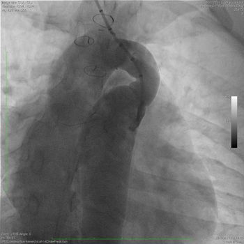
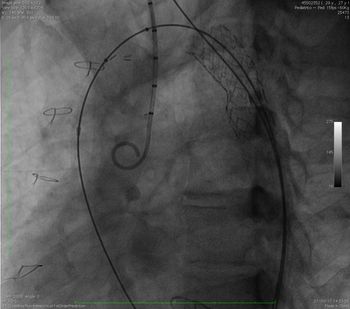
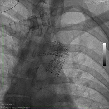
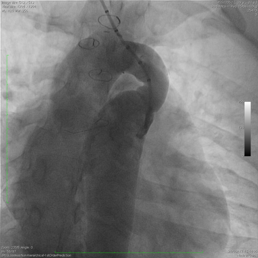
A girl born in 1990 and affected by aortic coarctation syndrome (bicuspid aortic valve, ventricular septal defect and CoAo) was treated with a two-step surgical approach in the first year of life: coarctectomy by subclavian flap (sec. Waldausen) and pulmonary bending at birth, followed by de-bending and ventricular septal defect closure one year later. She was regularly followed up with yearly echocardiography, 24 hour electrocardiogram Holter and stress test with pletismography. A cardiac magnetic resonance (MR) performed during follow-up in 2017 showed a tortuous aortic isthmus with left kinking (minim calibre 10 × 12 mm – Z score 2.02) and a low sisto-diastolic excursion of the abdominal aorta. At that time, 30-mmHg blood pressure gradient between upper and lower limbs was present at rest, increasing to 50 mmHg during maximal physical effort (with drop off to basal values during recovery phase). The heart catheterisation confirmed the magnitude of the re-coarctation as well as the tortuosity of the aortic isthmus (Fig 1). As a consequence, based on the length of the lesion, an uncovered ANDRA XL 48 mm stent mounted on an Atlas gold balloon 18 × 60 mm was placed by inflating the balloon at nominal pressure (6 Atm) (Fig 2). The procedure was well tolerated, and absence of gradient and stent fracture were detected. Eight months later, during a planned follow-up echocardiogram, a demodulated Doppler in abdominal aorta was detected. A stent evaluation by fluoroscopy showed a third-grade fracture (Fig 3).
Aortography showing the tortuosity of the aorta and the severity of aortic coarctation. The lesion length was 40 mm, the minimum diameter was 10 mm, and the ascending aorta diameter was 18 mm.
AndraStent® implantation. The stent was well deployed, and no fracture was detected after implantation.

Third-grade fracture of AndraStent® at fluoroscopic check performed for increased gradient at echocardiographic evaluation.

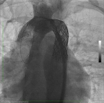
A second heart catheterisation was performed to stabilise the stent fracture and to relieve the isthmic gradient. The attempt to cross the stent was first made through the radial artery, but with this approach, it was difficult to pass through the second half of the stent. Conversely, the femoral approach was successful: the fragments were easily crossed by wire, and then a Mullins 12 fr introducer was passed through the AndraStent®. A covered Cheatham platinum (CP) stent 45 mm mounted on a 16 × 60 mm balloon was implanted to treat the lesion, to fix the stent fragments and to exclude possible aortic wall injury secondary to stent fragment mobilisation. The stent was then proximally shaped with a non-compliant balloon. Final angiography showed a good placement of the devices (Fig 4).
CP covered stent implanted over the fractured device.

Discussion
The most common complications after stent placement in CHD are restenosis by neo-intimal proliferation, embolisation or distal migration of the stent, erosion of the vessel, aneurism and/or pseudo aneurism formation, iatrogenic cardiac arrhythmias and hematoma at the site of cannulation.Reference Breinholt, Nugent, Law, Justino, Mullins and Ing1–Reference Carrozza4
Stent fracture is a rare complication with limited data available about its incidence and characteristics. Most reports of stent fracture involve right heart (right ventricle and pulmonary artery conduit) whose incidence ranges from 15% to 43%;Reference Breinholt, Nugent, Law, Justino, Mullins and Ing1 even more rare is the incidence of stent fracture in coarctation of the aorta, ranging from 0.01% to 0.08%.Reference Chakrabarti2,Reference Carrozza4–Reference Qureshi, McElhinney, Lock, Landzberg, Lang and Marshall8 The pathophysiological mechanisms of stent fracture are not well defined. Two causes are hypothesised: overloading, when the highest stretching force that the specific stent material can withstand is overwhelmed and fatigue, when the ability of the material to resist the pulsatile blood load is gradually lost. Different factors, intrinsic and extrinsic, can contribute to stent fracture: the material employed (stainless steel, platinum iridium, cobalt chromium), the geometry and the design of the stent (e.g., cut tube, welded wire, open versus closed cell, thickness of the struts, nodes, connecting segments), the type of delivery balloon and the structure into which the stent is implanted.Reference Chessa9 Covered stents have a lower rate of complications probably because of the direct coverage of the lesion (in fact, they are used in more complex and tighter aortic coarctation).Reference Butera5 In the literature, stent fractures were reported after aortic coarctation treatment with Palmaz stent (Johnson and Johnson), while the most reported complication during CP stent implantation (Cheatham Platinum-Numed Inc., Hopkinton, NY, USA) is aortic aneurism, dissection and rupture.Reference Butera5–Reference Qureshi, McElhinney, Lock, Landzberg, Lang and Marshall8 All of them share closed cell design. Palmaz stents are stainless steel devices, while platinum iridium is used to build CPs. The stent used in this clinical report (AndraStent® XL) is a bare Cobalt Chromium device with hybrid cell design (closed and open-cells design), whose employment in aortic coarctation is recent and data show favourable immediate and midterm outcome.Reference Butera5,Reference Golden10 Except for our experience, no other AndraStent® fracture has been reported.
Although a correlation between the pattern of fracture and stent type seems to be present, longitudinal pattern in Palmaz and radial pattern in Genesis and CP, it is not clear which stent better resists in vivo because methods of testing device strength rarely realistically simulate the forces to which they are exposed. It is likely that the compliance of the tissue where the stent is placed, the presence of surgical material and post-operative changes (e.g. calcification and fibrosis) play an important role in stent fracture.Reference Chessa9
In this case report, stent fracture was discovered because demodulated Doppler in abdominal aorta was found during follow-up. This highlights the importance of monitoring every patient who underwent stenting of aortic isthmus, even if stent fracture can be silent and the grade of fracture does not correlate with severity of symptoms.Reference Carrozza3 Only X-ray (XR) and CT are able to detect fractures and their severity, but guidelines about when and what method to perform during follow-up (XR, CT, MR) are lacking.
In conclusion, stent fracture is a rare event and a principal cause has not been determined; follow-up is important and at least one radiological test should be performed after the procedure.
Acknowledgments
None.
Financial support
This research received no specific grant from any funding agency, commercial or not-for-profit sectors.
Conflicts of interest
None.
Ethical standards
All procedures performed in this study involving human participants were in accordance with the ethical standards of the institutional and/or national research committee and with the 1964 Helsinki declaration and its later amendments or comparable ethical standards. Informed consent was obtained from all individual participants included in this study.