Introduction
The world poultry industry is under threat from infectious diseases which are responsible for substantial economic losses worldwide (Shah, Reference SHAH2013; Shah et al., Reference SHAH, XU, YAN, SONG and LI2010; Reference SHAH, XU, YAN, SONG and LI2011, Ullah et al., Reference ULLAH, RIAZ, UMAR and SHAH2013). Chicken infectious anaemia (CIA) is an important disease in major poultry-producing countries (Balamurugan and Kataria, Reference BALAMURUGAN and KATARIA2006), and economic losses due to this disease arises from poor growth, increased death rate, carcass condemnations and the cost of antibiotics used to control secondary bacterial infections (Dren et al., 2000). CIA is highly contagious disease. The disease is caused by the chicken anaemia virus (CIAV) which belongs to the genus Gyrovirus of the family Circoviridae (Schat, Reference SCHAT2009), and is recognised as an important avian pathogen worldwide (Todd, Reference TODD2000). CIAV is a non-enveloped, icosahedral, single stranded DNA virus. (Yamaguchi et al., Reference YAMAGUCHI, IMADA, KAJI, MASE, TSUKAMOTO, TANIMURA and YUASA2001). The virus can infect chickens of all age groups but only young chicks may develop clinical signs (Todd and McNulty, Reference TODD and MCNULTY2007). After two weeks of age, susceptibility to clinical disease decreases rapidly due to development of an effective humoral immune response (Todd, Reference TODD2000). Both sexes are affected; broilers are found to be more susceptible (Schat and Van Santen, Reference SCHAT and VAN SANTEN2008; Brentano et al., Reference BRENTANO, LAZZARIN, BASSI, KLEIN and SCHAT2005) compared to other poultry types. Vertical as well as horizontal modes of transmission are involved in the spread of CIAV among chickens, resulting in clinical and sub-clinical infections respectively (Yuasa and Yoshida, Reference YUASA and YOSHIDA1983). As it is transmitted through eggs, eradication of CIA is virtually impossible in the field. (Todd and McNulty, Reference TODD and MCNULTY2007; Tan and Tannock, Reference TAN and TANNOCK2005).
The virus replicates in erythroid and lymphoid progenitor cells, causing unapparent, sub-clinical infections that leads to depletion of these cells (Balamurugan and Kataria, Reference BALAMURUGAN and KATARIA2006). Receptiveness to disease can be extended by early exposure to other lymphocidal agents such as infectious bursal disease virus (IBDV), Marek's diseases virus (MDV), adenoviruses or certain avian reoviruses and others which interfere with the development of immunity (Senthilkumar et al., Reference SENTHILKUMAR, KATARIA, DHAMA, RAHUL and BARADHWAJ2006). Production or transfer stress might enhance susceptibility to CIAV infection (Carrie and Schat, Reference CARRIE and SCHAT2003). It is clinically characterised by anorexia, weakness, unthriftiness, weight loss, severe anaemia, (paleness of comb, wattle, eyelids and legs) and sudden death (Kuscu and Gurel, Reference KUSCU and GUREL2008).
Lesions consist of transient severe bone marrow aplasia and pancytopenia. Bone marrow characteristically changes from red to pale to a whitish colour and may have a fatty consistency (Todd and McNulty, Reference TODD and MCNULTY2007). The liver becomes pale, then discoloured and enlarged. At the peak of the anaemic phase there is intense atrophy of the lymphoid organs, including the thymus, bursa of Fabricius and spleen. Other visceral organs might show areas of congestion and haemorrhages (Dhama et al., Reference DHAMA, KATARIA, DASH, SENTHILKUMAR and TOMAR2002). Infected chickens appear to exhibit an increased manifestation of secondary bacterial infections and decreased responsiveness to vaccines, suggesting that the virus decreases immunity (Schat et al., Reference SCHAT, MARTINS, O'CONNELL and PIEPENBRINK2011). The effects of the virus on the immune system and the subsequent decrease in immunity have been studied in some detail and will be dealt with below.
Immunosuppressive effects of chicken infectious anaemia virus (CIAV)
CIAV mainly affects cellular immune system. B cells are not susceptible to CIAV infection and are not directly affected by the virus (Adair, Reference ADAIR2000). CIAV replicates in lymphocytes, causing destruction of thymic lymphocytes, and is directly cytotoxic for bone marrow (Adair, Reference ADAIR2000; Van Santen et al., Reference VAN SANTEN, JOINER, MURRAY, PETRENKO, HOERR and TORO2004). There is a decrease in the ratio between the weight of lymphoid organs, viz. thymus, bursa and spleen, and the whole-body weights. T and B cell proliferation activities reduces in immune organs, resulting in a significant decrease in immunoglobulin (IgG, IgM and IgA) levels in all body fluids of CIAV infected chicks which leads to decreased immunoprotection (Dhama et al., Reference DHAMA, KATARIA, DASH, SENTHILKUMAR and TOMAR2002).
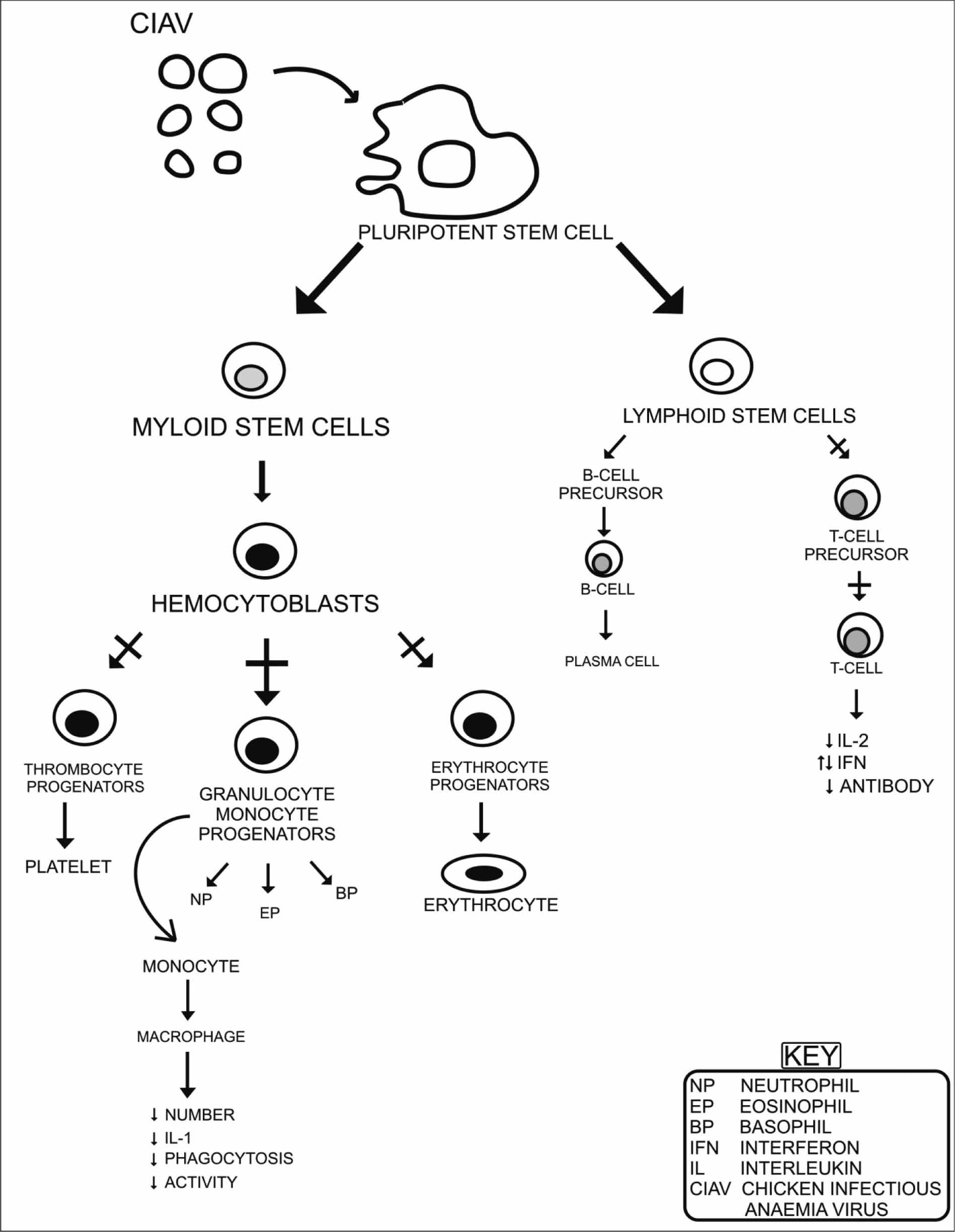
Destruction of precursor T cells results in depletion of mature cytotoxic and T-helper cells, with consequent effects on susceptibility and enhancement of the pathogenicity of secondary infectious agents and lower antibody responses (Adair, Reference ADAIR2000). It has been observed that there is a substantial reduction in macrophage function such as bactericidal activity, phagocytosis, cytokine (IL-1) production and Fc receptor expression (Tan and Tannock, Reference TAN and TANNOCK2005). Inhibition of interleukins (IL-1, IL-2, etc.) and IFN production adversely affects molecular immunoregulatory responses in the cytotoxic activities of macrophages, cytotoxic T-lymphocytes, natural killer (NK) cells and the expression of surface receptors (Figure 1) (Hu et al., Reference HU, LUCIO and SCHAT1993; De Herdt et al., Reference DE HERDT, VAN DEN BOSCH, DUCATELLE, UYTTEBROEK and SCHRIER2001). Cytokines and interleukins are an important part of the immune system and have been studied extensively (Shah, Reference SHAH2013; Shah et al., Reference SHAH, XU, YAN, SONG and LI2010; Reference SHAH, XU, YAN, SONG and LI2011; Song et al., Reference SONG, XU, YAN, HUANG, SHAH and LI2009; Xu et al., Reference XU, SONG, XU, YAN, SHAH and LI2008). Studies conducted using co-infection with IBDV and CIAV showed that highly virulent IBDV infection inhibited production of neutralising antibodies to CIAV in chickens at seven days post infection (DPI) (Kuscu and Gurel, Reference KUSCU and GUREL2008). This study first examined the persistence of CIAV in blood cells and the effective enhancement of primary CIAV infection as a result of low level of immunity caused by IBDV infection. Haematocrit, histopathology, intracellular calcium signalling, and lymphocyte sub-populations were analysed over a six week period in individual chicks inoculated with the isolate of CIAV. The mean lymphocyte proliferation stimulation index (SI) of the inoculated group was significantly lower than that of the control group. Percentages of CD3+, CD4+, CD8+ and NK cells decreased significantly (Bounous et al., Reference BOUNOUS, GOODWIN, BROOKS, LAMICHHANE, CAMPAGNOLI, CARDONA, LUCIO, O'CONNELL, JAGNE and SCHAT2000). CIAV exerts a destructive effect on both primary and secondary lymphoid tissues and especially suppresses the population of both helper (CD4+) and cytotoxic (CD8+) lymphocytes in the thymus (Adair, Reference ADAIR2000). Poor antibody response after CIAV infection at one day of age is a consequence of depressed T-helper responses in the early phase of infection (Kuscu and Gurel, Reference KUSCU and GUREL2008). There is marked damage to haematopoietic and lymphopoietic tissues, viz. stem cells in bone marrow and precursor T-lymphocytes in the thymus (Figure 1). The bursa, spleen and other lymphoid organs are also depleted of lymphoid cells (Dhama et al., Reference DHAMA, KATARIA, DASH, SENTHILKUMAR and TOMAR2002).
Decreased immunological activities were observed in CIAV-infected birds in the presence of concurrent infection with other viruses such as Marek's disease (Markowski-Grimsrud and Schat, Reference MARKOWSKI-GRIMSRUD and SCHAT2003), infectious Bursal disease (Hagood et al., Reference HAGOOD, KELLY, WRIGHT and HOERR2000) and adenoviruses (Balamurugan and Kataria, Reference BALAMURUGAN and KATARIA2006), leading to synergistic effects of both agents. High doses of aflatoxin resulted in elevated mortality, immunosuppression and possibly vertical transmission of CIAV (Kuscu and Gurel, Reference KUSCU and GUREL2008). CIAV infection causes decreased immune response against several vaccine viruses leading to vaccination failures or aggravation of the residual pathogenicity of attenuated vaccine viruses, and could even promote the emergence of variant viruses (Dhama et al., Reference DHAMA, KATARIA, DASH, SENTHILKUMAR and TOMAR2002; Adair and McNulty, Reference ADAIR and MCNULTY1997). As an example of this, birds immunised against coccidiosis in the presence of CIAV infection showed a lower level of protection (Dhama et al., Reference DHAMA, KATARIA, DASH, SENTHILKUMAR and TOMAR2002). The impairment of specific CTL after natural and experimental infections of chickens with CIAV and MDV has been reported (Markowski-Grimsrud and Schat, Reference MARKOWSKI-GRIMSRUD and SCHAT2003; Adair, Reference ADAIR1996). Similar assays for IFN-γ transcripts demonstrated a ten-fold increase in IFN-γ mRNA levels following infection with reticuloendothelial virus (REV) or REV with CIAV, while the latter alone caused a two- to four-fold increase (Smyth et al., Reference SMYTH, MOFFETT, CONNOR and MCNULTY2006; Davidson et al., Reference DAVIDSON, ARTZI, SHKODA, LUBLIN, LOEB and SCHAT2008). These results show a strong link between CIAV antibody status, replication, and the ability to generate REV-specific cytotoxic T lymphocytes. Because these viruses decrease immunity by subclinical infections, there is need for assessment of the immune status of chickens. Interference with induction of transcription of chicken IFN-α and IFN-γ was noted after sub-clinical infection with CIAV or IBDV (Balamurugan and Kataria, Reference BALAMURUGAN and KATARIA2006).
Depletion of thymocytes is caused by apoptosis, as evidenced from apoptosis-specific laddering patterns in DNA isolated from infected thymocytes, condensed chromatin and apoptotic bodies in the cytoplasm of epithelial cells (Miller et al., Reference MILLER, EALEY, OSWALD and SCHAT2003). The viral protein VP3 (apoptin) induces apoptosis in specific lymphoid cells, chicken thymocytes and lymphoblastoid cell lines (Hagood et al., Reference HAGOOD, KELLY, WRIGHT and HOERR2000), and this is an important phenomenon during the pathogenesis of CIAV (Hussein et al., Reference HUSSEIN, YOUSSEF, OSMAN, EL-EBIARY and SHALABY2003). The main cytopathogenic effects of CIAV infection are the induction of the apoptotic machinery (Noteborn, Reference NOTEBORN2004) and the appearance of viral protein-3 (VP3) which induced characteristic ‘doughnut-like’ structures similar to those observed in CIAV-infected cells (Noteborn and Koch, Reference NOTEBORN and KOCH1995). Apoptosis appears to be a feature of the lymphocyte depletion in the thymic cortex, which may be mediated by apoptin (VP3) (Dhama et al., Reference DHAMA, KATARIA, DASH, SENTHILKUMAR and TOMAR2002).
Proposed model of CIAV immunopathogenesis: haemocytoblasts in the bone marrow and T cell progenitors in the cortex of the thymus are main target cells of chicken infectious anaemia virus (CIAV).

Young chicks orally infected with CIAV had marked reduction in macrophage and T cell functions (Adair, Reference ADAIR2000; Kuscu and Gurel, Reference KUSCU and GUREL2008). These immunosuppressive effects of CIAV make it difficult for the exposed birds to develop an effective immune response against invading pathogens. The destruction of T cell precursors, granulocyte and macrophage progenitors, place adverse impacts on the bird's immune system (Schat, Reference SCHAT2009).Vaccine failure has resulted to several viruses under experimental conditions during severe CIAV induced immune-suppression. Depleted availability and supply of almost all T cells and reductions in essential cytokine production result in negative effects on other healthy cells which are not attacked by virus itself (Moeini et al., Reference MOEINI, OMAR, RAHA and YUSOFF2011). Thus, non-availability of T helper cells and cytokines affect the normal function of the B cell population and this drastically leads to reduced antibody responses (Adair, Reference ADAIR2000).
Haemocytoblast are specialised bone marrow cells from which the erythroid and myeloid series are derived. The haemocytoblasts in the bone marrow are among the first cells in which CIAV antigen is detected at around 3 ± 4 days after infection at day-old (Noteborn, Reference NOTEBORN2004). The destruction of haemocytoblasts results in severe decreases in erythroid and myeloid cells which ultimately leads to the anaemia which is characteristic of the disease (Drén et al., Reference DRÉN, KANT, VAN ROOZELAAR, HARTOG, NOTEBORN and KOCH2000). Thrombocytes are derived from haemocytoblats and the concurrent decrease in their number is probably the cause of intramuscular haemorrhages (Kuscu and Gurel, Reference KUSCU and GUREL2008). These destructive changes are observed in blood leading to decreased haematocrit levels and the number of circulating leukocytes from eight days post-infection (Adair, Reference ADAIR2000).
Neutralising antibodies of low titre are first detectable after three weeks post inoculation of CIAV to day-old chickens (Yuasa and Yoshida, Reference YUASA and YOSHIDA1983). The immune response and antibodies titre is more rapid and higher if older chicken of 28 or 42 days age are inoculated with CIAV. In older chickens the response is long lasting and the virus is cleared from blood as well as several other tissues, however, infectivity may still persist in some of the organs (Schat, Reference SCHAT2009; Hermann et al., Reference HERMANN, KOSKI, TAYLOR and GATEWOOD2012).
Complete protection against CIAV induced disease has been shown to be conferred by maternal antibodies. It is supported by many studies which showed CIAV infection and development of specific antibodies in chickens of eight and 12 weeks age as maternal antibodies disappear at two to three weeks of age. Such infections are the result of horizontal transmission and the disease does not develop (Adair, Reference ADAIR2000).
Future prospects in CIA vaccination
Vaccination, good poultry health and management are the proper control measures for CIA. During rearing, immunisation of breeder flocks against CIAV is thought to provide more passive immunity for young chicks during first weeks of life (Oluwayelu et al., Reference OLUWAYELU, OLALEYE and TODD2010) and will minimise vertical transmission of the virus. So far, inactivated CIAV vaccine immunisation has produced significant results for breeders. Progeny derived after such immunisation are less prone to CIAV infections and perform better based on body weight, liveability and feed conversion. Such vaccinations are economically more beneficial for broiler producers (Fussell, Reference FUSSELL1998). However, consideration should be given to the nature and immunopathogenesis of CIAV infection in relation to other agents before the introduction of a vaccination programme. Likewise, CIAV pathogenicity is enhanced with IBDV co-infection, so IBD control should be first priority in such cases (Oluwayelu et al., Reference OLUWAYELU, OLALEYE and TODD2010). Acquired immunity from the use of vaccines efficiently prevents vertical transmission in the flock (Gurunathan et al., Reference GURUNATHAN, KLINMAN and SEDER2000). Using chicken embryo-propagated Cux-1 isolates live vaccine, although it gave effective protection, harbours the risks of reversion to virulence (Sanjay et al., Reference SANJAY, KLINMAN and SEDER2000). Cell culture-propagated CIAV in an adequate titre can serve as an effective drinking-water vaccine; however, reversion to virulence, even after many passes, has been reported (Moeini et al., Reference MOEINI, OMAR, RAHA and YUSOFF2011). Vaccination should be performed three to four weeks prior to the onset of lay to avoid the hazard of spreading the vaccine virus through eggs (Schat et al., Reference SCHAT, MARTINS, O'CONNELL and PIEPENBRINK2011). Moeini et al. (Reference MOEINI, OMAR, RAHA and YUSOFF2011) developed an effective recombinant subunit vaccine (VP1 and VP2 proteins of CIAV) using a baculovirus insect cell culture system to provide an alternative safe and cost-effective vaccine. Yamaguchi et al. (Reference YAMAGUCHI, IMADA, KAJI, MASE, TSUKAMOTO, TANIMURA and YUASA2001) showed that a single amino acid change at residue 394 of VP1, a major determinant of pathogenicity, can generate a low-pathogenicity CIAV. These approaches can provide good potential candidates for development of a safe and stable genetically modified, live-attenuated CIAV vaccine, which could help in disease control.
In a recent improvement, plasmid DNA-based vaccines have emerged as one of the more promising applications of non-viral gene therapy. DNA vaccines are potentially safer than other traditional vaccines; have the potential for simultaneous immunisation against multiple antigens or pathogens; and more importantly, can stimulate both humoral and cell-mediated immune responses. This makes them attractive for the development of effective subunit vaccines against viral and bacterial pathogens which will likely require both neutralising antibody and cell-mediated immune responses for protection (Ullah et al., Reference ULLAH, RIAZ, UMAR and SHAH2013; Gurunathan et al., Reference GURUNATHAN, KLINMAN and SEDER2000). CIAV subunit vaccine expresses CIAV proteins in recombinant baculovirus (Moeini et al., Reference MOEINI, OMAR, RAHA and YUSOFF2011), and it has been shown that co-synthesis of VP1 and VP2 is required for the induction of neutralising antibodies that protect progeny chicks against CIAV challenge (Sommer and Cardona, Reference SOMMER and CARDONA2003). However, vaccination with plasmid DNA encoding such CIAV proteins has not been studied. The development of a DNA vaccine against CIAV using a eukaryotic co-expression vector encoding the VP1 and VP2 proteins of CIAV, simultaneously may be important.
Conclusions
Chicken infectious anaemia is a global problem and causes adverse effects on poultry health and production systems, especially in the broiler industry and for the production of specific pathogen free eggs. There is a need to conduct epidemiological studies of the disease especially in developing countries and develop suitable diagnostic and control measures. Policies for the control of the immunosuppressive behaviour of disease are not consistent between poultry companies. However, research and field observations are helping farmers to refine their strategies for controlling immunosuppression caused by CIAV, which are largely based on the vaccination programmes for breeders and their progeny, and good management practices to minimise stress. There is a strong likelihood that this viral infection will be recognised in additional avian species and thus poultry farmers will be challenged with new and prevailing immunosuppressive viral diseases. Both horizontal and vertical transmission of CIAV leads to its potential spread to other geographical areas. Nevertheless, there is ongoing research in this field and it is expected that many exciting developments will arise in the next decade. The poultry industry is refining their strategies for controlling immunosuppression based on research and field observations. Recombinant DNA-based methodologies will have major roles in both diagnosis and the development of vaccines for the control of CIAV. However, strategies to control immunosuppression are largely based on vaccination programs for breeders and their progeny. The best approach to minimise the prevalence of this disease is to use the best vaccines available, properly apply them and ensure an appropriate level of nutrition to enable the best possible response to these vaccines. New advances in avian immunology research progress the understanding of defence mechanisms of CIAV at a molecular level. Hyper-immunisation of birds using immune potentiating agents like cytokines can be a proficient approach to cope with the situation. There are still many gaps in our knowledge of the pathogenesis and epidemiology of CIAV. However, with the availability of reagents such as CIAV-specific monoclonal antibodies and DNA probes, it should be possible in the near future to answer questions about CIAV latency, the pathogenesis of subclinical infections, the role of CIAV in immunosuppression and its interaction with other viruses and infectious agents. Similarly the development of more sensitive and rapid diagnostic methods will more clearly define the importance of CIAV as a pathogen worldwide.