Introduction
In amphibians, mature oocytes are arrested in metaphase II due to the presence of high levels of active maturation promoting factor (MPF), a complex composed of cyclin B and cyclin dependent kinase (cdc2/p34). After fertilization, MPF activity decreases abruptly, which allows the oocyte to complete meiosis II (Gautier & Maller, Reference Gautier and Maller1991; Minshull et al., Reference Minshull, Murray, Colman and Hunt1991; Ookata et al., Reference Ookata, Hisanaga, Okano, Tachibana and Kishimoto1992; Galas et al., Reference Galas, Barakat, Doreé and Picard1993; Schultz & Kopf, Reference Schultz and Kopf1995; Sánchez Toranzo et al., Reference Sánchez Toranzo, Giordano, López and Bühler2007). In fertilization, the spermatozoon acts as a signal that triggers transient increases in free Ca2+ concentrations that are essential to trigger egg activation events (cortical granule exocytosis, meiosis resumption and pronuclear formation) and would be responsible for MPF inactivation (Watanabe et al., Reference Watanabe, Hunt, Ikawa and Sagata1991, Kline & Kline Reference Kline and Kline1992; Abrieu et al., Reference Abrieu, Doree and Fisher2001; Liu et al., Reference Liu, Chen, Cheng and Ju2002).
The increases in free Ca2+ concentrations originate predominantly from intracellular stores located in the endoplasmic reticulum (Miyazaki et al., Reference Miyazaki, Shirakawa, Nakada and Honda1993; Deguchi et al., Reference Deguchi, Osanai and Morisawa1996); and it is mainly mediated by one or both intracellular calcium channels: the inositol triphosphate receptor (IP3R) and the ryanodine receptor (RyR).
Our laboratory has reported evidence of the existence and functionality of IP3Rs and RyRs from in vitro matured R. arenarum oocytes. Both receptors play a role in Ca2+ release mechanisms during activation of R. arenarum oocytes (Ajmat et al., Reference Ajmat, Bonilla, Zelarayán and Bühler2011).
Great varieties of chemical substances or physical stimuli can also induce oocyte activation and are used in protocols in vitro fertilization and cloning, allowing the initiation of egg development. Ionomycin, a calcium ionophore, induces a peak in [Ca2+]i, it has been used in association with 6-dimethylaminopurine (6-DMAP), a protein kinase inhibitor; this procedure induces the formation of pronuclei and blastocysts in bovines, but the pregnancy rate is very low, which is attributed to polyploidy due to treatment with 6-DMAP (Bhak et al., Reference Bhak, Lee, Ock, Mohana Kumar, Choe and Rho2006). Therefore, the testing of new products to induce oocyte activation as well as the use of biological systems that affords methodological advances such as amphibian oocytes acquire great significance.
Dehydroleucodine (DhL), a sesquiterpene lactone belonging to the guaianolide group, is the active principle of a wild plant, Artemisia douglasiana Besser. This lactone has been demonstrated to induce cell cycle arrest in G2 in cultures of Allium cepa meristematic cells (López et al., Reference López, Giordano and López2002), in myocytes from the blood vessels of cultured rats (Cruzado et al., Reference Cruzado, Castro, Fernandez, Gomez, Roque, Giordano and Lopez2005, Polo et al., Reference Polo, Castro, Cruzado, Collino, Cuello-Carrión, Ciocca, Giordano, Ferrari and López2007) and in cancer cells (Siriwan et al., Reference Siriwan, Naruse and Tamura2011).
Our work team has demonstrated that DhL inhibits meiosis resumption in R. arenarum oocytes, preventing MPF activation (Sánchez Toranzo et al., Reference Sánchez Toranzo, Giordano, López and Bühler2007). It has also been observed that the inactivation of the alpha-methylene lactone function by DhL hydrogenation decreases but does not eliminate the biological effect of this lactone, which suggests that the lactone group would not be solely responsible for its activity (Sánchez Toranzo et al., Reference Sánchez Toranzo, López, Martínez, Bühler and Bühler2009).
The aims of this work are to test the ability of DhL and its hydrogenated derivative 2H-DhL to induce chemical activation in R. arenarum oocytes and to study the participation of calcium in the process.
Materials and methods
Animals
Adult specimens of Rhinella arenarum were collected in the northwestern area of Argentina and kept at 15°C until use, which generally took place 7 days after collection. Experimental manipulation and oocyte culture were conducted at room temperature (22–25°C) using Amphibian Ringer solution (AR): 6.6 g NaCl/l, 0.15 g KCl/l, 0.15 g CaCl2/l, containing penicillin G-sodium (30 mg/l), streptomycin sulphate (50 mg/l) and 0.005 M Tris–HCl buffer (pH 7.4). The activation assays were performed in calcium-free Tris-buffered saline solution containing 7.59 g NaCl/l, 2.40 g Tris/l; HCl added to pH 7.4. EGTA (0.01 mM) was added to ensure the absence of calcium in the culture medium (Ca2+-free AR). Reagents were added (5 μl) directly to the culture medium.
Hormones and reagents
Progesterone (Sigma) was dissolved in ethanol and added directly to the culture medium to give a final concentration of 2.5 μM. Heparin sodium salt was from Rivero. Ruthenium red (Ted Pella) was dissolved in ddH2O in order to obtain a suspension; then it was heated at 60°C for 5 min while shaking and centrifuged at 1600 rpm for 10 min. The supernatant resulted in a stock solution <1%. BAPTA/AM [1,2-bis(2-aminophenoxy) ethane-N,N,N′,N′-tetraacetic acid tetrakis (acetoxymethyl ester)] at ≥95% purity was dissolved in dimethylsulphoxide (DMSO) and added directly to the culture medium to give a final concentration of 10, 50 and 100 μM. Dehydroleucodine (DhL) at 93% purity and 11,13-dihydro-dehydroleucodine (2H-DhL) at 95% purity were obtained according to Giordano (Giordano et al., Reference Giordano, Guerreiro, Pestchanker, Guzman, Pastor and Guardia1990, Reference Giordano, Pestchanker, Guerreiro, Saad, Enriz, Rodriguez, Jauregui, Guzman, Maria and Wendel1992). DhL and its derivative were dissolved in DMSO and various doses were added to the culture medium.
Oocytes collection
Fully grown ovarian oocytes (1.7–1.8 mm in diameter) were obtained from adult female specimens of Rhinella arenarum. Oocytes were denuded by manually pulling off the follicle epithelium and theca layer using fine forceps under a stereoscopic microscope. Follicle cells were removed by shaking in AR for 5 min with gentle shaking (100 oscillations/min). Oocytes that remained only with the vitelline envelope were considered as denuded oocytes.
In vitro maturation of oocytes
Hormonal maturation was induced by treatment of denuded oocytes with progesterone. Oocyte maturation was assessed 18 h after follicle cell removal or hormone addition. Meiotic reinitiation was scored both by the presence of a transient white spot at the animal pole and by cytological examination. In our working conditions, oocytes reached metaphase II at 18 h after defolliculation or progesterone treatment.
Sperm injection procedures
Eggs were microinjected using a Leitz Wetzlar Micromanipulator apparatus equipped with an injection pipette. Glass micropipettes (Humagen™ Fertility Diagnostics) were filled by suction of a microdrop (30 nl) containing sperm suspension (final concentration 2 × 10⁶). All injections were carried out at room temperature. Microinjections were performed when eggs were correctly positioned with the animal pole facing outward and the injection pipette was pushed into the cytoplasm.
Sperm suspensions were obtained by gently disrupting the testes in 4 ml AR and centrifuging at 1085 g for 10 min; then, the pellet was resuspended in AR.
Cytological preparations
Oocytes were fixed in Ancel and Vintemberger's solution (10% formaldehyde, 0.5% acetic acid and 0.5% NaCl), embedded in paraffin and sliced into 5 μm thick sections. Then they were stained with Alcian Blue at pH 2.5 and counterstained with eosin. This method allowed us to observe cortical granules.
Statistical analysis
Results are expressed as means ± standard error of the mean (SEM). Comparisons among different treatments were carried out using Student's t-test at a 5% significance level.
Results
Effect of DhL on oocyte activation
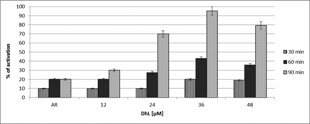
In order to determine the ability of DhL to induce oocyte activation, denuded ovarian oocytes matured in vitro with progesterone (2.5 μM) were exposed to different doses of DhL (12, 24, 36, 48 μM) and cultures during different times (30, 60, or 90 min). Results (Fig. 1) indicated that DhL at concentrations of 12–36 μM, induced oocyte activation in a dose- and time-dependent manner. We found a decrease in activation levels at higher concentrations of DhL. As shown in Fig. 1, DhL 36 μM was able to induce 95% activation after 90 min of treatment.
Dose–time response curve of DhL. Groups of 20 denuded fully grown ovarian oocytes matured in vitro with progesterone (2.5 μM) were incubated in AR alone or AR with different doses of DhL (12, 24, 36, 48 μM) and after different times (30, 60, 90 min) activation parameters were evaluated. Values are the mean ± standard error of the mean (SEM) (n = 5). Each experiment was performed on a different animal.

Effect of 2H-DhL on oocyte activation
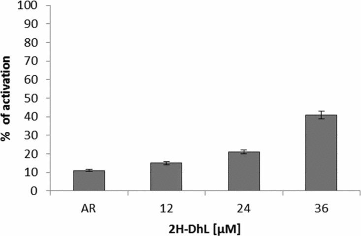
In order to determine whether a derivative of DhL inactivated for alpha-methylene lactone sesquiterpene lactones could induce activation in the same way as does DhL, different doses of 2H-DhL (12, 24 and 36 μM) were assayed in denuded ovarian oocytes matured in vitro with progesterone (2.5 μM). The signs of activation were scored after 90 min of culture (Fig. 2).
Dose–response curve 2H-DhL. Groups of 20 denuded fully grown ovarian oocytes matured in vitro with progesterone (2.5 μM) were incubated in AR alone or AR with different doses of 2H-DhL (12–36 μM) and after 90 min activation parameters were evaluated. Values are the mean ± standard error of the mean (SEM) (n = 5). Each experiment was performed on a different animal.

Results indicated that 2H–DhL is able to induce oocyte activation, but is less active than DhL; with the highest dose of 2H-DhL (36 μM) assayed, only 40% activation was obtained.
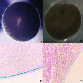
The external morphological features of activation induced by DhL and 2H-DhL were similar to those observed in normal fertilization. Lactone-treated oocytes exhibited flattening of the animal pole, disappearance of the white spot and elevation of the vitelline envelope (Fig. 3 A, B). The cortical granule exocytosis of oocytes activated by treatment with DhL or 2H-DhL was scored in cytological preparations stained with Alcian Blue. As shown in Fig. 3 D, oocytes activated with DhL or 2H-DhL showed negative Alcian Blue, indicating a complete extrusion of cortical granules. Matured oocytes cultured in AR for 90 min were used as Alcian Blue positive controls (Fig. 3 C). All these morphological parameters are similar to the ones observed in sperm-induced oocyte activation.
Oocytes activation. (A) Oocyte matured in vitro with progesterone exhibiting a white spot on the animal pole. Note the vitelline envelope close to the plasma membrane. (B) Oocytes treated with DhL or 2H-DhL. Note the disappearance of the white spot, the elevation of the vitelline envelope and the flattening of the animal pole. (C) Section of a mature oocyte stained by Alcian Blue–eosin. There is a continuous line of cortical granules under the plasma membrane (×400 magnification). (D) Section of an oocyte activated with DhL treatment. Exocytosis of the granules has occurred. Alcian Blue–eosin (×600 magnification).

Pronucleus formation
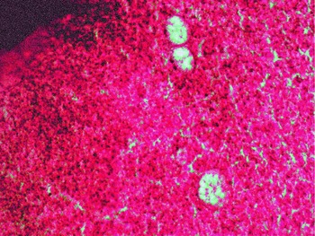
To ascertain the ability of DhL-activated oocytes to induce pronuclear formation, they were microinjected with homologous sperm approximately 2 min after DhL (36 μM) treatment. Pronuclear formation was monitored by cytological examination of oocytes fixed at various times after sperm injection. Two hours after sperm injection, oocytes exhibited well developed pronuclei (Fig. 4).
Pronuclear formation. Section of an oocyte treated with DhL 36 μM and microinjected with 30 nl of sperm suspension. Pronuclear formation was scored 2 h after microinjection in cytological preparations. There are three well developed pronuclei. Hematoxylin–eosin (×1000 magnification).

Involvement of extracellular calcium in DhL-induced activation
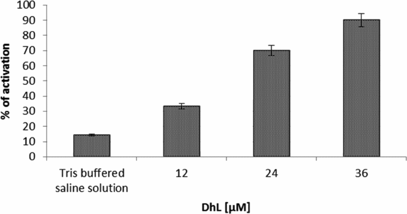
In order to determine the participation of extracellular calcium in the activation induced by different doses of DhL, mature oocytes were cultured in medium with and without Ca2+ for 90 min. The results obtained (Fig. 5) indicate that the absence of Ca2+ in the culture medium did not significantly affect the percentage of DhL-induced activation.
Involvement of extracellular calcium in DhL-induced activation. Groups of 20 denuded fully grown ovarian oocytes matured in vitro with progesterone (2.5 μM) were incubated calcium-free Tris-buffered saline solution with different doses of DhL (12–36 μM). Activation parameters were evaluated after 90 min of culture. Values are the mean ± standard error of the mean (SEM) (n = 4). Each experiment was performed on a different animal.

Effect of BAPTA/AM on DhL-induced activation
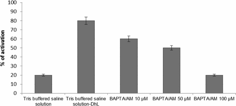
In order to analyse whether or not intracellular free calcium was needed for DhL-induced activation, matured oocytes were pretreated for 30 min with the calcium chelator BAPTA/AM (10, 50, 100 μM) before addition of DhL (36 μM). The signs of activations were monitored after 90 min of incubation considered from the moment when the inducer was added. The results in Fig. 6 show that treatment with BAPTA/AM at the concentrations tested significantly inhibited DhL-induced activation. As control we used oocytes treated only with DhL (36 μM). This result suggests that intracellular calcium is critical in DhL-induced oocyte activation.
Effect of BAPTA/AM on DhL-induced activation. Groups of 20 denuded fully grown ovarian oocytes matured in vitro with progesterone (2.5 μM) were pre-incubated for 30 min in calcium-free Tris-buffered saline solution and different doses of BAPTA/AM (10–100 μM) and activation was induced by DhL (36 μM). Activation parameters were evaluated after 90 min of culture. Values are the mean ± standard error of the mean (SEM) (n = 4). Each experiment was performed on a different animal.

Participation of IP3Rs and RyRs in DhL-induced oocyte activation
In order to determine the participation of IP3Rs and RyRs in intracellular Ca2+ release during DhL-induced activation, we evaluated the effect of heparin, an IP3R antagonist, and ruthenium red, a known RyR antagonist.
According to the report of Ajmat et al. (Reference Ajmat, Bonilla, Zelarayán and Bühler2011), both heparin and ruthenium red induce inhibition of activation when they are microinjected or added to the culture medium. On the basis of these data, we used the doses with the greatest effect on the activation of this species when added to the culture medium.
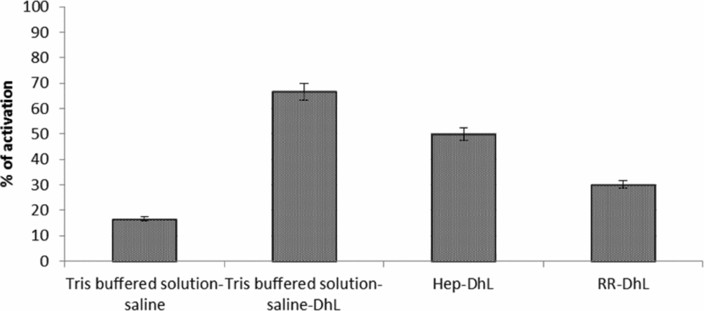
Denuded ovarian oocytes matured in vitro with progesterone (2.5 μM) were pre-incubated in calcium-free Tris-buffered saline solution for 30 min (control) and in calcium-free Tris-buffered saline solution with ruthenium red (100 μM) or heparin 10 μg/ml before the addition of DhL (36 μM). Oocyte activation was scored after 90 min of culture. The results presented in Fig. 7 show that heparin inhibited 20% DhL-induced activation while ruthenium red inhibited 40% compared to control.
Effect of heparin and Ruthenium Red on DhL-induced oocyte activation. Groups of 20 denuded fully grown ovarian oocytes matured in vitro with progesterone (2.5 μM) were pre-incubated in calcium-free Tris-buffered saline solution with the addition of heparin (10 μg/ml) or ruthenium red (100 μM) for 30 min before inducing activation with DhL (36 μM). Activation parameters were evaluated after 90 min of culture. Values are the mean ± standard error of the mean (SEM) (n = 5). Each experiment was performed on a different animal.

Discussion
Our results indicated that dehydroleucodine (DhL) and its hydrogenated derivative 11,13-dihydro-dehydroleucodine (2H-DhL) were able to induce oocyte activation in a dose-dependent manner and that the process is independent of extracellular Ca2+ but dependent on intracellular free Ca2+.
2H-DhL is less active than DhL, showing that the inactivation of the alpha-methylene lactone function by the hydrogenation of DhL decreases but does not eliminate the biological effect of this lactone since higher doses were required to obtain a significant effect. These results agree with the reports of Sánchez Toranzo et al. (Reference Sánchez Toranzo, López, Martínez, Bühler and Bühler2009) concerning the effect of lactones as inhibitors of oocyte maturation and suggest that the effect of DhL on meiosis progression does not depend only on the activity of the alpha-methylene lactone function.
The experiments with sperm injection indicate that the activation induced by DhL is genuine, as oocytes are capable of inducing pronucleus formation.
Considering that in the process of oocyte activation MPF must be inactivated to allow the end of meiosis, it is possible that the lactones assayed could act by inhibiting the activity of this factor, which would also account for their effect as maturation blockers in this species (Sánchez Toranzo et al., Reference Sánchez Toranzo, López, Martínez, Bühler and Bühler2009). It is known that the fertilizing sperm induce an increase in free-Ca2+ concentration in oocytes that cause the inactivation of MPF, thus allowing the completion of meiosis and the start of the activation process. In these species, oocytes have an important pool of sequestered Ca2+ that supports the activation induced by DhL with no need of external Ca2+. Similar results were reported by Zelarayán et al. (Reference Zelarayán, Toranzo, Oterino and Bühler2004) and Sánchez Toranzo et al. (Reference Sánchez Toranzo, Bühler and Bühler2012) with respect to the maturation process of this species.
Transient Ca2+ release from intracellular stores is modulated through both agonist-gated channels, IP3Rs and RyRs. Both receptors have been generally detected by functional criteria. IP3 induces repetitive or single calcium release in the oocytes of all species studied, while RyR is expressed only in the oocytes of some species. In Rhinella arenarum the presence of RyRs in the oocytes has been described as well as its participation in the process of activation (Ajmat et al., Reference Ajmat, Bonilla, Zelarayán and Bühler2011). Results achieved by Ajmat et al. (Reference Ajmat, Bonilla, Zelarayán and Bühler2011, Reference Ajmat, Bonilla, Hermosilla, Zelarayán and Bühler2013) show that ruthenium red 100 μM and heparin 10 μg/ml in the culture medium as well as the microinjection of ruthenium red 100 μM and heparin 1 μM blocked Ca2+ output from the intracellular reservoirs in R. arenarum oocytes and strongly inhibited activation induced by insemination with homologous sperm.
Although these two receptors may have different functional roles in the regulation of calcium release during meiotic maturation and fertilization, it appears that both IP3Rs and RyRs contribute to the significant increase in intracellular calcium during fertilization and activation in the bovine oocyte (Wang et al., Reference Wang, White, Reed and Campbell2005).
Experiments with antagonists of the RyRs (ruthenium red) and IP3Rs (heparin) revealed that both Ca2+-mobilizing systems were activated by lactones, and the blockage of either of the systems alone was not sufficient to prevent total activation induced with DhL. In this species, RyR appears to be the largest contributor to Ca2+ release in DhL-induced activation through the calcium-induced calcium-release mechanism (CICR). The differences in the percentages of activation induced by DhL (controls) obtained in these experiments could be because these experiments were conducted in the months of July and August when oocytes are less sensitive to treatment.
It is possible that these lactones activate a PLC with the consequent production of IP3, which would induce the release of Ca2+ from the endoplasmic reticulum. This increase in calcium would also activate RyRs, thus increasing Ca2+ release toward cytosol. This increase in Ca2+ would be responsible on the one hand for inhibiting the cytostatic factor that keeps MPF active, allowing the end of meiosis, and on the other for inducing the exocytosis of the cortical granules, which establishes a block to polyspermy.
Although more studies are needed on the mechanism of action by which these lactones induce oocyte activation in Rhinella arenarum, the results of this study open interesting perspectives for the use of these lactones as chemical activators in in vitro fertilization and cloning.
In summary, our results indicate that a sesquiterpene lactone, dehydroleucodine (DhL), and its hydrogenated derivative 11,13-dihydro-dehydroleucodine (2H-DhL) were able to induce oocyte activation in a dose-dependent manner. The effect of these lactones is independent of extracellular Ca2+ but dependent on intracellular Ca2+ release through IP3Rs and RyRs.
Acknowledgements
This work was supported by a grant from the Science Council of the National University of Tucumán (CIUNT) and the National Agency for the Promotion of Science and Technology (FONCYT).