Introduction
Inflammatory myofibroblastic tumour is a rare neoplasm which occurs in the soft tissues of young adults and children.Reference Weiss and Goldblum1 The most recent World Health Organization classification defined these lesions as ‘tumours of uncertain malignant potential’, due to their tendency to recur locally and the very small risk of distant metastasis.Reference Coffin, Fletcher, Fletcher, Unni and Mertens2 Inflammatory myofibroblastic tumour occurs most commonly in the lungs, where it presents as a solitary lung lesion. The lesion was first described at this location, and was originally thought to represent an inflammatory process rather than a neoplastic one.Reference Umiker and Iverson3 However, this tumour has been reported throughout the body; the most common extrapulmonary sites are the omentum and mesentery, while head and neck cases are considered rare.Reference Coffin, Watterson, Priest and Dehner4, Reference Batsakis5 The majority of head and neck inflammatory myofibroblastic tumours occur in the larynx, oral cavity, upper oesophagus and salivary glands.Reference Batsakis5 Only three cases of inflammatory myofibroblastic tumour of the tonsil have been described in the literature, in patients aged 41, 62 and 63 years.Reference Gangopadhyay, Mahasin, Kfoury and Ali6–Reference Weilbaecher and Sharma8
Here, we present the first reported case in the English language literature of inflammatory myofibroblastic tumour of the tonsil in a young, pregnant woman.
Case report
A 28-year-old, pregnant woman was referred to the emergency department with a 10-day history of sore throat, fever and malaise, which had failed to respond to antibiotics.
Examination was consistent with bilateral tonsillitis, with the right tonsil appearing larger than the left.
Despite intensification of antimicrobial therapy, the patient's symptoms failed to resolve, and she re-presented to the ENT department two weeks later.
She was a non-smoker and only an occasional drinker of alcohol, and had no past medical history of note.
Examination at this time revealed a clinically suspicious, grade II, erythematous right tonsil, with ulceration on the upper pole.
A computed tomography scan confirmed the presence of an enlarged right tonsil along with reactive, level II, right-sided nodes.
Since the patient was nine weeks' pregnant, a local biopsy was arranged.
Initial histological evaluation was suggestive of a spindle cell carcinoma. However, this diagnosis was revised after immunohistochemical staining confirmed the lesion to be an exceptionally rare inflammatory myofibroblastic tumour.
Carbon dioxide laser excision of the right tonsil was conducted. Histologically clear margins were obtained. There were no complications of surgery, and the patient progressed on to have an uneventful pregnancy.
Thirteen months after surgery, the patient remained free of disease.
Pathology
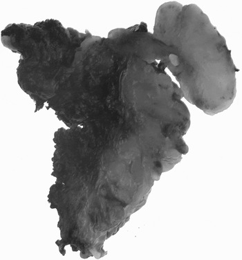
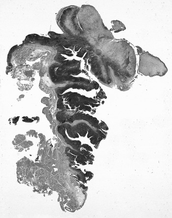
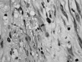
Macroscopically, an ulcerated, polypoid tumour was observed, (Fig 1, Fig 2) which microscopically comprised atypical large spindle cells mixed with inflammatory cells (Fig. 3). The tumour expressed smooth muscle actin, vimentin and cyclin D1 antigens. There was no expression of cytokeratin. Furthermore, both the biopsy and the main specimen were negative for anaplastic lymphoma kinase 1.
Sectioned surgical tonsil specimen, showing an exophytic, 2 cm maximal diameter mass on the surface. The tonsillar surface is ulcerated, with inflammatory slough.

Haematoxylin and eosin staining of the whole surgical specimen, showing an ulcerated, exophytic mass on the surface of the tonsil.

High power photomicrograph showing scattered spindle cells with vesicular nuclei admixed with plasma cells and eosinophils. (H&E; ×200)

Discussion
Inflammatory myofibroblastic tumour is an uncommon lesion. The term ‘inflammatory pseudotumour’ is a generic label applied to a wide range of inflammatory and neoplastic lesions. However, over the last two decades inflammatory myofibroblastic tumour has come to be considered as a distinct entity due to its characteristic histological and molecular features.Reference Coffin, Patel, Perkins, Elenitoba-Johnson, Perlman and Griffin9
Although these tumours were widely considered to be benign, Meiss and Enzinger reported considerable aggressive behaviour, including 10 cases of local recurrence and three of distant metastasis.Reference Meiss and Enzinger10 Although the lesion in these cases was termed ‘inflammatory fibrosarcoma’, there was considerable clinical and histological overlap with the lesion reported in Coffin and colleagues' 1995 paper under the name ‘extrapulmonary inflammatory myofibroblastic tumour’ (in this study, recurrence rates were lower and no metastases were documented).Reference Coffin, Watterson, Priest and Dehner4 As a result, these two synonyms have come to represent two ends of a neoplastic spectrum, with inflammatory myofibroblastic tumour being relatively benign and inflammatory fibrosarcoma its more aggressive counterpart.
It is also important to note that approximately 50 per cent of inflammatory myofibroblastic tumours demonstrate a chromosomal rearrangement of the anaplastic lymphoma kinase gene.Reference Coffin, Patel, Perkins, Elenitoba-Johnson, Perlman and Griffin9 This distinct molecular feature of inflammatory myofibroblastic tumours further supports the neoplastic nature of these lesions and their distinction from other inflammatory pseudotumours.
A review of the English language literature revealed only three previous reports of inflammatory myofibroblastic tumour of the tonsil.Reference Gangopadhyay, Mahasin, Kfoury and Ali6–Reference Weilbaecher and Sharma8 These three patients were aged 62, 63 and 41 years, and two of the three were taking immunosuppressants. The patient reported by Gangopadhyay et al. was a renal transplant recipient and therefore required long term immunosuppressants, including prednisolone. The patient described by Newman and Shinn had also taken long term prednisolone for asthma and retroperitoneal fibrosis.Reference Newman and Shinn7 It is therefore worthy of note that our patient was pregnant at the time of presentation; pregnancy is known to be an immunomodulated state, even during the earliest gestational period. Admittedly, the extent to which this immunomodulation leads to recognisable clinical immunocompromise is as yet not fully elucidated.Reference Lattuada, Mangioni, Viganò, Ntrivalas, Rossi and Palotti11–Reference Honig, Rieger, Kapp, Sutterlin, Dietl and Kammerer14
Inflammatory myofibroblastic tumour was first described in the lung, although similar lesions have subsequently been reported at various extrapulmonary sites, the most common being the mesentery and omentum. Although these tumours affect a wide age range, they show a predilection for young adults and children, with females being affected slightly more often than males.Reference Weiss and Goldblum1
The presenting symptoms of inflammatory myofibroblastic tumour depend on the site of tumour origin, but symptoms may also be systemic and non-specific and therefore suggestive of a neoplastic process. This was reflected in our case, with presenting symptoms including suspicious ulceration of the tonsil and non-specific malaise. Diagnosis is therefore dependent upon histology. The histological profile of inflammatory myofibroblastic tumour is very distinctive, being composed of a mixture of spindle cells together with a variable inflammatory component, which most commonly involves fibroblastic cells. Cytokeratin is expressed in 36–77 per cent of cases.Reference Weiss and Goldblum1 Unlike typical carcinomas, the expression is focal.
• Inflammatory myofibroblastic tumour very rarely involves the tonsil; this has previously been reported in only three, immunosuppressed patients in their fifth and seventh decades
• Inflammatory myofibroblastic tumour is known to be locally aggressive; the standard treatment at other sites is local excision
• The presented patient represents the first reported case in the English language literature of inflammatory myofibroblastic tumour involving the tonsil in a pregnant woman
• Recommended treatment comprises multidisciplinary consultation to establish the diagnosis, and subsequent complete local excision
Inflammatory myofibroblastic tumours are known to be locally aggressive, and recurrence rates vary according to anatomical site, being less than 2 per cent for tumours confined to the lung and increasing up to 25 per cent for extrapulmonary lesions.Reference Coffin, Fletcher, Fletcher, Unni and Mertens2, Reference Coffin, Watterson, Priest and Dehner4 Thus, we advocate complete surgical excision with careful follow up.
Conclusion
Inflammatory myofibroblastic tumour rarely occurs in the tonsil. Clinically and pathologically, this entity is known to be locally aggressive and can present in a manner not dissimilar to a high grade tonsillar carcinoma. As a result, a multidisciplinary approach should be taken in order to arrive at the correct diagnosis. Due to the risk of local recurrence, the recommended treatment is complete local excision with careful follow up. Although a rare lesion, inflammatory myofibroblastic tumour should be considered in the differential diagnosis of an enlarged tonsil, especially in an immunocompromised patient.