INTRODUCTION
Knowledge about diversity and distribution of leptostracans (Crustacea, Phyllocarida) has improved substantially in the last three decades (e.g. Dahl, Reference Dahl1985, Reference Dahl1990; Martin et al., Reference Martin, Vetter and Cash-Clark1996; Moreira et al., Reference Moreira, Díaz-Agras, Candas, Señarís and Urgorri2009), and a number of new taxa, including new genera, have been described all around the world, from marine caves to the deep sea (Hessler, Reference Hessler1984; Bowman et al., Reference Bowman, Yager and Iliffe1985; Haney et al., Reference Haney, Hessler and Martin2001; Haney & Martin, Reference Haney and Martin2004; Song et al., Reference Song, Moreira and Min2012), as well as in a variety of shallow-water habitats and substrates: sponges (Ortiz et al., Reference Ortiz, Winfield and Cházaro-Olvera2011), seagrasses (Dahl, Reference Dahl1985), algae (Haney & Martin, Reference Haney and Martin2005) and sediments (Vetter, Reference Vetter1996a, Reference Vetterb; Ledoyer, Reference Ledoyer1997; Moreira et al., Reference Moreira, Koçak and Katagan2007), or in baited traps (Olesen, Reference Olesen1999; Lee & Bamber, Reference Lee and Bamber2011).
The genus Nebalia Leach, 1814 is by far the most speciose to date, with more than 30 recognized species (Bochert & Zettler, Reference Bochert and Zettler2012). Most Nebalia species have been described from European coasts and the north-eastern Pacific Ocean, but in the last years a number of new species have been described, for instance, from south-eastern Asia, Gulf of Mexico and South Africa (e.g. Ortiz et al., Reference Ortiz, Winfield and Cházaro-Olvera2011; Bochert & Zettler, Reference Bochert and Zettler2012; Song et al., Reference Song, Moreira and Min2013). In the Mediterranean Sea, six species of Nebalia have been reported to date (Koçak et al., Reference Koçak, Moreira and Katagan2011) of which three are present in the eastern Mediterranean and possibly another two undescribed species as well (Koçak et al., Reference Koçak, Moreira and Katagan2010; Moreira et al., Reference Moreira, Sezgin, Katagan, Gönülal and Topaloglu2012).
New findings of leptostracan specimens from the Turkish Aegean Sea and Cyprus have revealed the presence of a new species of Nebalia, which is closely related to other sympatric species in the eastern Mediterranean Sea, N. strausi Risso, 1826 and N. kocatasi Moreira, Koçak & Katagan, Reference Moreira, Koçak and Katagan2007, and which is described herein as N. mediterranea sp. nov.
MATERIALS AND METHODS
Sampling was done in four subtidal stations at depths of between 27 and 67 m at Izmir Bay and Ildırı Bay, Aegean Sea (Turkey) and Famagusta Bay (Northern Cyprus). Samples were taken from 2003 to 2009 using a Van Veen Grab and then sieved through a 0.5 mm mesh. Specimens were fixed in 5% formaldehyde and later rinsed with fresh water and then preserved in 70% ethanol. Line drawings were done with the aid of a camera lucida connected to a compound microscope. Drawings were made from the holotype and one dissected paratype female. Measurements were done by using an ocular micrometer. Total length (TL) was measured from the articulation between rostrum and carapace to the posterior end of the caudal furca excluding setation; dorsal carapace length (DCL) was considered as the distance between the articulation of the rostrum and the margin of the posterodorsal cleft; lateral carapace length (LCL) was considered as the longest distance parallel to body axis along lateral surface between the anterior and the posterior margin; carapace height (CH) was measured as the longest perpendicular distance to body axis between the dorsal and the ventral margin; rostrum length (RL) was measured along the midline. Description of specimens was done following the model of Dahl (Reference Dahl1985) and other recent studies (e.g. Martin et al., Reference Martin, Vetter and Cash-Clark1996; Olesen, Reference Olesen1999; Haney & Martin, Reference Haney and Martin2005). The type series is deposited in the Museum of the Faculty of Fisheries, Ege University, Izmir (ESFM).
RESULTS
SYSTEMATICS
Order Leptostraca Claus, 1880
Family Nebaliidae Samouelle, 1819
Genus Nebalia Leach, 1814
Nebalia mediterranea sp. nov.
(Figures 1–7, Table 1)
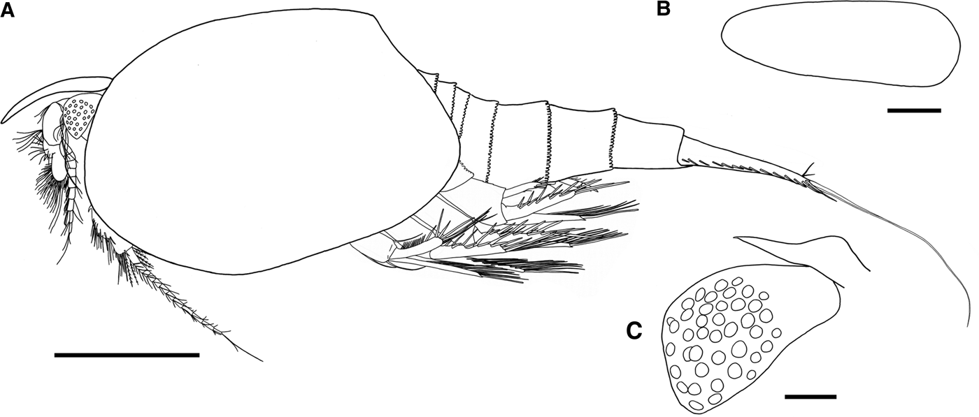
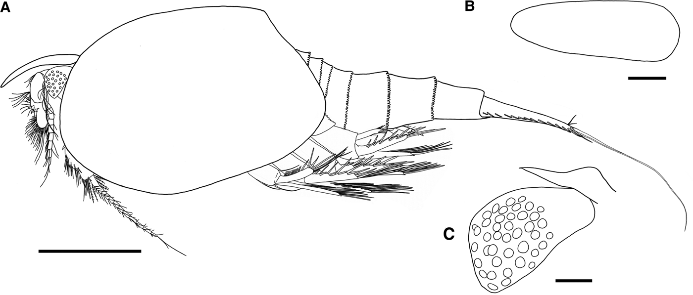
Nebalia mediterranea sp. nov., holotype, postovigerous female (ESFM-MAL/2009-004): (A) lateral view; (B) rostrum, dorsal view; (C) eye and supraorbital scale, lateral view. Scale bars: A, 1 mm; B, 0.2 mm; C, 0.1 mm.
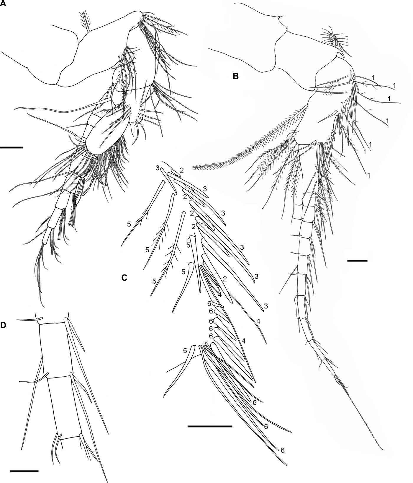
Nebalia mediterranea sp. nov., paratype, postovigerous female (ESFM-MAL/2009-006): (A) antennule, lateral view; (B) antenna, lateral view; (C) antenna, peduncle third article, lateral view; (D) antenna, peduncle, detail. Numbers on (B) and (C) refer to rows of setae described in the text. Scale bars: A–C, 0.1 mm; D, 50 μm.

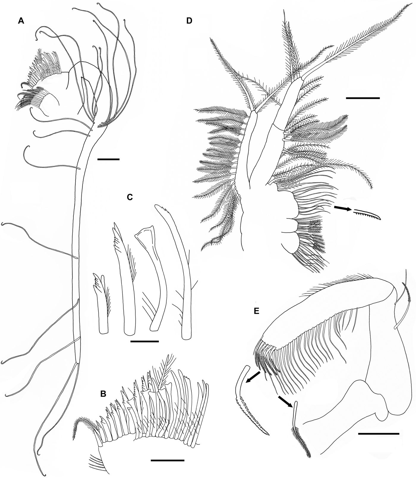
Nebalia mediterranea sp. nov., paratype, postovigerous female (ESFM-MAL/2009-006): (A) first maxilla, endites and palp; (B) first maxilla, second endite, detail; (C) first maxilla, second endite, setae types; (D) second maxilla; (E), mandible, mandibular palp and detail of setae of third article. Scale bars: A, D–E, 0.1 mm; B, 50 μm; C, 20 μm.
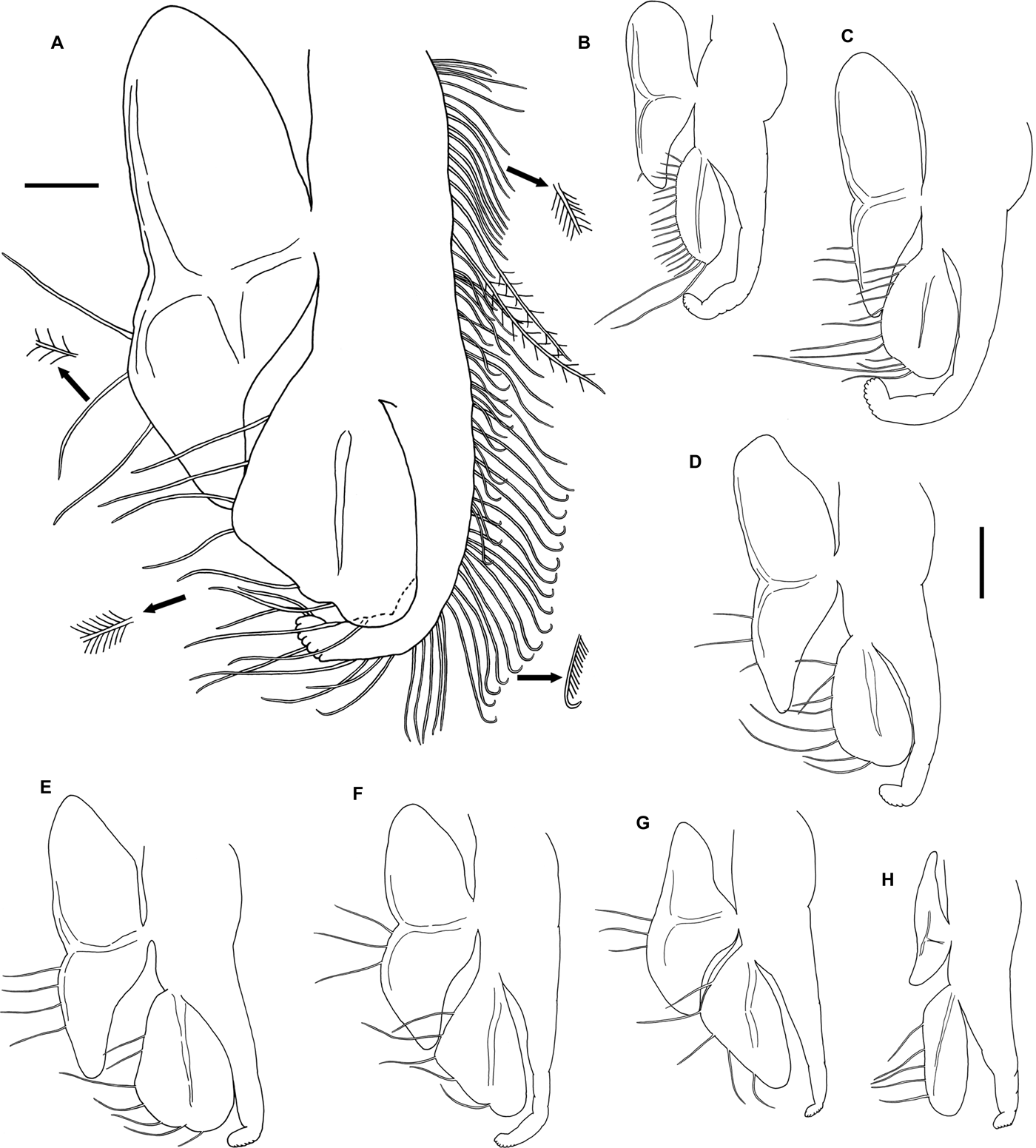
Nebalia mediterranea sp. nov., paratype, postovigerous female (ESFM-MAL/2009-006): (A) thoracopod 5, showing setation; (B) thoracopod 1; (C) thoracopod 2; (D) thoracopod 3; (E) thoracopod 4; (F) thoracopod 6; (G) thoracopod 7; (H) thoracopod 8. Scale bars: A, 0.1 mm; B–H, 0.2 mm. B–H, same scale bar.
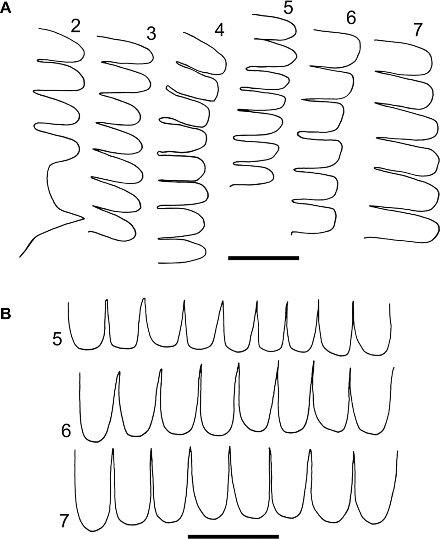
Nebalia mediterranea sp. nov., paratype, postovigerous female (ESFM-MAL/2009-006): (A) pleonites 2–7, posterior lateral border, denticles; (B) pleonites 5–7, posterior dorsal border, denticles. Scale bars: A & B, 50 μm.
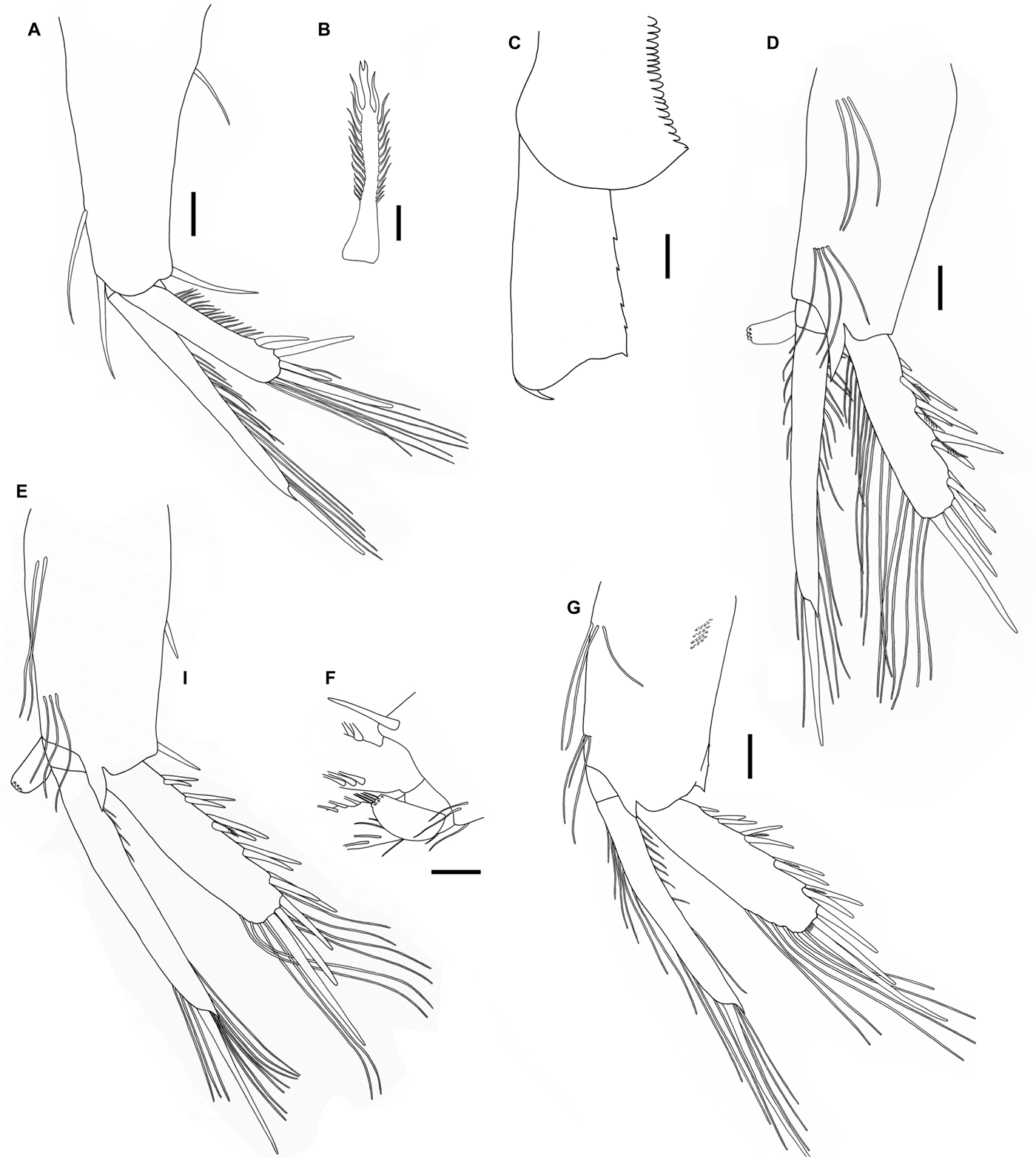
Nebalia mediterranea sp. nov., paratype, postovigerous female (ESFM-MAL/2009-006): (A) first pleopod, lateral view; (B) first pleopod, spine of external lateral border; (C) fourth pleopod, protopod, lateral view; (D) second pleopod, lateral view; (E), third pleopod, lateral view; (F) third pleopod, detail of protopod distal end and appendix interna; (G) fourth pleopod, lateral view. Scale bars: A, C–G, 0.1 mm; B, 10 μm.
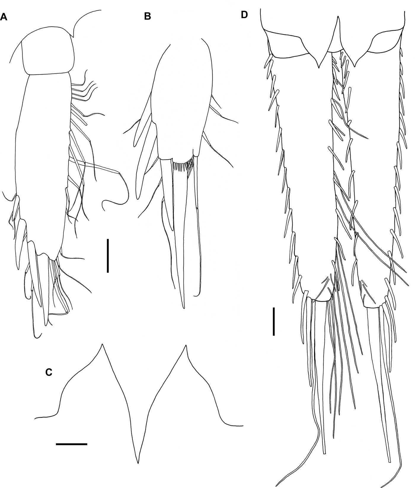
Nebalia mediterranea sp. nov., paratype, postovigerous female (ESFM-MAL/2009-006): (A) fifth pleopod, ventral view; (B) sixth pleopod, ventral view; (C) anal plates; (D) caudal furca and anal plates, ventral view (setules not figured for plumose setae on inner side of furcal rami; plumose setae not figured in left ramus). Scale bars: A–C, 50 μm; D, 0.1 mm. A & B, same scale bar.
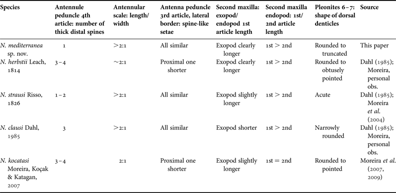
Comparison of relevant diagnostic characters among Nebalia mediterranea sp. nov. and similar Mediterranean species of Nebalia.
Nebalia sp. 2: Koçak et al., Reference Koçak, Moreira and Katagan2010.
TYPE MATERIAL
Holotype: postovigerous ♀, RL 0.80 mm, DCL 1.85 mm, LCL 2.50 mm, CH 1.82 mm, TL 5.17 mm (Foça, Izmir Bay; coordinates: 38°38′07″N 26°40′29″E; water depth: 67 m; mud; 12 November 2009) [ESFM-MAL/2009-004].
Allotype: subadult ♂, RL 0.62 mm, DCL 1.12 mm, LCL 1.62 mm, CH 1.30 mm, TL 3.17 mm (Foça, Izmir Bay; coordinates: 38°39′42″N 26°41′22″E; water depth: 27 m; sand; 12 November 2009) [ESFM-MAL/2009-005].
Paratypes: 1 postovigerous ♀ (dissected), same data as allotype [ESFM-MAL/2009-006]. 1 preovigerous ♀, same data as allotype [ESFM-MAL/2009-007]. 1 juvenile (Famagusta Bay, Northern Cyprus; coordinates: 35°07′30″N 33°57′00″E; water depth: 46 m; sandy-muddy sediment with fragments of Caulerpa sp.; 23 October 2003) [ESFM-MAL/2003-003]. 1 preovigerous♀ (Ildırı Bay; coordinates: 38°23′25″N 26°27′12″E; water depth: 50 m; sand-mud; 25 June 2003) [ESFM-MAL/2003-004].
DIAGNOSIS OF FEMALE
Average LCL 2.37 mm (N = 4). Rostrum about 2.5 times as long as wide. Antennular scale about 2.15 times as long as wide. One short distal spine on antennule peduncle fourth article. Three similar spine-like setae on external lateral face of antenna peduncle third article. Second maxilla endopod first article about 1.2 times as long as second article. Second maxilla exopod clearly longer than endopod first article. Fourth pleonite epimeron forming posterolateral subtriangular pointed process. Denticles along posterior dorsal borders of pleonites 5–7 rounded to slightly truncated. Pleopod 4 protopod with 4 serrations along posterior border; posterolateral corner acutely pointed. Uropods as long as pleonite 7 and anal somite combined. Terminal seta of caudal furca about 1.8 times as long as rami.
DESCRIPTION OF ADULT FEMALE
Carapace (Figure 1A): oval, DCL 1.82 mm, LCL 2.42 mm, CH 1.77 mm (N = 2), covering partially lateral sides of pleonites 3 and 4.
Rostrum (Figure 1A, B): long, 0.32 times as long as LCL, extending beyond eyestalk, about 2.5 times as long as wide, with parallel margins, distally tapering to a rounded apex. Ventral keel protruding posteriorly, with medial depression where supraorbital plates fit in.
Compound eye (Figure 1C): oval, with ommatidial part covering slightly more than two-thirds of total length of eye, dark pigmentation covering entire ommatidial part. Eyestalk neither lobed nor subdivided. Supraorbital scale tapering distally to acute tip, about half length of eyestalk.
Antennule (Figure 2A): peduncle four-segmented. First article shorter than eyestalk. Second article with (1) one plumose seta arising from anterior third, (2) cluster of about nine smooth and plumose setae arising from posterolateral surface, (3) subterminal cluster of about 10 short and long simple setae and one plumose seta. Third article shorter than the second, widest distally, with cluster of 6–7 simple setae on superior distal margin, one long simple seta on inferior distal margin. Fourth article shorter than third, with (1) medial row of seven simple setae, (2) one thick short spine distally, (3) lateral row of four simple setae, (4) one simple seta arising from inferior distal margin, longer than antennular scale. Antennular scale oval, about 2.15 times as long as wide; anterior margin convex, with (1) row of setae provided with teeth along margin of distal half, (2) numerous simple setae of different lengths, (3) one longer and thicker simple seta arising from inferior distal margin. Flagellum long, well-developed, with nine articles, about as long as the peduncle; each article with (1) 2–3 aesthetascs, (2) four simple setae on anterodistal margin, one longer than the others, oriented backwards.
Antenna (Figure 2B–D): peduncle three-segmented. First article with anterodistal process ending in small acute spine. Second article more or less rectangular, with distal acutely pointed process. Third article longer than second; with several rows of setae along medial anterior margin: (1) seven sparsely plumose setae, (2) six spines increasing in length along proximal half, (3) six simple setae increasing in length, (4) four simple setae thinner than those of (3), two short and two long, (5) three slightly plumose setae and three similar spines along lateral border, third spine associated to four longer simple setae, (6) terminal row of eight spines, increasing in length distally; long plumose seta on posterior margin; cluster of about 12 plumose setae along distal interior margin; two short setae on posterodistal margin. Flagellum well-developed, composed of 11 articles; each article with four terminal setae, one shorter than the others.
Mandible (Figure 3E): Mandibular palp three-segmented. Second article about 1.8 times as long as first; two simple setae on distal third: (1) one on lateral face, plumose along distal third, (2) one simple seta subterminally on superior margin. Third article slightly longer than second; proximal half of superior margin with row of short setae; inferior margin with three rows of setae (1) about five plumose setae on ending of proximal quarter, (2) longer plumose setae than those of (1) extending along distal half, (3) about six curved, dentate setae along distal margin; one simple seta on superior distal margin.
First maxilla (Figure 3A–C): first endite (proximal) with rounded medial margin bearing row of plumose setae. Second endite (distal) larger than first; medial margin with plumose seta and three rows of setae: (1) about 10 setae of increasing size with 1–2 large teeth along distal inferior margin, accompanied by several smaller teeth, one large tooth on distal superior margin, (2) about seven spatulate setae, (3) row of six setae with several teeth along distal superior margin. Palp well-developed, about five times the length of protopod, with long and spaced setae along entire length; setae with apex recurved distally.
Second maxilla (Figure 3D): Protopod with four endites bearing setae with setules, endites 1 and 3 being the largest and fourth endite the smallest. Fourth endite with 5 plumose setae, the distalmost being the longest. Endopod two-segmented, longer than exopod, proximal article 1.2 times as long as distal one; lateral margin with spaced plumose setae; second article with three distal terminal setae, one of them longer than endopod. Exopod longer than proximal article of endopod; medial margin with plumose setae, two distalmost setae as long as or slightly longer than exopod.
Thoracopods (Figure 4): Endopod longer than exopod; with numerous setae along anterior margin. Distal article of endopod slightly enlarged, more or less directed backwards, possibly provided with about 6–7 setae. Endopod segmentation weak. Exopod with 5–18 plumose setae along posterior margin, more numerous on anterior thoracopods. Epipod with 0–4 plumose setae along posterior margin. Thoracopod 8 epipod smaller than those of thoracopods 1–7.
Pleonites (Figures 5 & 6C): Pleonites 2–7 with denticles at least along posterior dorsal border; denticles with more or less parallel sides, distally rounded to truncated. Pleonite 2 bearing distally rounded denticles along dorsal and dorsolateral posterior border. Pleonite 3 with distally rounded denticles along dorsal and lateral posterior border. Pleonite 4 with distally rounded to truncated denticles along dorsal and lateral posterior borders; epimeron posterolateral corner forming slightly pointed subtriangular process. Pleonites 5–7 with distally rounded to slightly truncated denticles along dorsal and lateral posterior borders.
Pleopods 1–4 (Figure 6): Pleopods 1–4 stenopodous, composed of protopod, endopod and exopod. Pleopod 1 protopod with posterior margin even; three basal simple setae: (1) long seta arising subdistally, (2) long seta between the two rami, (3) long seta near the base of exopod, surpassing proximal half of exopod; posterior margin with simple seta on proximal third. Endopod two-segmented, longer than exopod; lateral and medial margins of distal article each with about 15 plumose setae, distal margin with acute process at apex and long, robust terminal seta. Basal article shorter, appendix interna provided with three short recurved hooks. Exopod about 0.6 times as long as protopod; with row of about 16 short serrate spines along lateral border (‘spine-row’), each spine with tridentate tip, central tooth bifid; four stout smooth spines on distolateral border, the distalmost being the longest; about 18 long plumose setae along medial margin.
Pleopods 2–3 similar in appearance. Protopod with (1) one simple setae on posterior margin proximal third, (2) 2–3 long simple setae on anterior proximal margin, (3) three long simple setae subdistally near base of endopod, and (4) one slightly thicker seta near base of exopod. Acute triangular process between endopod and exopod; posterior margin even. Endopod longer than exopod, two-segmented; proximal article short, provided with appendix interna; lateral and medial borders of distal article each with about 17–20 setae, most of them long and plumose, ending in one long spine. Exopod without a row of stout spines, instead with a row of 4–5 spine pairs, each pair consisting of one long and one shorter spine, with short plumose seta in between; medial margin with about 20 long plumose setae, three distal spines, the terminal one being the longest.
Pleopod 4 protopod anterior margin with three long simple setae along proximal third and cluster of three long simple setae subdistally near the base of endopod; four simple setae and one spine along posterior margin on proximal third; posterior border with four serrations; posterolateral corner acutely pointed. Distribution pattern of setae and spine pairs on margins of endopod and exopod similar to that of pleopods 2–3.
Pleopods 5–6 (Figure 7A, B): Fifth pleopod uniramous, two-segmented; distal article about 3.6 times as long as wide, with four stout conical spines along distolateral and terminal border, increasing in length distally; about 25 simple setae along medial and distal border, setae appear jointed at midlength. Sixth pleopod uniramous, one-segmented; with four stout conical spines along distolateral and terminal border, the distalmost being the longest, slightly longer than article, with circlet of acute teeth basally. Medial and terminal borders with ‘jointed’ setae similar to those of fifth pleopod. Both pleopod pairs with broad, acute triangular process between bases of rami, extending posteriorly.
Anal somite, anal plates and uropods (Figures 1C & 7C, D): Anal somite (pleonite 8) short, slightly longer than pleonite 7. Anal plates with broad bases, nearly equilateral in shape, acutely tapering distally. Uropods elongate, as long as pleonite 7 and anal somite combined. Each uropod with about 17 robust setae along lateral margin increasing distally in size, the terminal about 1.8 times length of uropod; about 11 robust setae and 12 plumose setae on lateral inner margin, cluster of four simple setae subterminally.
Colour: Fixed animals whitish or transparent with apparent dark pigment in eyes.
MALE
Only one subadult male found, similar to female, differing in having an antennal flagellum with more than 15 articles.
HABITAT
This species has been collected from sandy, muddy-sandy to muddy-sandy sediments with fragments of Caulerpa sp., at depths of between 27 and 67 m.
DISTRIBUTION
To date, Nebalia mediterranea sp. nov. is only known from Izmir Bay and Ildırı Bay (Aegean Sea, Turkey), and Famagusta Bay (Northern Cyprus) (this work); Gerence Bay (Aegean Sea; Koçak et al., Reference Koçak, Moreira and Katagan2010).
ETYMOLOGY
The species name refers to the Mediterranean Sea, where the new species was found.
REMARKS
Nebalia mediterranea sp. nov. is the fourth species of leptostracan reported so far from the eastern Mediterranean Sea. The new species differs from the other known species of Nebalia in the following unique combination of characters: the rostrum is about 2.5 times as long as wide, the antennular scale is clearly more than twice as long as wide, the fourth article of the antennule peduncle has only one short thick distal spine, the third article of the antenna peduncle has three spine-like setae on external lateral face which are similar in appearance, the first article of the endopod of the second maxilla is about 1.2 times as long as the second article, the exopod of the second maxilla is clearly longer than the first article of the endopod, the posterior dorsal borders of pleonites 6–7 are provided with distally rounded to truncated denticles, the protopod of pleopod 4 has four serrations along posterior border and the posterolateral corner is acutely pointed, and, finally, the terminal seta of the caudal rami is about 1.8 times the length of the entire ramus. The two specimens described by Koçak et al. (Reference Koçak, Moreira and Katagan2010) as Nebalia sp. 2 undoubtedly belong to this species according to the combination of the aforementioned characters, in particular when considering the features of the setae on the peduncle of antennule and antenna, the endopod and exopod of the second maxilla and the shape of the denticles on pleonites 6 and 7 (cf. Figures 4C, E & 5A, H in Koçak et al., Reference Koçak, Moreira and Katagan2010).
Nebalia mediterranea sp. nov. is similar in external appearance to other eastern Mediterranean species namely N. strausi Risso, 1826 and N. kocatasi Moreira, Koçak & Katagan, Reference Moreira, Koçak and Katagan2007, as well as to N. herbstii Leach, 1814 and N. clausi Dahl, Reference Dahl1985, also reported from the western Mediterranean (Table 1). The new species, however, can easily be distinguished from most of other eastern Atlantic and Mediterranean taxa by first examining the shape and length of the spine-like setae of the external face of the peduncle third article of the antenna. For instance, in N. troncosoi Moreira, Cacabelos & Domínguez, 2003 and N. reboredae Moreira & Urgorri, 2009 all those setae are similar in appearance (i.e. all spine-like), while in N. mediterranea sp. nov., N. herbstii, N. strausi, N. clausi and N. kocatasi the 3–4 proximal setae are slightly plumose and the distalmost three are spine-like. However, in N. herbstii and N. kocatasi the two distalmost spine-like setae are clearly longer than the proximal one while in N. clausi, N. strausi and N. mediterranea sp. nov. the three setae are similar in length and thickness. Nebalia strausi can be distinguished from N. mediterranea sp. nov. in having acutely pointed denticles along posterior dorsal borders of pleonites 6–7. Furthermore, in N. herbstii and N. clausi the fourth article of the antennule bears more thick distal spines (≥3); in N. clausi the second maxilla exopod does not surpass the level of articulation of the two articles of the endopod and the rostrum of N. herbstii is comparatively wider than that of N. mediterranea sp. nov. Other species from nearby geographic areas, i.e. N. marerubri Wägele, 1983 (Red Sea) and N. dahli Kazmi & Tirmizi, 1989 (Arabian Sea), mostly differ from N. mediterranea sp. nov. in the shape of the denticles of pleonites 6–7.
The finding of this new species raises interesting questions about the coexistence of several Nebalia species in similar substrata at subtidal depths. Thus, the scarce ecological data available suggest that N. strausi, N. kocatasi and N. mediterranea sp. nov. may appear in similar sandy-muddy sediments. On the contrary, Moreira et al. (Reference Moreira, Díaz-Agras, Candas, Señarís and Urgorri2009) pointed out that up to six leptostracan taxa may coexist in the same environment (i.e. embayments in NW Iberian Peninsula), but any given species seems to appear mostly in one type of sediment. Previous experimental work also pointed to preferences or differential survival of leptostracans according to the sedimentary features (e.g. granulometry, organic matter content; Vetter, Reference Vetter1996a). Therefore, it would be desirable to obtain more quantitative data about distribution, abundance and habitat features of the Mediterranean leptostracan taxa; this will be necessary to determine, for instance, whether competitive interactions may allow spatial segregation among such morphologically similar taxa, or, alternatively, if this is due to subtle differences in features of appendages (e.g. setation). This may ultimately help to understand the regional patterns of diversity of extant leptostracans.
ACKNOWLEDGEMENTS
We would like to thank our colleagues for their help in obtaining benthic samples. Comments from one anonymous reviewer are greatly appreciated.