Introduction
All species experience a reaction to a given action; be it physical, chemical, and/or physiological. The reaction could be a visible change, or something occurring at the molecular level. The nervous system is the platform that communicates, transfers, and integrates the details for regulation and control of each of their biologic actions, reactions, and intermediate events. This occurs via a dedicated set of cells (e.g., neurons and glial cells) that together constitute the neuron fibers, fascicles, nerve ramifications, and overall nervous system that interconnects everything in the body. A nerve may be damaged due to severe nerve injury (neurotmesis or axonotmesis)[Reference Griffin, Hogan, Chhabra and Deal1] or neurodegenerative diseases (peripheral neuropathy),[Reference Resnick, Stansberry, Harris, Tirivedi, Smith, Morgan and Vinik2] both requiring medical intervention. Currently, healing and recovery of the nerves’ fundamental performance remains limited: even with a successful nerve reconstruction resulting from an autologous nerve transplantation. Autologous nerve transplantation is the first line therapy (the “gold standard”) as it is ready to address complicated nerve gaps resulting from a serious wound. It is non-immunogenic, and provides an immediate nerve bridge that contains viable Schwann cells (SCs) and crucial signaling molecules involved in axonal renewal. However, there are multiple limitations in the use of autografts, including the potential need for a second surgery, the scar and/or neuroma formation, the inferiority against the original nerve, and functional loss (only 51.6% may gain a motor recovery, and 42.6% may obtain sensory healing).[Reference Grinsell and Keating3–Reference Taylor, Braza, Rice and Dillingham5]
In the USA, damage to the nervous system is impacting approximately 20 million people,[4] and implicates around US$150 billion in annual healthcare expenses.[Reference Taylor, Braza, Rice and Dillingham5] Therefore, significant efforts and resources for the studies of nerve regeneration strategies have been invested, including a deep fundamental understanding of the regeneration process using cellular models.
Platforms for 3D cell culture have been developed to mimic the native peripheral nerve system (PNS) cell growth and function, in order to study tissue repair or diseases of the nervous system (e.g., Parkinson and Alzheimer).[Reference Choi, Kim, Hebisch, Sliwinski, Lee, D'Avanzo, Chen, Hooli, Asselin, Muffat, Klee, Zhang, Wainger, Peitz, Kovacs, Woolf, Wagner, Tanzi and Kim6–Reference Ravi, Paramesh, Kaviya, Anuradha and Paul Solomon8] There is a need to introduce better approaches for therapeutic nerve regeneration. 3D cell culture models provide a practical and empirical way of observing cell activity and enable the quantification of the possible outcomes under different controlled conditions. The properties and functions of every cell in a distinct body are already encoded within the cells, at a level of detail that organizes and supports the complete sequence of biologic events for the development of cells to their mature stage with a specific role.[9] In the case of a damaged cell and/or tissue, it is expected that with the appropriate conditions the cells will activate and respond to specific signals to perform their role in the reconstruction and regeneration of damaged tissue.[Reference Kempermann and Gage10, Reference Simons and Clevers11] 3D cell culture provides models to study the underlying biology of peripheral nerve remodeling, and gives insight on the response to novel therapies or strategies for tissue repair. Although the focus is 3D cellular model for peripheral nerve regeneration, the study of 2D models can be advantageous as they are mostly composed of flat and rigid surfaces and are more manageable for cell observation thus facilitating the study of the effect of topographical cues.[Reference Edmondson, Broglie, Adcock and Yang12–Reference Mobasseri, Faroni, Minogue, Downes, Terenghi and Reid14] The main drawback of 2D models is that they do not represent the naturally 3D environment of cells, leading to non-predictive data for in vivo experiments.[Reference Edmondson, Broglie, Adcock and Yang12, Reference Behan, DeWitt, Bogdanowicz, Koppes, Bale and Thompson13]
Experimental strategies are focused on modeling the ideal settings to provide the appropriate environment for neurons in an effort to restore a level of performance comparable prior with any injury. Research in regenerative medicine and tissue engineering uses a variety of polymeric devices to offer a suitable 3D cellular arrangement that supports neuronal growth and maturation to produce modern healthcare alternatives.[Reference Tian, Prabhakaran and Ramakrishna15] 3D cell culture models will precede in vivo developments. The general approaches for 3D cellular models are relevant as they are closer to in vivo systems, and enable a more profound perspective of the cell development and fate characteristics (e.g., via imaging, molecular biology and genomics/transcriptomics techniques, and other analytical techniques); thus, ensuring the proposed designs, treatments, and potential benefits are well understood before any test subject is exposed to measured risks, or otherwise unwanted effects.[Reference Ravi, Paramesh, Kaviya, Anuradha and Paul Solomon8]
For example, many authors choose the sciatic nerve as the model test tissue after in vitro testing using a 3D cell culture model. Sciatic nerve is the biggest nerve in the human body,[Reference Shane Tubbs, Rizk, Shoja, Loukas and Barbaro16] which facilitates management and visibility of the studies. Sciatic nerve is associated with multiple pain-related diseases, such as sciatica, which is caused by irritation of the nerves in the lower spine in consequence of lumbar spinal stenosis (narrowing of the spinal canal in the lower back), degenerative disc disease, among others.[Reference Freeman17–Reference Johnson, Lancaster, Zhen, He, Gupta, Kong, Engel, Krick, Ju, Meng, Enquist, Jia and McAlpine21] Also, the fact that it extends from the spinal nerve in the lower back via many ramifications into peroneal and tibial nerves all the way to the toes, makes it ideal to model different types of nerve tissue regeneration. Rat serve as the primary animal model to study the sciatic nerve.[Reference Angius, Wang, Spinner, Gutierrez-Cotto, Yaszemeski and Windebank22]
Designing 3D cellular models used for peripheral nerve regeneration is challenging due to the complexity of the nervous system.[Reference Evans23] The role of the nervous system cannot be underestimated because it interconnects many different tissues throughout the body (as the sciatic nerve example). Neurons contain critical signals that will affect further systems, sensory and motor functions, and overall health.[Reference Yongqiang24] Growth factors (GFs) are key elements on the cascade of events to promote axonal advancement because of the interactions with surrounding cells and extracellular matrix components.[Reference Georgiou, Bunting, Davies, Loughlin, Golding and Phillips19, Reference Griffith and Naughton25, Reference Xm, Guénard, Kleitman and Mb26] Neurotrophic factors (NTs), promote nerve cell proliferation, neurite outgrowth, or neuronal cell differentiation.[Reference Rajaram, Chen and Schreyer27] Some NTs are: nerve growth factor (NGF), brain-derived growth factor (BDNF), glial cell-dervied neurotrophic factor (GDNF), NT-3 and NT-4/5.[Reference Walker28]
In this prospective article, we reviewed recent works on 3D cell culture models for nerve regeneration, and present some of the knowledge gained, including materials, fabrication techniques, and analytical methods. We will begin this review with an overview of neuronal biology, followed by a literature review of recent works on 3D cell culture models.
Peripheral nerve biology
There are two (2) areas defining the nervous system, the central nervous system (CNS) consisting of the brain and the spinal cord; and the PNS, which is distributed along a large network of neurons connected to the tissues, organs, and anatomical systems in the body that sustain life. The nervous system is the channel for inputs and outputs (stimulus and a response) that enables organisms to become aware of their environment. The nervous system manages all the actions in a body, either voluntary or involuntarily. For this, different types of neurons are continuously active: the afferent neurons, which obtain information from the five senses, and pass it to the CNS; the multipolar interneurons, which relay messages between other neurons within the CNS; and the efferent neurons, which drive our motor skills via the PNS. If a patient suffers an efferent fiber injury, they may not be able to move the innervated tissue because the brain impulse would not relay to the muscle. Likewise, if the impairment happens in an afferent fiber, the brain will not sense stimulus within the environment. As nerves carry both types of neuron fibers, a combination of efferent and afferent fibers are affected by injury.
There are many levels of complexity among voluntary and involuntary reactions that are made possible by the nervous system. Identifying a situation, deciding what to do next, and performing an action is an example of the communication of the brain and the body, and the velocity of this process will vary on other factors not in the scope of this discussion. However, there are some “closed loops” where afferent fibers are directly connected to efferent fibers, and an impulse does not necessarily reach the brain (e.g., reflexes, equilibrium, and other involuntary actions).
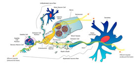
Nerves are formed as a cascade of conduits inside other conduits.[Reference Kiernan and Rajakumar29] The nerve is surrounded by a protective tissue known as the epineurium. Inside of it are the blood vessels and neuron bundles along with conduits called fascicles. As illustrated in Fig. 1, the neurons can carry impulses or action potentials to the soma, and then propagate the signals along the axon and axon terminals. The axons are protected by a myelin sheath produced by SCs located within the endoneurium. Early regeneration of the axon is what prompts the restitution of the nerve function.[Reference Griffin, Malahias, Hindocha and Wasim30] There is some regenerative ability in the nervous system,[Reference Huebner, Strittmatter and Koenig31] and this is true specifically in the PNS as it is exposed to macrophages that clear damaged tissue; and later assisted by SCs, which guide axon regeneration and provide the myelin sheath to support the re-connection between any gap.[Reference Griffin, George and Ho32]
Basic structure of a nerve.

The PNS nerves branches out of the CNS to sense and collect input signals, and it is also responsible for transmitting the commands that controls muscle movement. The biochemical explanation of neuronal impulses, also known as potential actions, is described as an electrochemical wave propagated within the surface membrane of a neuron. The initial stimulation of neuron opens channels leading to sodium (Na+) influx that depolarizes the membrane. The signal propagates as the adjacent section of membrane allows Na+ influx and depolarization. This wave of depolarization continues at a rapid rate down the neuron, resulting in a nerve impulse traveling inbound (afferent, sensory) and outbound (efferent, motor) to or from the brain in milliseconds.[Reference Kiernan and Rajakumar29]
After a severe nerve injury, the axon separates from the soma and, cytokines/GFs surrounding the injury will recruit and activate macrophages, which then infiltrate the site of injury to clean-up axon and myelin debris—this is known as Wallerian degeneration.[Reference Shane Tubbs, Rizk, Shoja, Loukas and Barbaro16, Reference Burnett and Zager33] Wallerian degeneration is a controlled phase that extends to the following node of Ranvier, which is a gap in the myelin sheath that is exposed to the extracellular matrix (ECM). Wallerian degeneration stops the synapse and communication between the neural networks leading to atrophy of the associated muscle or gland, which could become permanently deteriorated if the axon fails to re-establish connectivity.
The neuron gap is defined as two parts: the proximal stump closest to the soma and distal stump that will degenerate. After macrophages remove the degenerated debris, the soma grows as the nucleus migrates toward the cell boundaries, and the local non-myelinating SCs at the endoneurium align to guide axon regeneration. A regenerated axon sprouts from the proximal end, and grows to join the two ends to reestablish the network connection. To ensure the proper reconnection, surgery helps to establish a guide for these sprouts leading to eventual re-innervation, either by using autologous nerve transplant or an engineered scaffold.
During nerve regeneration, the native ECM must be reconstructed. The major components of the ECM in the nervous system can be divided into four groups: collagens and related molecules as structural parts (e.g., types I, II, III, IV, and V); non-collagenous glycoproteins as binding agents of cells to mediate their effects or induce cell binding to other molecules [e.g., laminin (LN), fibronectin, entactin, and vitronectin]; glycosaminoglycans (GAGs) to facilitate cell migration, adhesion, and organization, as well as support the formation of proteoglycans by providing the chains of several functional groups (e.g., hyaluronic acid, dermatan, chondroitin, keratan, and heparan); and finally proteoglycans, which have been closely associated with fundamental cellular processes, cell attachment and growth, and cell receptor signaling/binding (e.g., chondroitin sulfate and heparin sulfate).[Reference Rutka, Apodaca, Stern and Rosenblum34, Reference Carbonetto35]
Selection of cells
The use of in vitro cellular models have advantageous features over in vivo models, particularly those aspects regarding technical complexity, costs, and ethical concerns.[Reference Geuna, Raimondo, Fregnan, Haastert-Talini and Grothe36] The appropriate selection of cells for the in vitro cellular models will support projections and expectations for potential in vivo models. Moreover, in vitro cellular models are not necessarily subjected to regulations as with animal or human models. The goal is to obtain representation of the subject tissues and translate the research towards clinical developments. Yet, the biologic characterizations made at the laboratory barely exemplify what is to be expected in normal conditions, regardless of the source. For this, researchers can use primary cells (cells isolated directly from human or animal tissue), or cell lines (cells that have been continually passaged over a long period of time). Cell lines are cost-effective option, but may be less physiologically representative of native cells because cell lines have been immortalized. Primary cells may be more representative of the native tissue; however, they need to be isolated from animals leading to increased oversight. Within the PNS, there is increased accessibility to obtain primary cells, such as SCs from sciatic nerve and neurons or mixed cultures from dissociated or whole dorsal root ganglia (DRG), compared with CNS cells that typically need to undergo several cell-specific purification steps. Researchers should select their test subjects strategically considering the resources available, and in a step wise approach per the progress obtained, and future developments envisioned.
As an actual cellular model for neurons, whole DRG or dissociated DRG neurons are commonly used. The DRG is obtained from a cluster of nerve cell bodies located in the dorsal root of the spinal cord, constituting afferent fibers. Their outgrowth have been demonstrated both alone or when co-cultured with SCs.[Reference Geuna, Raimondo, Fregnan, Haastert-Talini and Grothe36–Reference Yin, Zhang, Bo and Gao38] DRGs are a pivotal test subject for experiments using growth and/or regulatory factors.[Reference Liu, Gao, Wang, Zhang, Liu and Li39]
Other neural cells used for nerve regeneration studies include primary neurons sourced from the CNS, including neurons from the hippocampus, and cerebral cortex. For research purposes, these cells are mainly derived from rodents and, are very effective to profile the basic physiological properties and behavior of neurons at the experimental conditions, including those for the PNS. Some of the physiological properties include cell reactions to scaffold composition and stiffness.[Reference Gattazzo, Urciuolo and Bonaldo40–Reference Koch, Rosoff, Jiang, Geller and Urbach42] These cells are commonly used for the evaluation of the neurotoxicity of pharmacologic compounds, since the brain areas sources for these cells are susceptible for neural toxins.[Reference Lein, Barnhart and Pessah43, Reference Rahmani, Shoae-Hassani, Keyhanvar, Kheradmand and Darbandi-Azar44]
Alternatively, back in 1976, Greene and Tischlert established the PC12 cell line (not a neural cell line, but derived from a neuroendocrine tumor of the adrenal medulla) as it has a reversible response to NGF. PC12 are a commonly used cell line for investigating cell/material interactions as well as pharmacologic studies since there is ample knowledge regarding their behavior, proliferation, and differentiation. Also, PC12 cells have a high throughput as a first-level assessment tool; they are robust, and substantially easier to culture than primary or stem cells. Nowadays, the PC12 cells are a versatile resource that are continuously used for the research of polymeric scaffolds and improved 3D cell culture strategies.[Reference Greene, Tischlert and Kuffler45–Reference Shi, Gao, Feng, Ding, Cao, Kuga, Wang, Zhang and Cai48]
Glial cells are the main neuronal support cells of the nervous system with the SCs (e.g., S42 cell line or primary SC isolated from the sciatic nerve) being the most widely studied for PNS regeneration.[Reference Georgiou, Bunting, Davies, Loughlin, Golding and Phillips19, Reference Johnson, Lancaster, Zhen, He, Gupta, Kong, Engel, Krick, Ju, Meng, Enquist, Jia and McAlpine21, Reference Li, Zhang, Wang, Zhao, Zhu, Zheng, Wang, Zhao and Yang49–Reference Kim, Hwang, Aguilar, Park and Kim52] These cells contribute to neuron pathfinding by guiding their axons between the proximal to distal stumps, and by producing myelin sheath to protect them. SCs are so important to nerve regeneration that further alternatives for them have been explored by using differentiated stem cells (ADSC or iPSC) to SC-like cells[Reference Mobasseri, Faroni, Minogue, Downes, Terenghi and Reid14, Reference Xu, Zhang, Chen, Li, Li and Feng20, Reference Schuh, Morton, Banerjee, Grasl, Schima, Schmidhammer, Redl and Ruenzler53, Reference Wang, Tang, Park, Zhu, Patel, Daley and Li54] or bone marrow mesenchymal stem cells (BMSC) to effect nerve regeneration, either by direct and/or indirect paracrine signaling to express glial markers.[Reference Oliveira, Mostacada, de Lima and Martinez55, Reference Tohill, Mantovani, Wiberg and Terenghi56] SCs can be induced from ADSCs cells by addition of glial GF and co-culture with SCs. A key factor when working with cell differentiation and proliferation is cell density, since it has been reported that higher cell density equals an increase of cell–cell interactions, which is also related to the influence of glial GF.[Reference Najafabadi, Bayati, Orazizadeh, Hashemitabar and Absalan57]
Olfactory ensheathing cells (OECs) are a distinct type of glial cells found in the CNS exclusively supporting the olfactory tract nervous system. Their function is to provide myelin sheath to the unmyelinated axons of the olfactory neurosensory cells, as SCs does for PNS. Important facts about the OEC uniqueness and properties for using them in nerve repair studies is because, in nature, they continuously sustain a regenerative environment.[Reference Kiernan and Rajakumar29, Reference Higginson and Barnett58] Olfactory neurons are regularly replaced from a population of precursor stem cells (about every 7 weeks), and OECs become more actively phagocytic to discard axonal debris and dead cells (in cell culture studies, they are capable of phagocytose bacteria).[Reference Nazareth, Lineburg, Chuah, Tello Velasquez, Chehrehasa, St John and Ekberg59, Reference Panni, Ferguson, Beacham, Mackay-Sim, Ekberg and St John60] OECs, together with other glial cells available at the olfactory bulb, encourage the corresponding axonal growth. During these cycles, OECs can move through glial scar tissue (important feature for damaged nerve reconnection) producing NTs (e.g., BDNF and NGF) and expressing key markers (e.g., S100 and p75);[Reference Silva, Cooke, Tam, Sousa, Salgado, Reis and Shoichet61] allowing researchers multiple perspectives to understand nerve regeneration via the glial cells interactions.[Reference Ruitenberg, Vukovic, Sarich, Busfield and Plant62]
Some researchers have concentrated their attention into macrophages,[Reference Mokarram, Merchant, Mukhatyar, Patel and Bellamkonda63, Reference Niemi, DeFrancesco-Lisowitz, Roldán-Hernández, Lindborg, Mandell and Zigmond64] which precede the accomplishments of SCs. It has been documented that the extent of macrophage presence at the site of injury (M2, anti-inflammatory phenotype), grants a favorable influence in nerve regeneration.[Reference Chen, Piao and Bonaldo65–Reference Cattin, Burden, Van Emmenis, MacKenzie, Hoving, Garcia Calavia, Guo, McLaughlin, Rosenberg, Quereda, Jamecna, Napoli, Parrinello, Enver, Ruhrberg and Lloyd67] In the native nerve regeneration process, macrophages together with fibroblasts provide the initial platform and route for the undifferentiated SCs to form clusters that eventually guide the growing axons across the gap, all the way back toward their target.[Reference Parrinello, Napoli, Ribeiro, Digby, Fedorova, Parkinson, Doddrell, Nakayama, Adams and Lloyd66, Reference Cattin, Burden, Van Emmenis, MacKenzie, Hoving, Garcia Calavia, Guo, McLaughlin, Rosenberg, Quereda, Jamecna, Napoli, Parrinello, Enver, Ruhrberg and Lloyd67] For 3D cell culture models, such types of platforms may be provided by polymeric scaffolds.
Among the preferred cells selected to investigate nerve regeneration as previously discussed, recent development uses cellular co-culture systems. Briefly, a cellular co-culture system allows further visibility of a more complex system whether to study specific interactions (synergetic or antagonist), or to evaluate multiple factors controlling cellular behavior. Co-culture can be comprised heterogeneous cells to cells; cells with specific GFs or other pharmacologic agents; and cells to drive the behavior of other cells.[Reference Georgiou, Bunting, Davies, Loughlin, Golding and Phillips19, Reference Xu, Zhang, Chen, Li, Li and Feng20, Reference Pateman, Harding, Glen, Taylor, Christmas, Robinson, Rimmer, Boissonade, Claeyssens and Haycock50, Reference Daud, Pawar, Claeyssens, Ryan and Haycock51, Reference Goers, Freemont and Polizzi68–Reference Hu, Wu, Gou, Tao, Zhang, Liu, Kang, Jiang, Huang, He, Chen, Du, Gou, Faroni, Mobasseri, Kingham, Reid, Chhabra, Ahlawat, Belzberg, Andreseik, Xie, Gu, Ding, Williams, Bell, Haycock, Kehoe, Zhang, Boyd, Koshy, Ferrante, Lewin, Mooney, Chang, Chen, Yue, Qi, Yan, Huang, Melerzanov, Du, Gamez, Nichol, Suri, Li, Kolar, Kingham, Heinemeyer, Reimers, Johnson, Pateman, Lin, Murphy, Atala, Morrison, Pati, Zhu, Hu, Widgerow, Salibian, Lalezari, Evans, Khalifian, Angius, Huang, Niu, Xu, Hsueh, He, Xue, Fu, Park, Jung, Kang, Cho, Lee, Dunn, Wu, Yao, Bender, Bennett, Waddell, Doctor, Marra, Yao, Billiar, Windebank, Pandit, Cheng, Lu, Fu, Kokai, Lin, Oyster, Marra, Chhabra, Williams, Wang, Dellon, Carrino, Wolf, Yamada, Cukierman, Liu, Santiago, Clavijo-Alvarez, Brayfield, Rubin, Marra, Shen, Yang, Liu, Kingham, di Summa, Lee, Choi, Wu, Lee, Tseng, Hsu, Bain, Mackinnon and Hunter73] Any use of these cells as models are possible if some type of suitable platform is available that will enable them to attach and perform their function. Therefore, the 3D cell culture success depends on the matrix to be seeded, such as the polymeric scaffolds that offer the 3D structures.
Polymeric materials in use
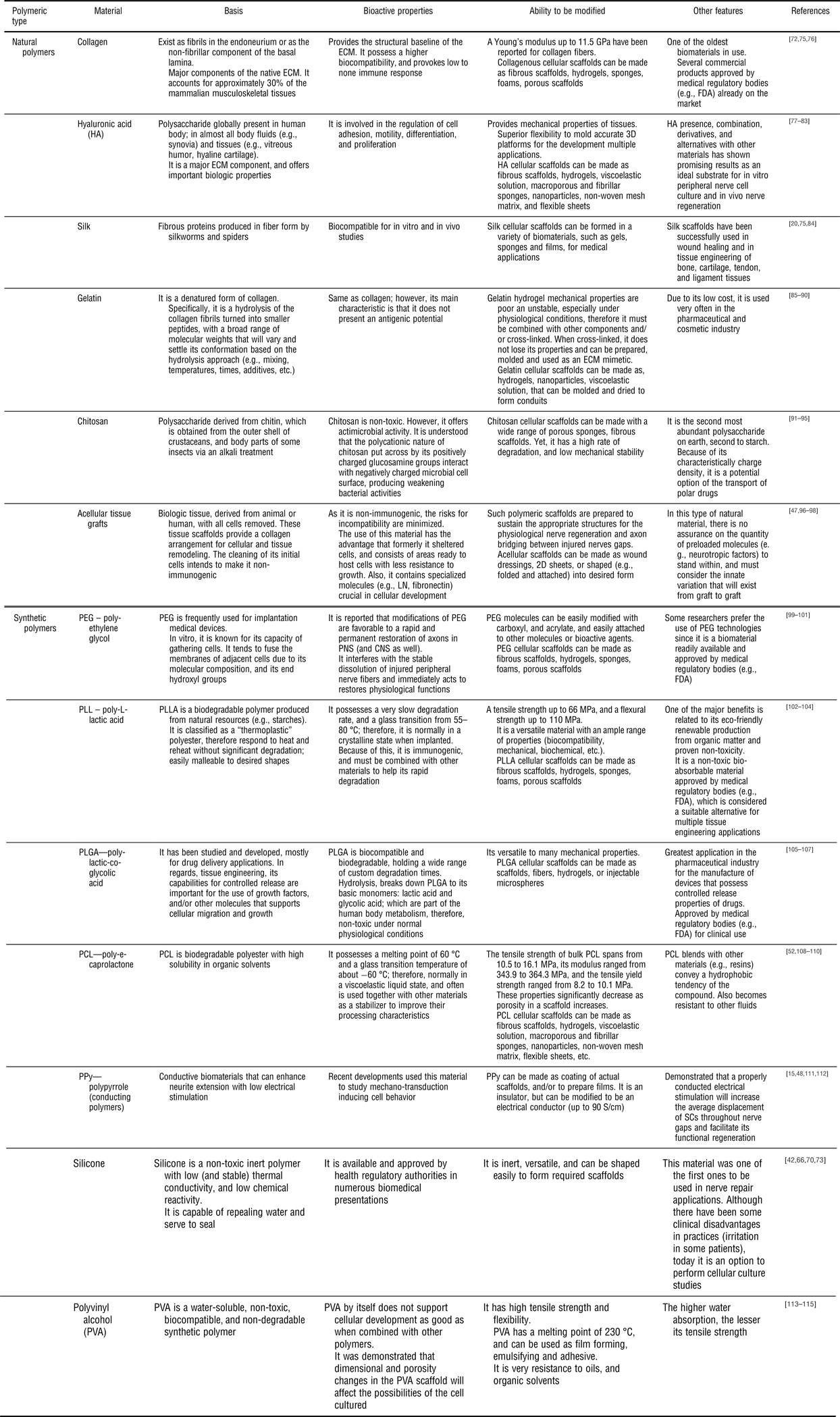
Polymeric materials can be divided into two major groups: natural and synthetic.[Reference Painter and Coleman74] For scaffolds, those that can orchestrate nerve regeneration and are available for use in 3D cell culture are presented in Table I. Mechanical properties of the scaffold can be customized per intended application depending on the molecular weight and polymerization reaction (and/or copolymerization ratio) for a given polymer. Another key element for the constitution of polymers to serve their intended purpose is their cross-linking. Cross-linking involves the anchoring of polymer chains to reinforce the final construct, influencing cellular behavior. Cross-linking sets a controlled way to inhibit degradation of the polymer by enzymatic activity, or cellular remodeling, and offers a longer duration to investigate other aspects of research interest, or as long as needed for biomedical applications. For example, polymeric scaffolds (within an animal or patient) would be designed to degrade over time. Its purpose is to facilitate tissue ingrowth, and timely degradation will allow the cells to replace the entire scaffold.
Polymeric materials for 3D scaffolds.

Many other polymeric biomaterials could be explored, as presented by relevant reviews preceding this one.[Reference Daly, Yao, Zeugolis, Windebank and Pandit116, Reference Bell and Haycock117]
Common cross-linking methods include chemical (e.g., aldehydes) or via radiation [e.g., ultraviolet (UV) and heat] enabling different levels of polymer bonding, therefore affecting cells development as their microenvironment changes.
Three-dimensional cell culture techniques
An appropriate environment is attained when the platform provided mimics the native ECM attributes as much as possible to support cell migration, proliferation and differentiation. It is known that the ECM regulates cell development via biochemical cues and signaling. A polymeric scaffold entails the mechanical properties to support cell attachment and its progress in a 3D space. Various preparation techniques are described:
Flat (films)
One of the most important reasons to continue developing options for nerve tissue regeneration is to overcome the drawbacks of the current gold standard techniques such as the autograft, for example donor site morbidity or the need for multiple surgeries. Polymeric structures may replace the use of autografts, and thus serve as ideal platforms for 3D cell culture. Flat films are often employed to study PNS regeneration due to their simplicity in manufacturing. Raimondo and colleagues,[Reference Fregnan, Ciglieri, Tos, Crosio, Ciardelli, Ruini, Tonda-Turo, Geuna and Raimondo118] developed a flat chitosan cross-linked scaffold that later was rolled and glued with biomedical cyanoacrylate glue, obtaining a 3D structure that served as a conduit for in vivo test as a bridge between two nerve stumps with a 10 mm gap of the median nerve. The main component is the polysaccharide chitosan, which is very attractive for its unique physicochemical properties along with good biocompatibility, biodegradability, and antibacterial features.[Reference Fregnan, Ciglieri, Tos, Crosio, Ciardelli, Ruini, Tonda-Turo, Geuna and Raimondo118, Reference Hui Hsu, Ho and Tseng119] Given its poor mechanical strength,[Reference Yang, Gu, Tan, Hu and Wang95] a cross-linking technique, which supported glial cell proliferation, was applied using dibasic sodium phosphate (DSP) and glycodoxypropyltrimethoxysilane (GPTMS). To validate this, the tests were divided into two main groups: an in vitro test by culturing the RT4-D6P2T cell line (a schwannoma cell line) and DRG explants over the flat surface, and an in vivo test using 3D scaffold rolled as a conduit on long rat median nerve defect. In the case of cultured cells, 84% of the population took an elongated form and DRG showed a high neurite growth and expression of Bcl2 gene was observed after four days of culture. The adaptation of the cells to the substrate was demonstrated by expression of Bcl2 (which regulates cell survival) that was lower for 3 days, compared with the control, and after day 6 it reached a similar value to control (glass). This proves the successful combination of chitosan and cross-linking techniques to develop a competent substitute for nerve autografts. In vivo results for the rolled 3D conduit showed that, chitosan cross-linked with only DSP induced a functional recovery at week 6 and almost comparable results with autografts at week 9, oppose to chitosan cross-linked with DSP and GPTMS that detached from suturing site due to the fragility of the scaffold.[Reference Fregnan, Ciglieri, Tos, Crosio, Ciardelli, Ruini, Tonda-Turo, Geuna and Raimondo118]
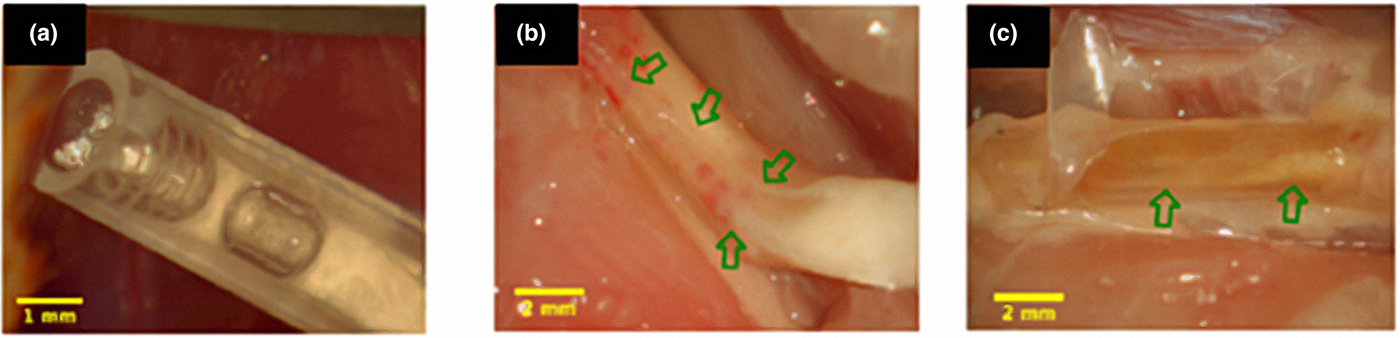
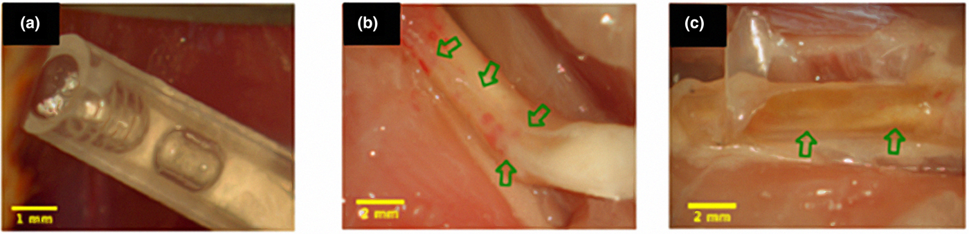
Axonal regeneration is highly desired,[Reference Hsu, Kuo, Chen, Yen, Chen, Chen, Huang and Cheng46] and work contributes through the bioengineered LN-coated chitosan multi-walled conduit containing BMSCs was tested in vivo to serve as a bridge between a 10 mm long gap in the sciatic nerve of Sprague–Dawley rats. The chitosan component has been very attractive due to its biocompatible and biodegradable properties.[Reference Hui Hsu, Ho and Tseng119, Reference Busilacchi, Gigante, Mattioli-Belmonte, Manzotti and Muzzarelli120] An overview of the chitosan film preparation consisted of lyophilized chitosan, re-dissolved on 2% acetic acid and placed on coverslip to allow the solvent to evaporate. The chitosan film was modified with plasma treatment to increase the hydrophilicity, followed by LN covalently bonded by surface loading. Three groups were tested: empty silicone conduit, LN-modified chitosan scaffolds in silicone conduit (multi-walled) and LN-modified chitosan scaffold with BMSCs in silicone conduit (multi-walled plus BMSC). The BMSC resulted to be key element during in vivo experiment to down-regulated the extent of inflammation, reducing hyperplasia, and for proper neuronal growth [Figs. 2(a)–2(c)]. This is also corroborated with recovery index for 74.31 ± 26.51% for the conduit of BMSC, 17.31 ± 5.89% for the chitosan group and 31.52 ± 10.43% for the empty group.[Reference Hsu, Kuo, Chen, Yen, Chen, Chen, Huang and Cheng46]
Multi-walled chitosan channel inner silicone conduit (a); the connective tissue-like surrounding the outer tube of the laminin (LN)-conjugated chitosan scaffolds and nerve regeneration along with the outer wall of the conduit (b); nerve grow back through the inner channels of the BMSC-treated chitosan scaffold conduit (c). The arrow indicated a regenerated nerve. Reprinted from Hsu et al.[Reference Hsu, Kuo, Chen, Yen, Chen, Chen, Huang and Cheng46] Copyright (2013), with permission from Elsevier.
Chitosan was also the focus of the work by Meyer et al.,[Reference Meyer, Stenberg, Gonzalez-Perez, Wrobel, Ronchi, Udina, Suganuma, Geuna, Navarro, Dahlin, Grothe and Haastert-Talini121] in which the main concern was the capacity of artificial nerve guides to reconstruct critical length of sciatic nerve defects (15 mm) in vivo using healthy Wistar and diabetic Goto–Kakizaki rats. The diabetic rats were included to evaluate if the effect is the same under a relevant increase of blood glucose level that resembles type 2 diabetes patients, the above is done as a control because diabetic animals are less sensitive to nerve injury and they have a slow regeneration capacity.[Reference Stenberg and Dahlin122] The innovation is the introduction of the chitosan film to chitosan nerve tubes in order to achieve recovery of a critical defect length of 15 mm, since it was proven that the hollow chitosan tubes were capable to repair defects of 10 mm.[Reference Kruse, Luebbers, Grätz and Obwegeser123] The chitosan film promoted SC attachment and supported DRG neurite outgrowth in vitro,[Reference Wrobel, Serra, Ribeiro-Samy, Sousa, Heimann, Barwig, Grothe, Salgado and Haastert-Talini124] and this motivated combining the fine-tuned chitosan nerve guides (CNGs) tubes with the chitosan film. A novel outcome in diabetic rats is the presence of substantial regenerative matrix after 56 days of surgery, due to an increased number of activated SC, which attracts re-growing axons. Improvement on functional and morphological results of nerve regeneration, compared with hollow CNG, states that the role of chitosan of modifying an existing product introduces a new generation of medical devices for peripheral nerve reconstruction.
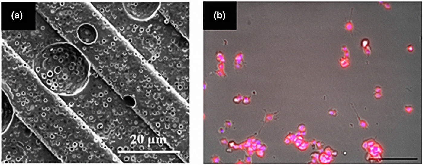
Poly lactic-based co-polymers (PLLA) has also been used to develop multifunctional films due to its biodegradability, low toxicity, and functional end groups.[Reference Nectow, Marra and Kaplan125, Reference Uz, Sharma, Adhikari, Sakaguchi and Mallapragada126] The word multifunctional applies since the film possess surface micropatterns for topographical cues along with surface gradients of NGF, which it is released in a controlled manner using nanoparticles. The multifunctional films were tested in vitro using PC-12 cells as a model of peripheral nerve regeneration. An overview for the engineering of the multifunctional films consisted on a dry-phase inversion technique to develop the porous topography that allowed the distribution within the film of the micro particles CPEG:CPH (composed of random copolymers: CPTEG, CPH) that controlled the release of the GFs. The surface gradients were developed using longitudinal micropatterns through ester–amine interactions and physical self-assembly. Figure 3 shows a scanning electron microscopy (SEM) image of the topography of the multifunctional films (A); and the bioactive release of β-NGF promoting neurite extension (B). Neurite extension was quantified through the device outgrowth module software used for the collection of cellular images in two different β-NGF conditions: β-NGF released from microparticles and soluble β-NGF directly added to PC-12 cells in culture media. The film promoted guided neurite extension of cells of 10 µm total per cell in 2 days.[Reference Hui Hsu, Ho and Tseng119]
(a) SEM images of porous, OVA encapsulated 50–50: CPTEG-CPH particles incorporated in patterned PLLA films; (b) Bioactivity of β-NGF released from 50 : 50 CPTEG: CPH microparticles (125 µg/mL) on the neurite extension of PC-12 cells. PC12 cell density: 2 × 10⁴ cells/cm2. Incubation time: 2 days. Neurites of the cells in every condition were immunostained with βIII-tubulin along with Cy3 secondary (shown in red), and DAPI for nuclei (shown in blue). Reprinted from Uz et al.[Reference Uz, Sharma, Adhikari, Sakaguchi and Mallapragada126] Copyright (2016), with permission from Elsevier.

Similarly, Torigoe et al., used a film model to study the regenerative effects of hyaluronan tetrasaccharide (HA4) on axons applied to common peroneal nerves cut from mice. Although HA is considered as a regenerative inhibitory agent in the CNS, HA can be depolymerized to specific forms of oligosaccharides containing different properties. For example, HA4, has been shown to decrease the death of K562 bone marrow cells and, increase the growth of PC-12 cells.[Reference Torigoe, Tanaka, Ohkochi, Miyasaka, Yamanokuchi, Yoshidad and Yoshida127] The proximal end of the nerve was maintained in vivo between two 3 × 4 mm2 sheet of fluorine resin films sewn together and accompanied by the addition of a few drops of Ringer's Solution, thus observing the growth of regenerating axons. For the purposes of this investigation, instead of the Ringer's solution, HA4 drops were applied in the surface of the film at concentrations of 10, 100, or 1000 µg/mL and the neurite behavior was evaluated at 6 h, 2, 3 and 4 days later. In all groups (HA4 10, 100, 1000, and control) after 6 h, terminal bulbs could be seen at the proximal tip of the injured nerve, but only in the HA4 100 and HA4 1000 groups were extensions of terminal sprouts observed and on day 2 the regenerating axons were spread over the film. HA4 100 was cited as promoting increased axon length with a growth rate of 268 µm/day, approximately three times faster than controls. From day 3, the appearance of SCs occurs, which accelerate the growth of regenerating axons. At this stage, the growth of HA4 100 group was equal to that of the control and it was possible to determine that the SCs were distributed along the axons. This improvement in regeneration using HA4 is due to the reduction of the time of onset of terminal sprouting and the accelerated growth of axons, which manifests itself in response to an increase in the metabolism of neurons.[Reference Torigoe, Tanaka, Ohkochi, Miyasaka, Yamanokuchi, Yoshidad and Yoshida127]
Three-dimensional Printing
The 3D printing is a novel technique that is transforming science and education and has emerged since the 1980s as an additive manufacturing technology with fundamentals based on stereolithography and is also known as RP (rapid prototyping) or additive manufacturing (AM).[Reference Lin and Peng128–Reference He130] The 3D bio-printing is the use of 3D printing equipment combined with printing processing of biocompatible materials and cells through layer-by-layer. This technology was introduced in 1993 by Langer and Vancanti, with broad applications on tissue engineering and regenerative medicine fields.[Reference Langer and Vacanti131] In general, the process consists of: imaging, design, selection of materials and cells, and printing of the product.[Reference Murphy and Atala129] The printing materials are what assigns the classification of the developed scaffolds as: solid, soft, or free scaffold.[Reference Lin and Peng128] In the case of 3D scaffolds for peripheral nerve regeneration related with soft tissues and organs, these are classified as soft and free scaffolds.[Reference Lin and Peng128]
One of the main advantages of 3D bio-printing technology is the capacity to develop complex structures of custom-tailored shapes and sizes as well as internal shapes, and pores that can be controlled to adjust the diffusion of oxygen, nutrients, and waste of cells.[Reference El-Ayoubi, Eliopoulos, Diraddo, Galipeau and Yousefi132, Reference Zhang, Zhao, Zhu, Huang, Zhu, Tao and Zhang133] 3D bio-printing technology promises to elaborate structures that foment cellular function, provide mechanical support and generates patient-specific products.[Reference Lin and Peng128, Reference Langer and Vacanti131] As part of current challenges, 3D bio-printing technology involves high working temperatures to achieve melting of materials that compromises scaffolds bioactivity, this is specifically seen on nozzle-based 3D printing technologies as well as FDM (Fused Deposition Modeling) and its variants.[Reference Lin and Peng128, Reference Murphy and Atala129] Additional drawbacks are present when the equipment involves nozzle technologies that only accepts fluids with low viscosity and low cell density because of the high shear force and issues with clogging. These challenges and limitations are overcome with laser printing, a nozzle-free system, which has a wide range of working viscosity values (1–300 mPa/s) along with high cell density solutions with negligible effect on cell viability during the process.[Reference Koch, Kuhn, Sorg, Gruene, Schlie, Gaebel, Polchow, Reimers, Stoelting, Ma, Vogt, Steinhoff and Chichkov134] However, laser technologies also have its drawback such as: time consuming because of the necessary low overall flow rate needed for the rapid gelation, and high cost. Upon the many 3D bioprinting strategies the most common are inkjet, micro-extrusion, and laser.[Reference Murphy and Atala129] 3D bioprinting represents a risen technology for the development of patient-specific products.
The 3D bioprinting uses 3D printing equipment, an example is the use of a commercially inexpensive desktop 3D printer to tailored custom architecture of bio-conduits for peripheral nerve regeneration.[Reference Hu, Wu, Gou, Tao, Zhang, Liu, Kang, Jiang, Huang, He, Chen, Du, Gou, Faroni, Mobasseri, Kingham, Reid, Chhabra, Ahlawat, Belzberg, Andreseik, Xie, Gu, Ding, Williams, Bell, Haycock, Kehoe, Zhang, Boyd, Koshy, Ferrante, Lewin, Mooney, Chang, Chen, Yue, Qi, Yan, Huang, Melerzanov, Du, Gamez, Nichol, Suri, Li, Kolar, Kingham, Heinemeyer, Reimers, Johnson, Pateman, Lin, Murphy, Atala, Morrison, Pati, Zhu, Hu, Widgerow, Salibian, Lalezari, Evans, Khalifian, Angius, Huang, Niu, Xu, Hsueh, He, Xue, Fu, Park, Jung, Kang, Cho, Lee, Dunn, Wu, Yao, Bender, Bennett, Waddell, Doctor, Marra, Yao, Billiar, Windebank, Pandit, Cheng, Lu, Fu, Kokai, Lin, Oyster, Marra, Chhabra, Williams, Wang, Dellon, Carrino, Wolf, Yamada, Cukierman, Liu, Santiago, Clavijo-Alvarez, Brayfield, Rubin, Marra, Shen, Yang, Liu, Kingham, di Summa, Lee, Choi, Wu, Lee, Tseng, Hsu, Bain, Mackinnon and Hunter73] The choice of material is crucial on 3D printing technology specially on inkjet bioprinting, since it needs to be printed liquid and then form a solid 3D structure. 3D structure is achieved by the use of materials that can be cross-linked after the deposition by chemical, pH, or ultraviolet mechanisms.[Reference Murphy and Atala129] This specific material requirement is the reason for the modification of gelatin, which adds properties such as photo cross-linking and radical polymerization. Gelatin methacryloyl (GelMA), is one of the chosen material for the desktop 3D printer application.[Reference Hu, Wu, Gou, Tao, Zhang, Liu, Kang, Jiang, Huang, He, Chen, Du, Gou, Faroni, Mobasseri, Kingham, Reid, Chhabra, Ahlawat, Belzberg, Andreseik, Xie, Gu, Ding, Williams, Bell, Haycock, Kehoe, Zhang, Boyd, Koshy, Ferrante, Lewin, Mooney, Chang, Chen, Yue, Qi, Yan, Huang, Melerzanov, Du, Gamez, Nichol, Suri, Li, Kolar, Kingham, Heinemeyer, Reimers, Johnson, Pateman, Lin, Murphy, Atala, Morrison, Pati, Zhu, Hu, Widgerow, Salibian, Lalezari, Evans, Khalifian, Angius, Huang, Niu, Xu, Hsueh, He, Xue, Fu, Park, Jung, Kang, Cho, Lee, Dunn, Wu, Yao, Bender, Bennett, Waddell, Doctor, Marra, Yao, Billiar, Windebank, Pandit, Cheng, Lu, Fu, Kokai, Lin, Oyster, Marra, Chhabra, Williams, Wang, Dellon, Carrino, Wolf, Yamada, Cukierman, Liu, Santiago, Clavijo-Alvarez, Brayfield, Rubin, Marra, Shen, Yang, Liu, Kingham, di Summa, Lee, Choi, Wu, Lee, Tseng, Hsu, Bain, Mackinnon and Hunter73] The developed cryoGelMA, named after the conduit preparation via cryopolymerization, is turned into a bio-conduit by the introduction of multipotent adipose-derived stem cells (ADSCs) to generate local environment to foment the regeneration of axons, which is possible with the use of 3D printing. The versatility of 3D printing is taken as an advantage to engineer conduits of different geometries such as multichannel or bifurcating and even personalized structures.[Reference Griffith and Naughton25, Reference Langer and Vacanti131] To validate the conduits, an in vitro test was performed using ADSCs, and in vivo testing was performed using a rat model with a 10 mm sciatic nerve gap. The positive respond of the ASCs to the bio conduits is assured through the deposition and accumulation of extra cellular components along with the expression of NTs such as BDNF and GDNF, all to assure axonal regeneration. It is important to highlight that 3D bioprinting does not alter the properties of the materials. The cryoGelMA conduits meet the standards of an average degradation time frame, which are complete degradation at 2–4 months, along with an inflammation time line of no more than a 3-week period of acute/inflammatory, which will lead to infection.[Reference Anderson, Rodriguez and Chang135] This approach is an example of the potential of 3D printing for the fabrication of a precise structure. The use of a desktop 3D printer using “lock and key” molds, emphasizes the simplicity, flexibility and low-cost application with projections to reconstruct complex personalized nerve conduits to mimic physiological conditions and fomenting cell function to achieve axonal regeneration.
An example of microextrusion, one of the most used techniques of 3D printing, is through the development of a silicone 3D scaffold that under the same printing process offers control over the geometrical, physical, and biochemical cues.[Reference Johnson, Lancaster, Zhen, He, Gupta, Kong, Engel, Krick, Ju, Meng, Enquist, Jia and McAlpine21] The geometry is showcased by the development of a two-branched device to mimic bifurcating nerve, the physical cues via printing patterns (microgrooves), and biochemical cues through the printing of GFs gradient, NGF for sensory path and glial cell derived neurothrophic factor (GDNF) for motor path, on gelatin methacrylate hydrogel droplets that encapsulated the GFs. As one of the advantages of 3D printing, this work uses the original tissue structure as template for the pathway geometry with the use of the SLS (structured light scanning) technique that eventually leads to the 3D nerve model scaffold. The choice of material for the 3D neuro scaffold is silicone mainly because is a neuro compatible material, very stable with promising biodurability, and biocompatible, ideal for 3D printing microextrusion technique.[Reference Henstock, Canham and Anderson136] The 3D conduit developed through microextrusion shows a multifunction conduit with physical, biochemical, and doubled branched device. To showcase the 3D printing capacity implementing physical cues, an in vitro testing showed that primary SCG (superior cervical ganglion) neurons followed the physical cues as measured by fast Fourier transform (FFT). In addition, it is shown that the NGF gradient serve as chemoattractant for sensory axons and the GDNF gradient increased migratory velocity of SCs from 5.1 to 12.6 μm/h. The end product consisted of a silicone hollow conduit with gradient patterns of GF hydrogel droplets that serve as biochemical cues, and also physical cues by the addition of microgrooves, which mimics the 3D structure of a real bifurcating nerve, all possible with the use of 3D printing technology, which elevates the versatility of this technique.
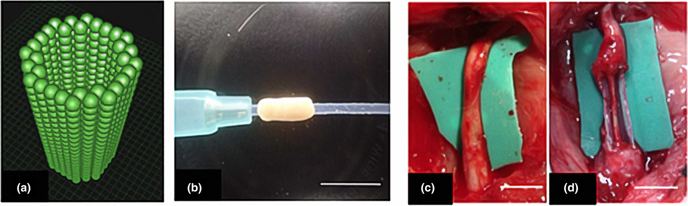
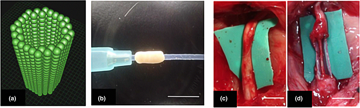
As mentioned above, the materials used to perform 3D printing are the ones that classify the type of scaffold, and an example of a free scaffold is one in which is only composed of fibroblast multicellular spheroids (MCSs) developing a biologic tubular structure.[Reference Yurie, Ikeguchi, Aoyama, Kaizawa, Tajino, Ito, Ohta, Oda, Takeuchi, Akieda, Tsuji, Nakayama and Matsuda137] Bio 3D printing is characterized by developing a complete biologic tubular structure, in this case using homogeneous fibroblast MCSs. A pre-designed 3D tube-like structure represents the position of the spheroids that are robotically placed into skewers of a 9 × 9 needle array [Figs. 4(a) and 4(b)].[Reference Yurie, Ikeguchi, Aoyama, Kaizawa, Tajino, Ito, Ohta, Oda, Takeuchi, Akieda, Tsuji, Nakayama and Matsuda137] This “needle-array” arrangement promotes MCSs to fuse and after 1 week are removed and cultured on bioreactor to obtained a structurally scaffold-free construct. The Bio 3D conduit was evaluated in vivo on rat right sciatic nerve, with a 5-mm gap that was bridged using an 8 mm Bio 3D conduit and control group with silicone tube. After eight weeks post-surgery, the Bio 3D conduits promoted peripheral nerve regeneration confirmed with successfully bridged through the nerve gap. In the case of silicone conduit, a very thin regenerated nerve was observed [Figs. 4(c) and 4(d)]. This is a novel example of how a 3D structure for peripheral nerve regeneration is developed without the use of a foreign material by 3D bioprinting.
(a) The pre-designed 3D tube-like structure. The green spheres represent homogeneous multicellular spheroids that were developed using only human normal dermal fibroblasts. (b) The Bio 3D conduit according to the pre-designed 3D model. (c) Regenerative nerve after eight weeks of surgery in the Bio 3D. (d) Regenerative nerve after eight weeks of surgery in the silicone tube. Reprinted from Yurie et al.[Reference Yurie, Ikeguchi, Aoyama, Kaizawa, Tajino, Ito, Ohta, Oda, Takeuchi, Akieda, Tsuji, Nakayama and Matsuda137]
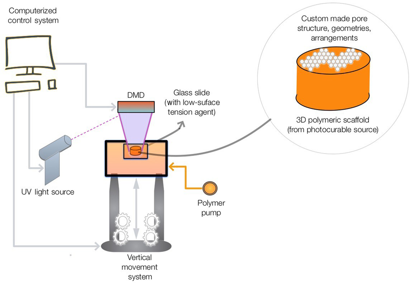
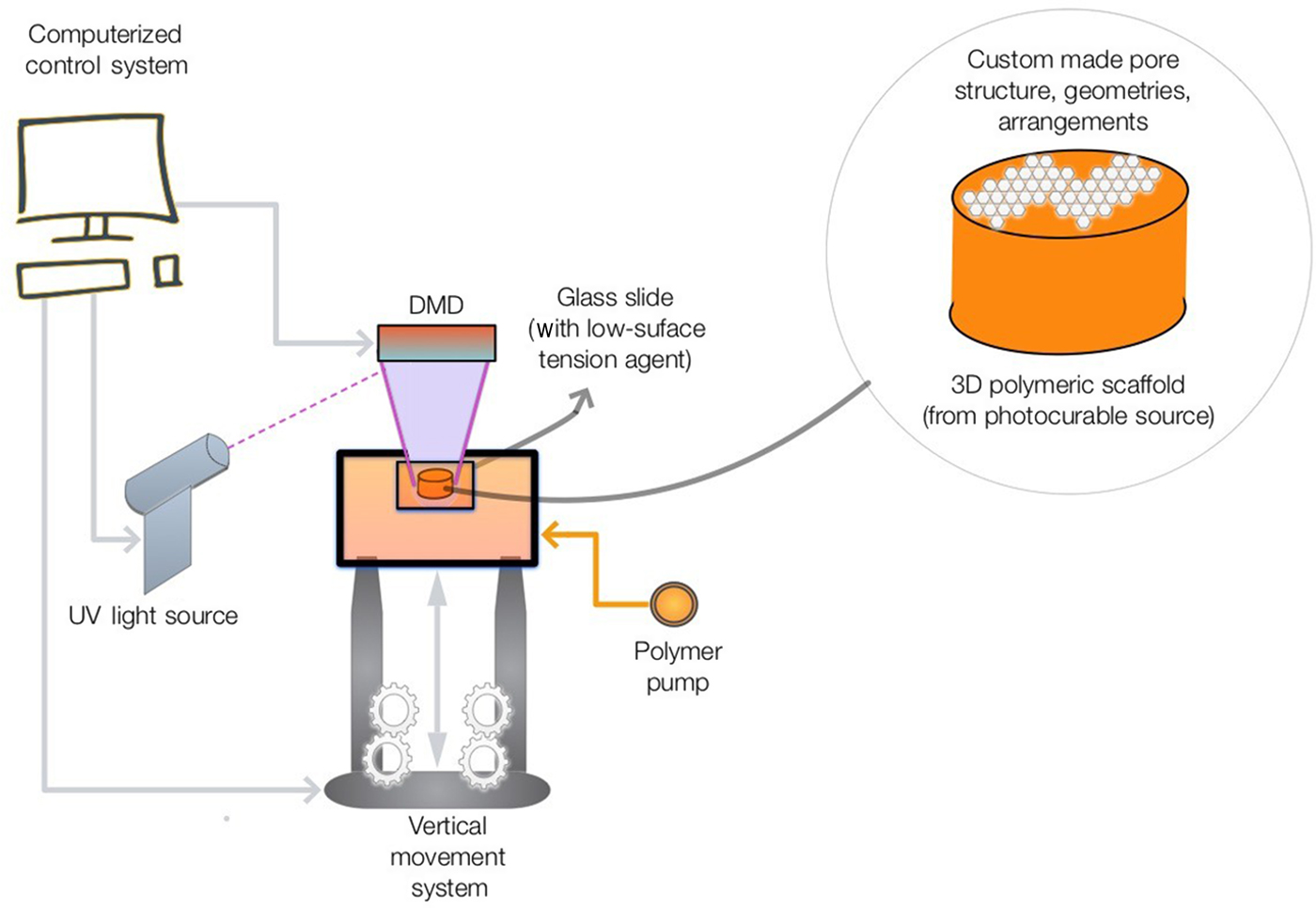
As part of the 3D printing emerging technologies to offer more custom made specialized scaffolds, the digital micro-mirror array devices microfabrication technique (DMD) provides an important space for innovation. DMD is a technique that takes advantage of free radical polymerization of photo-curable compounts to form/print solid 3D structures. Instead of doing so via a point-by-point scanning, the DMD system does it in a layer-wise mode. In fact, Pateman et al. combined together a microstereolithography setup including a DMD system.[Reference Pateman, Harding, Glen, Taylor, Christmas, Robinson, Rimmer, Boissonade, Claeyssens and Haycock50] The DMD system implements a chip that puts together micro-mirrors (1024 × 768) to reflect UV light shapes into a pre-polymer preparation and make entire layers at once (Fig. 5). These layers have high precision and resolution; achieve accurate designs (pore sizes, contours) and can be made to fit any possible model.[Reference Danquah and Mahato138]
Schematic representation of the DMD process (adapted from Ref. [Reference Soman, Tobe, Lee, Winquist, Singec, Vecchio, Snyder, Chen, Hall, Jolla, Winquist, Hall, Jolla and Snyder140]).
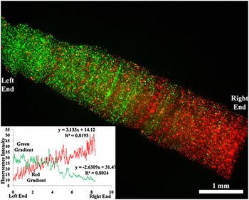
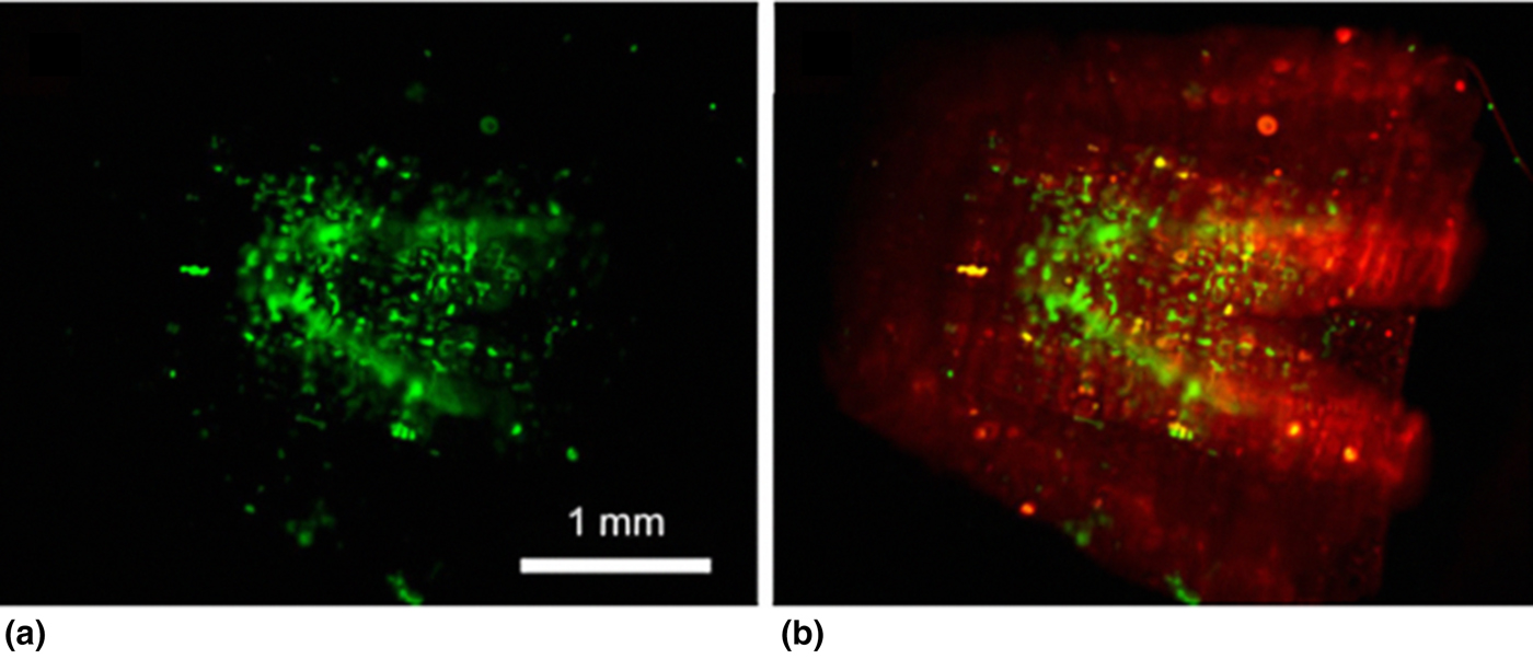
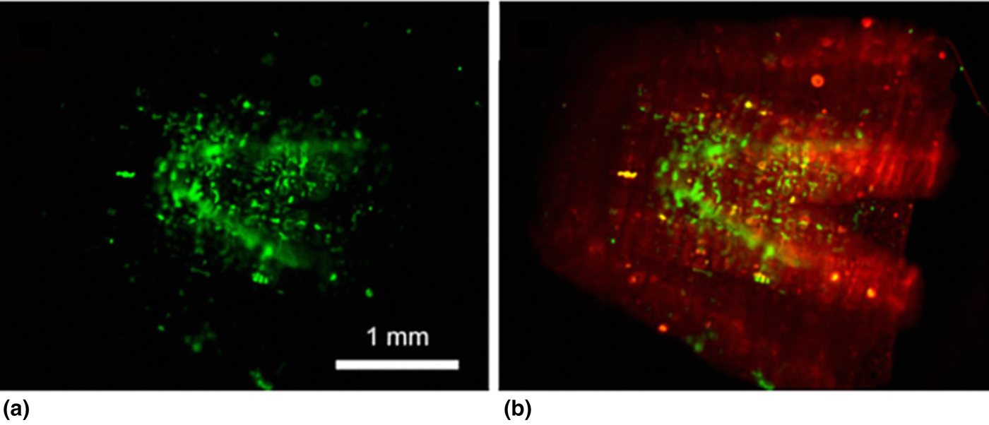
Not only does this technique allow freeform fabrication with unlimited alternatives to construct and mimic the nerve structure tissue, but some authors have found other breakthrough uses. For example, Suri et al. [Reference Suri, Han, Zhang, Singh, Chen and Schmidt77] used DMD to build a gradient of fluorescent microparticles in a HA scaffold (Fig. 6), this as a proof-of-concept showed that DMD can be applied to create gradients of biomolecules. A gradient of biomolecules like NGF favors an organized and controlled nerve regeneration.[Reference Kapur and Shoichet139] Also Fig. 7 presents the succesful culture of SCs inside the 3D scaffold composed of HA, developed using the DMD technique. Cells were seeded in the scaffold lumen, to demonstrate the viability of the cells in the custom design shapes. The cells were able to spread inside the geometries and expressed S100 proteins, which indicates that the adhered cells expanded over the 3D polymeric scaffold surfaces.[Reference Suri, Han, Zhang, Singh, Chen and Schmidt77]
Scaffold with two gradients running in opposite directions. Red and green fluorescent micro particles were mixed in a prepolymer solution and the scaffold was fabricated. The graph indicates the intensity profile of the fluorescence and thus the micro particle concentration (scale bar, 1 mm). Suri et al.[Reference Suri, Han, Zhang, Singh, Chen and Schmidt77] © Springer Science+Business Media, LLC 2011. With permission of Springer.

Fluorescence micro-graphs of the fabricated scaffolds. (a) and (b) show the SCs seeded inside the scaffold. The cells remained viable after 24 h when stained with calcein. Red fluorescent particles were added in the prepolymer solution to better visualize the scaffolds and the internal structure post-fabrication (scale bar, 1 mm). Suri et al.[Reference Suri, Han, Zhang, Singh, Chen and Schmidt77] © Springer Science+Business Media, LLC 2011. With permission of Springer.
It is important to make the reader aware that the virtue of the photo curable polymer demands special considerations.The main challenge confronted with the DMD techniques is related to UV light penetration and scattering. UV light induces the free radical polymerization; however, its sensitivity is enough to induce changes in the 3D scaffold structure. If light goes to uninteded areas, then the final product will not meet expectations. As a countermeasure to abberant UV-induced photo polymerization, Suri et al. controlled the exposure of UV at each layer, and used a free-radical quenching agent to limit the activation of polymer chains.[Reference Suri, Han, Zhang, Singh, Chen and Schmidt77]
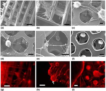
The work of Soman et al. demonstrated that DMD facilitated the cell culturing of neurons derived from human embryonic stem cells, and promoted the succesful differentiation and guided growth of neurites to reach its target at the distal stump. This was made possible because DMD enabled the custom geometries that help the 3D interconnectivity via the scaffolds’ pores of a PEG compound, which supported the alignment of neurite connections (Fig. 8). The studies were done with soft and stiff non-biodegradable PEGDA and biodegradable gelatin-based biomaterials scaffolds.[Reference Soman, Tobe, Lee, Winquist, Singec, Vecchio, Snyder, Chen, Hall, Jolla, Winquist, Hall, Jolla and Snyder140]
SEM images of PEG scaffolds with (a, b) log-pile and (c–f) hexagonal internal architecture support survival and transition of neurospheres to neuronal morphology, spreading from adherent cell clusters, and forming 2D and 3D intercellular projections. Neurospheres evenly penetrate and adhere to hexagonal PEGDA scaffolds. Larger neurospheres are largely excluded from the interior of the scaffold. (g–i) Neurons form 3D lattices on log-pile and hexagonal PEGDA scaffolds labeled for Tuj1. White arrow in (h) denotes a cell cluster adhered to the side of the log-pile scaffold with neural connections along the struts. Scale: 100 µm. Soman et al.[Reference Soman, Tobe, Lee, Winquist, Singec, Vecchio, Snyder, Chen, Hall, Jolla, Winquist, Hall, Jolla and Snyder140] © Springer Science+Business Media, LLC 2012. With permission of Springer.

Freeze-drying/porous
Freeze-drying or lyophilization is a useful technique to transform biomaterial solutions, which in many cases is composed of delicate proteins, into a dry solid form. The main underlying principle of this technique is sublimation: the polymer to be casted into a 3D structure is dissolved into an emulsion, then the removal of water and volatile substances is done by developing ice crystals at a very low temperature—depending on the solutions’ characteristic euthectic point the target temperature will vary. Then, by exposing the frozen solution to a vacuum enables the formed crystals to pass from its solid state to a vapor state directly resulting in a porous 3D polymeric structure. In this manner, drying happens without an excesive heating that could affect the involved biomolecule. The pore size of the 3D scaffold will depend on the crystals formed while the water/solvent within the polymer solution freezes. If the freezing rate is too fast, then crystals will be smaller and after the sublimation, the resulting pores will be smaller. Otherwise, a slow freezing rate will enlarge the ice crystals, and consequently bigger pores.[Reference Ratner, Hoffman, Schoen and Lemons141]
Some of the polymeric materials and cell lines that have been recently studied using the freeze drying fabrication technique include works with collagen and stem cells.[Reference Murphy, Matsiko, Haugh, Gleeson and O'Brien142–Reference Hortensius and Harley145] Here, the researchers reported the influence that stiffness, pore sizes architecture, and composition of GAG types (e.g., HA and condroitin sulfate) had over stem cell differentiation. This was performed to confirm the importance of the ECM components to increase or reduce the biomolecular signals in the cellular activity. Different approaches were used to enhance control stiffness for medical applications.
There are many biomaterials that usually would be a challenge to combine together, and obtain the desired biomolecular effects by design; ideally, systems of various macromolecules are thought to complement each other.[Reference Her, Wu, Chen, Chen, Chang and Wang146, Reference Kang, Liao, Wester, Leeder and Pearce147] However, there are practical issues such as incompatible solvents, and/or conflicting mixing conditions (temperature) that will not facilitate their merging. For these reasons, Niu et al. were able to demonstrate that one of the main advantages of the freeze-drying technique is to blend in a homogeneous emulsion of natural and synthetic polymers forming Chitosan (CS)–PLLA, and ultimately create a suitable method to develop a 3D scaffold with novel features to succesfully perform cell culture. Using the MC3T3-E1 osteoblast precursor cell line Niu et al. was able to demontrate the cell viability, survival, and proliferation on the freeze-dried CS–PLLA scaffold. Other researchers have used the same polymeric combined CS–PLLA scaffold loaded with recipient BMSCs to succesfully promote nerve regeneration and functional recovery of the sciatic nerve in animal models; however, in this instance the scaffold was not freeze dried.[Reference Niu, Li, Liu, Zhou, Feng, Cui and Fan148] The work of Zhang and Cui showed that chitosan with lactic acid (PCLA) combined via the freeze-drying technique resulted in a scaffold with better histocompatibility properties for BMSC than each material by itself.[Reference Zhang and Cui149]
Other practical issues of a co-polymeric combination prepared by freeze-drying are: biodegration, swelling, and loss in mechanical properties. In vivo degradation is expected as part of the scaffold design; furthermore, it must be considered as part of the potential medical device application; that once its objective is achieve (after the tissue is regenerated) the body continues its natural lifecycle. Polymeric degradation can be induced by pH change, hydrolysis, and immune biocompatibility. Combined co-polymers may have different degradation rates, and 3D cell culture models will enable researchers to understand and improve materials and conditions for the different healthcare needs. For that, the advanced controls that the freeze-drying technique permits regarding pore size and its interconnectivity within the scaffold matrix are favorable for studying cross-linking alternatives.[Reference Kozłowska and Sionkowska150]
Another technique that integrates freeze drying, is the freeze casting. Freeze casting benefits from the ice crystallization phenomena, and its freezing gradient/direction to develop a microstructure architecture for the scaffolds, whether porous, layered, or custom oriented. This happens as ice growths, carries the polymer dispersion along, and form separate clusters of the polymer particulates in the space between the ice crystals, therefore forming lamellar shapes. Later, via freeze drying, the ice sublimates leaving the designed scaffold. Preparation method, solvent used, polymer concentration, and freeze-drying cycle will determine the mechanical properties of the layered material. For example, particles locked in the ice crystals will convey and arrange connections between lamellae formations. The speed at which the ice grows in the freeze-drying cycle (particularly at the freezing stage) will determine the pore sizes of the future scaffold.[Reference Gaudillere and Serra151, Reference Wegst, Schecter, Donius and Hunger152] Francis et al. investigated how to enhance axonal regeneration after spinal cord injury. They fabricated a porous chitosan-alginate scaffolds with longitudinally oriented channels using the freeze-casting method, with the objective of enabling a guidance/path for the regenerating axons toward proximal and distal stump. They used DRG explants, these neural cells adhered and extended neurites through the scaffold in parallel alignment with the channel direction. Additional molecules were added (e.g., LN) to enhance the nerve guide conduit (NGC) properties, and characterize its potential applications.[Reference Francis, Hunger, Donius, Riblett, Zavaliangos, Wegst and Wheatley153]
Electrospun fibers
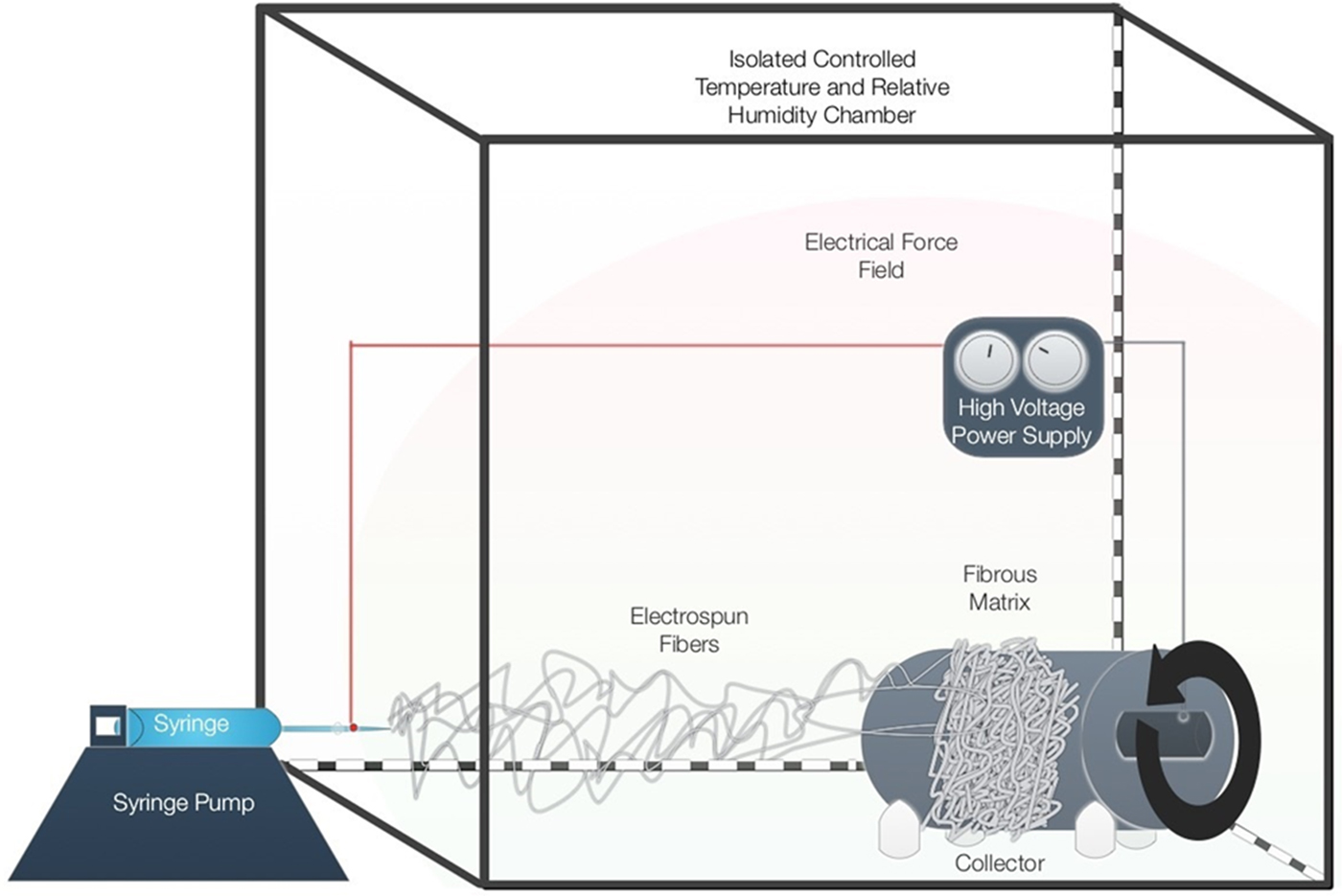
Several authors have worked on demonstrating that nanofibers can guide neurite outgrowth along the direction of fiber alignment.[Reference Xie, Liu, Macewan, Bridgman and Xia154, Reference tae Kim, Haftel, Kumar and Bellamkonda155] Electrospinning technique is a good alternative for this objective because nanofibers of several polymers can be prepared with an aligned structure that can be used to guide tissue regeneration.[Reference Huang, Zhang, Kotaki and Ramakrishna156] In electrospinning, a polymer solution is pumped through a needle that is exposed to a high voltage (Fig. 9). The solution must be received in a metallic collector, which is connected to the other end of the power supply and this generates an electric field that conducts the fiber for its deposition.[Reference Castilla-Casadiego, Ramos-Avilez, Herrera-Posada, Calcagno, Loyo, Shipmon, Acevedo, Quintana and Almodovar157] The main advantage of this method is that other elements such as fiber diameter, elastic modulus, morphology, and other mechanical properties can be adjusted thanks to the versatility of the technique by addition or variation of factors in the process and also, these characteristics can be determined through various techniques.[Reference Castilla-Casadiego, Maldonado, Sundaram and Almodovar158] One of the main limitations is the strict humidity control inside the chamber (which also leads to temperature control). High humidity implies the fiber formation with very diverse and unstable properties or may even prevent their formation. Another important point is the viscosity of solutions prepared for electrospinning: a very viscous solution may block the needle or the induced electromagnetic field may fail to break the surface tension of the solution. If the solution is not very viscous, it flows very quickly and leaves the tip of the needle like a “spray”, which implies that the solution flow must be decreased and therefore, the elaboration of the membrane is made very slowly. The use of strong and toxic solvents becomes the main disadvantage of the method, since it is subjected to strict regulations and procedures for their disposition and waste management. Also, it is important to emphasize that electrospinning uses high voltages, which puts at risk the health of the operator and must have high safety measures like the use of protection elements (in many cases increase the cost of the process).
Schematic representation of the electrospinning equipment and process at our home lab.

Linked to tissue regeneration, many authors have used electrospun nanofibrous scaffolds as structures for 3D cell cultures for a wide range of nerve cell lines, e.g., highly aligned PLLA fibers have been studied with primary neurons (DRG explants), human neuroblastoma cells (SH-EP and SH-SY5Y cells), SCs, PC-12 cells, among others. PLLA is a material widely used for its known biocompatibility, degradability, and aligned structures to support neuronal growth. Han et al.,[Reference Wang, Mullins, Cregg, Hurtado, Oudega, Trombley and Gilbert159] obtained aligned electrospun fibers made from PLLA, which allowed the proliferation of neurites and the communication of SCs. Electrospun scaffolds were applied in the culture of embryonic stage nine (E9) chick DRGs and rat SCs. With respect to the cells, they could perform the quantification of the density and length of the neurite outgrowth. E9 chicken DRGs cells were seeded into high and low density PLLA fibers to evaluate the effect on neurite outgrowth (cells were stained for neurofilaments). They observed the aligned fibers guided growth in a directed way, and by increasing fiber density, it increases growth by approximately 83% compared with 65% for low density fiber. It has been shown that the neurites grew once the DRG explants are attached to the fibers, and upon contact with the aligned configuration of the fiber, the neurites changed their direction to grow parallel to them. Even in experiments of more than 10 days, when cells bodies have been dissociated from the fiber, neurites continues to grow in the direction of fiber. Also, highly aligned fibers favor increased neurite growth compared with less aligned fibers (20% above to randomly aligned fibers). For the SCs, they attach to the fibers and grow in a directed manner for both high- and low-density fibers, but higher growth rates were observed in high-density fibers and their morphology becomes elongated and narrow. In the case of explants, SCs move away from them to join behind the neurites. Neuroblastoma cells were also shown to be elongated and narrow in aligned fibers oriented parallel to the fibers.[Reference Kang, Liao, Wester, Leeder and Pearce147, Reference Corey, Lin, Mycek, Chen, Samuel, Feldman and Martin160] Many literatures suggest aligned fibers are important for peripheral nerve studies since it enhances the regeneration and maturation of SCs.[Reference Koudehi, Fooladi, Mansoori, Jamalpoor, Amiri and Nourani87–Reference Liu89]
The influence of electrospun fiber size on SCs behavior has been studied through the manufacture of cross-linked gelatin electrospun fibres having different diameters in the range of 300–1300 nm in which SCs were cultured. Gelatin is highly biocompatible, biodegradable, not immunogenic, and maintains the collagen properties that favor cell growth. GPTMS or γ-glycidoxypropyltrimethoxysilane were used as cross-linking agents. By increasing the gelatin concentration and flow rate during electrospinning, fibers with greater diameter (340–1306 nm) and porosity were obtained. The increase in fiber diameter did not affect cell adhesion but lead to a decrease in cell extension area, so that the cells became more elongated. Cells are highly influenced by physical stimuli, and it is known that fiber diameter alter cellular morphology and proliferation (diameter can be controlled through modification of the electrospinning parameters). In general terms, the increase in fiber diameter affects other aspects, such as less organization of the actin cytoskeleton, increased filopodia formation (and cell migration), reduced proliferation, and even improvement in axonal growth.[Reference Pateman, Harding, Glen, Taylor, Christmas, Robinson, Rimmer, Boissonade, Claeyssens and Haycock50, Reference Gnavi, Fornasari, Tonda-Turo, Ciardelli, Zanetti, Geuna and Perroteau161] NG108-15 neuronal cells and primary rat SCs were evaluated under various diameter of PCL aligned electrospun fibers, individually, and as co-culture and with rat dorsal ganglion cells. It was shown that cell length on narrow fiber (1 µm) results on better neural outgrowth specially when evaluate as co-culture, meaning that this is a promising model since it mimics neural tissue properties. Although it was not measured, the positive response of SCs on the neuronal cell culture extension is attribute to the excretion of NT factors such as NGF and BDNF and also expression of ECM proteins.[Reference Daud, Pawar, Claeyssens, Ryan and Haycock51]
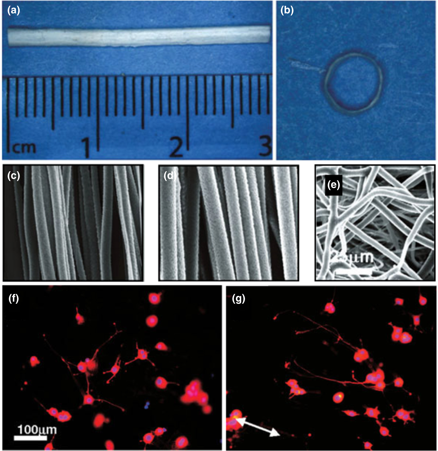
Yao et al., made an electrospun cylindrical nerve guide conduit using polycaprolactone (PCL) and designed it so that in the inner part of the scaffold the fibers would be aligned and thus determine which is the optimal diameter to favor the directional growth and neurite extension. PCL is characterized by being a slow degrading polymer and its application to a slow process such as nerve regeneration is favorable. The cylindrical scaffolds were fabricated on 1.4 mm diameter stainless steel wire to simulate the mouse sciatic nerve and it was possible to obtain random fibers on the outer side and aligned fibers on the inner surface. Alternatively, fiber flat scaffolds were also made with aligned or random orientation to analyze the guided neurite growth and images were obtained using SEM and FFT function were used to determine fiber orientation (Fig. 10). In this study, PC-12 cells were seeded with NGF as supplement. After 5 days of cell culture, the neurite grew parallel in an oriented way in aligned fibers with growth close to 190 µm compared with a 155 µm and randomly growth to random fibers thus showing that the NGCs provide a means to facilitate regeneration of axons.[Reference Yao, O'Brien, Windebank and Pandit162]
Analysis of the fiber alignment. (a) Sample of electrospun PCL conduit. (b) Cross-sectional image of the conduit. (c, d) SEM images of aligned fibers with varying fiber diameters. (e) Randomly oriented fibers. (f) Neurites of PC12 cells on randomly oriented and (g) Aligned neurite growth on aligned PCL fibers. From Yao et al.[Reference Yao, O'Brien, Windebank and Pandit162] Copyright © 2009 by John Wiley & Sons, Inc. Reprinted by permission of John Wiley & Sons, Inc.

A novel type of silk electrospun fiber with gold nanoparticles (GNP), shows the versatility of the method for different applications. Degummed silk fibroin fibers were immersed in a GNP solution and then subjected to freeze dried. The resulting powder was dissolved in formic acid (98% pure) at a concentration of 100 mg/mL and mixed with 1 mg of polyethylene oxide (PEO) per milliliter of solution. This final solution was electrospun for the development of a NGC by rolling the fiber onto a stainless-steel needle. This NGC was pretreated with SCTM 41 (rat SC) for 5 days before being tested for sciatic nerve regeneration in vivo. SCTM 41 showed a favorable response when there was cell adhesion and growth on the internal and external conduit surfaces and NGC with GNP showed very similar results to those exhibited by a normal nerve after 9 and 18 months of implementation. At structural level, all NGCs were intact and maintained adhesion to the sutured ends.[Reference Das, Sharma, Saharia, Sarma, Sarma, Borthakur and Bora163] In a similar manner, GF-containing particles can be designed and incorporated into the general process of manufacturing the nanofibers.[Reference Zuidema, Provenza, Caliendo, Dutz and Gilbert164]
Fiber alignment seems to promote axonal regeneration as shown in the research of Sperling et al.,[Reference Sperling, Reis, Pozzobon, Girardi and Pranke165] where mESCs interacting with 3D scaffolds composed of PLGA were studied.[Reference Kapur and Shoichet139] The target was to evaluate the response of cell survival, morphology, and differentiation over factors such as surface acceptance of polymer PLGA and fiber orientation, using random and aligned fibers. Immunocytochemistry was used to corroborate the expression of neural markers cultured on the PLGA scaffold. The marker for neural precursor cells, nestin, was highly expressed on aligned oriented fiber when compared with random. Expression of β 3-tubulin helped to demonstrate the typical neuronal morphology, including neurite branching and outgrowth processes. Moreover, expression of these markers was higher on 3D PLGA scaffolds as compared with flat scaffolds, highlighting the importance of 3D models. In a more recent work, Schuh et al., obtained homogeneous electrospun fibrin-PLGA fibers under established conditions in which they seeded cells and their results were favorable for proliferation along the fiber. MSCs were isolated from rat adipose tissue, which were in vitro differentiated to SCLs and SCs were isolated from rat sciatic nerves. Proliferation and cell viability assays were used to evaluate cell–scaffold interaction. Electrospun fibrous scaffolds were made with a parallel orientation and the seeded cells showed a positive response to the biomaterial as it grew along the fiber after 3 and 7 days of culture. They also propose the use of their nanofibers for in vivo studies by seeding cells inside the scaffold, modifying it to a self-shaping fibrin tubular structure with the incorporation of a hydrogel in the electrospun fiber.[Reference Schuh, Morton, Banerjee, Grasl, Schima, Schmidhammer, Redl and Ruenzler166]
Three-dimensional cell encapsulation
In this section, the focus is not only the technique for the development of the 3D structures but rather to showcase how these 3D arrangements are incorporated with cells monoculture and co-culture to mimic the cell microenvironment, via 3D cell encapsulation. Cell encapsulation is an approach in which hydrogels play an important role, along with scaffolds, both serving as platforms for monoculture and co-cultures. Hydrogel is a technique applied to a variety of polymers such as: poly (vinyl alcohol) (PVA), poly (glycolic acid) (PGA), poly (lactic acid) (PLA), and naturally derived biologic components such as cellulose, collagen, chitosan, hyaluronic acid, and basement membrane extract, having applications on tissue engineering and drug delivery fields, to name a few.[Reference Basu, Reddy, Doppalapudi, Domb, Khan and Peg167–Reference Hilderbrand, Ovadia, Rehmann, Kharkar, Guo and Kloxin169]
Once the proper techniques are performed to develop hydrogels, they turned into platforms with ideal conditions to mimic the microenvironment that surround cells. Hydrogel unique features are attractive for biomedical applications which are: high water content, rubbery structure, biocompatibility and high capability to incorporate significant amounts of functional groups, all combining to mimic living tissue.[Reference Hilderbrand, Ovadia, Rehmann, Kharkar, Guo and Kloxin169, Reference Lin, Wang, Wertheim and Coombes170]
A challenge with hydrogels is the control of matrix stiffness and biologic cues, which is not completely achieved with the use of natural hydrogels, mainly because of permanent protein structure and product variation, since they are derived from a natural source. These challenges are overcome with the modification of functional groups to form synthetic and hybrid hydrogels, being capable to control gel formation and degradation, supporting the fact that hydrogel techniques have a wide range of applications.
In many applications, hydrogels are combined with other techniques such wet spinning to produce complex structures, for example a macroporous alginate hydrogel fibrous scaffold for nerve repair.[Reference Lin, Wang, Wertheim and Coombes170] In brief, this structure was produced by wet spinning a solution composed of gelatin particles, alginate solution, and dorsal root ganglion cells (DRGs) into 0.5 M CaCl2 cross-linking solution. This approach highlights the use of hydrogels to develop 3D structures with cell encapsulation, more specific a co-culture since DRGs are a mixed of SCs and neurons. Crosslinking of hydrogels is one of the most important factors to produce a 3D, hydrophilic, insoluble polymeric network. It can be physical or chemical, and gives hydrogels stability.[Reference Peppas, Bures, Leobandung and Ichikawa171] Also, alginate was used since it is a polysaccharide that has a positive effect promiting axonal development, nerve repair, and ease of gelation property.[Reference Georgiou, Bunting, Davies, Loughlin, Golding and Phillips19, Reference Lee and Mooney172]
A scaffold's porosity is key for the diffusion of nutrients and oxygen, waste elimination, and is directly related to cell proliferation and tissue development. An example of how to control the porosity in a hydrogel is by the introduction of gelatin particles porogens, with a particle size range <90 µm, to the 1.5% w/v alginate solution, and then followed by extraction using distilled water at 37 °C, leaving a 3D assembly composed of alginate fibers with pores. The target variable to prove the effectiveness of the 3D macroporous alginate scaffold, was neurite outgrowth from the encapsulated DRGs. The results showed neurite outgrowth over a distance of 150 µm on day 11 on 3D macroporous structure, compared with no significant outgrowth over 15 days on non-macroporous. The reason for no significant neurite outgrowth is because of absence of pores and channel structure needed for the neurite extension. The combination of wet spinning with hydrogel principles, along with gelatin particles represent a prominent example of a 3D macroporous complex that serves as a structure to support neurite extension.
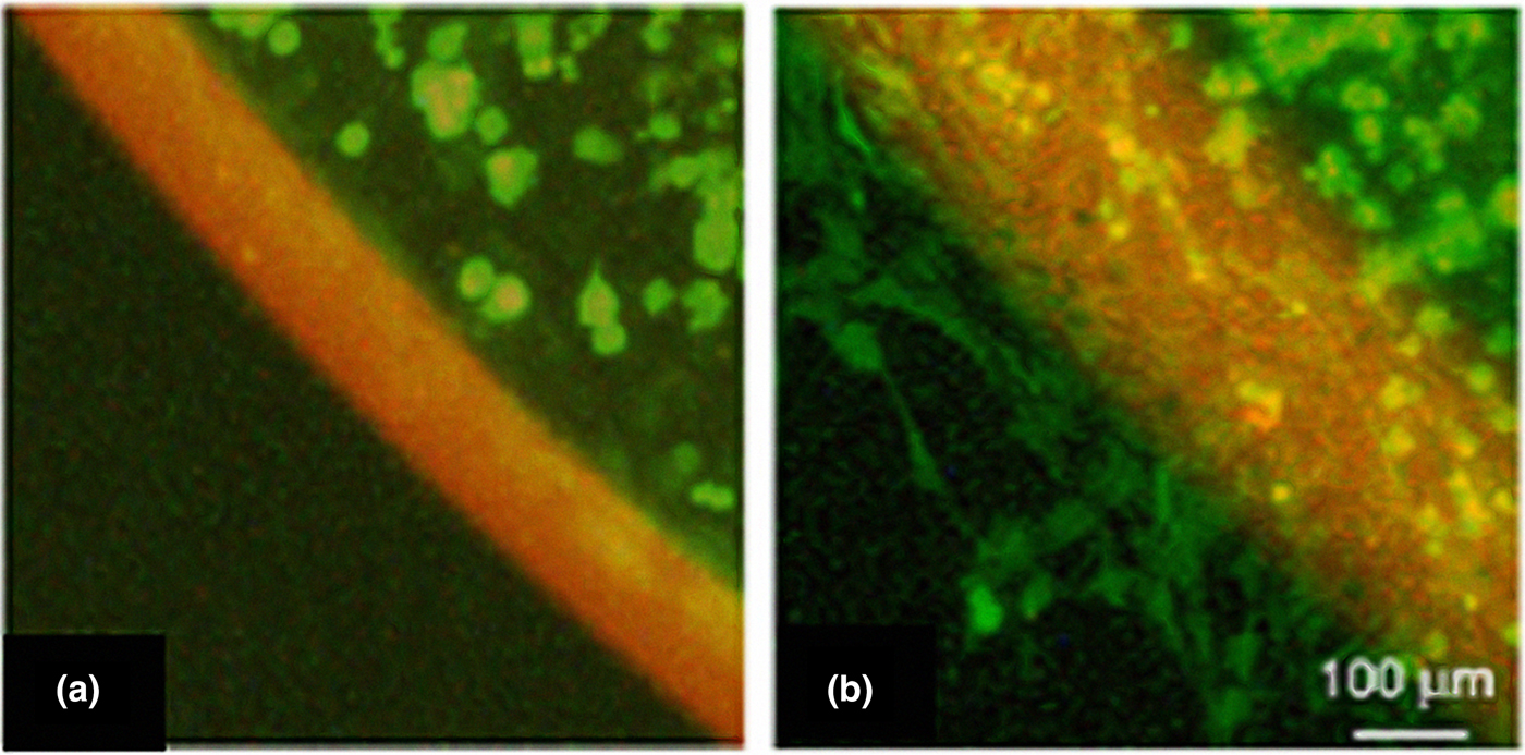
As mentioned above, the versatility of hydrogels can be seen by the combination of nerve conduits with hydrogel filling making a more stable and 3D arrangement that had a positive effect to promote cell adhesion and proliferation.[Reference Tonda-Turo, Gnavi, Ruini, Gambarotta, Gioffredi, Chiono and Ciardelli173] The hydrogel composition consisted of agar–gelatin at a 20 : 80 weight ratio (wt/wt) blend, crosslinked by the addition of genipin and the conduit was a porous PCL guide of 1.3 mm diameter and 15 mm length. A total of 20 µL of hydrogel was inserted using a syringe. The performance of the composite conduit was tested in vitro to prove the capacity of SCs to adhere, proliferate, and migrate. Also, a 3D migration assay, using neonatal olfactory bulb ensheating cells (NOBECs), proves no obstruction through the hydrogel solution, Figs. 11(a) and 11(b). Hydrogel presence on NGC supported cell, migration, adhesion, and proliferation. Hydrogels have the potential to continue enhancing conduits function for peripheral nerve regeneration.
Three-dimensional migration assay. Representative images of migrated neonatal olfactory bulb ensheathing cells (NOBECs) 0 (a), and 72 h (b) after incorporation in A/GL-GP (agar and gelatin cross-linked with genipin) hydrogel. Cells were stained with cell tracker green (CTG) and visualized in green; biomaterial autofluorescence is visualized in red. Reprinted from: Tonda-Turo et al.[Reference Tonda-Turo, Gnavi, Ruini, Gambarotta, Gioffredi, Chiono and Ciardelli173]

Cell Encapsulation of co-cultures offers to study cell–cell and natural ecosystem interactions.[Reference Goers, Freemont and Polizzi68, Reference Albrecht, Underhill, Wassermann, Sah and Bhatia174] The immediately advantage of this technique is a more detailed representation of physiological environment compared with monoculture, by the control of several factors like, type of medium, cell concentration, time scale, and the addition of degree of contact between cell populations. The degree of separation can be achieved through semi-permeable membranes, gels, microfluidic devices, and others.[Reference Goers, Freemont and Polizzi68, Reference Kraus, Boyle, Leibig, Stark and Penna69, Reference Lin and Chang175] The way in which the cell-culture degree of separation is arranged defines the experimental set-up, for this case 3D arrangements with scaffolds and hydrogels will be highlighted. Challenges that bound this technique is the high level of complexity mainly because of the multiple cell interactions generating unpredictability and instability, which leads to another challenge of poor knowledge of data acquisition tools since it involves multiple interactions and even ones that are unknown.[Reference Goers, Freemont and Polizzi68, Reference Albrecht, Underhill, Wassermann, Sah and Bhatia174, Reference Lin and Chang175]
Co-culture experimental setup depends upon the separation and suspension techniques or materials in which the cell populations will be exposed.[Reference Goers, Freemont and Polizzi68] It is important to highlight that the applied technique defines whether it will be a two or 3D structure. An example of a co-culture technique that generates a 3D structure is spheroidal co-culture, also known as MCS, which are developed by a number of techniques, including hanging drops culture, single-cell culture on non-adhesive surface, micromolding techniques, spinner flask culture, rotary cell systems, porous 3D scaffolds, and centrifugation pellet culture.[Reference Lin and Chang175] The co-culture spheroids were suspended in a 3D collagen matrix and together represented a 3D structure for peripheral nerve regeneration.[Reference Kraus, Boyle, Leibig, Stark and Penna69, Reference Cargill, Dee and Malcolm176] The co-cultured consisted of SCs (500 cells/spheroid) and NG108-15 a hybrid cell line (NG, 50 cells/spheroid). The cell ratios indicates that each spheroids contains ten times more quantity of SCs cells than NG cells, this could be attributed to enhance the spheroid environment with GFs secreted by SCs cells. The reason for the choice of cells, is that SCs and NG108-15 cells (which are neuroblastoma cross glioma cell line) complement well based upon the abilities of SCs to provide physical guidance axons and NG108-15 cells’ ability to differentiate toward mature motor neuron phonotype.[Reference Korff and Augustin177] The technique to developed the spheroids was hanging drops. The process starts with cell being centrifuged on non-adhesive wells, containing cell culture medium and 20% of methocel stock solution which prevents adhesion of cells and acts as an inert viscosity modulation substance. Methocel concentration is a parameter used to control de average size of the spheroids.[Reference Korff and Augustin177] A clear collagen gel was develop and serve as the matrix in which the pellets containing the spheroids were introduce, finally obtaining the 3D co-culture as hanging drops on the collagen matrix, as shown in Fig. 12.
3D co-culture, model of experimental setup. Spheroidal formation hanging drops and transfer in 3D collagen matrix. “Reprinted from Kraus et al.[Reference Kraus, Boyle, Leibig, Stark and Penna69] Copyright (2015), with permission from Elsevier”.

Neurite length was the response variable showed that in 2D culture is around 65 µm and the 3D model ranged from 115.56 to 176.6 µm, for 2.3× longer neurites.[Reference Kraus, Boyle, Leibig, Stark and Penna69] The main reason for this behavior is that the model was able to increase the interactions between both cell types. To minimized cultivation differences between 2D and 3D cell culture both had a collagen layer. It is clear that no neurite growth into the collagen layer was noticed on 2D cell culture, which confirms that 3D setup was capable to increase cell–cell contacts and promote neurite growth.
A co-culture of SCs and ADSCs seeded on silk fibrin/collagen scaffold is an approach to develop a tissue-engineered nerve conduit (TENC).[Reference Xu, Zhang, Chen, Li, Li and Feng20] The explanation for the choice of cells is that ADSCs are adult SCs that can be induced to neuropheres and neuronal-like cells when exposed to neurothrophic media, and this specific media is created by the presence of SCs. SCs have the capacity to secrete a variety of NTs, NGF, BDNF,GDNF, and others that help differentiate stem cells, making an ideal complement to promote and accelerated axonal growth. The 3D structure was develop by blending a solution of silk fibroin and collagen, at a ratio of 4 : 2, followed by cast molding and finished by lyophilization. It is important to mention that the conduit structure, stability, and biocompatibility, are dependent upon the choice of materials, for this case silk fibroin is added because of its excellent mechanical properties, which complements well with the biomcompatible properties of collagen.
The silk fibroin/collagen struture is seeded with the co-culture obtaning a TENC. Tissue regeneration was achieved on 1 cm long sciatic nerve defect in rats, which had results similar to autologous nerve grafts, but superior when compared with plain (non-co-culture scaffold). The conduit seeded with the co-culture mimics the native microenviroment, not only promoting the nerve regeneration, but also accelarating axonal growth as confirmed by the transdifferentiation of ADSCs when co-cultured with SCs. 3D models such as spheroidal co-culture and seeded co-culture on three dimensional scaffolds, projects to fill the gap between the monoculture assays and in vivo studies in order to represent cell–cell and cell–ECM complex interactions, offering a more detailed representation of physiological structures and conditions.[Reference Parikh, Hao, Hosseinkhani, Patil, Huntley, Tessier-Lavigne and Zou37, Reference Yang, Gu, Tan, Hu and Wang95]
DRG cells are a mixed of SCs and neurons, and are categorized as a co-culture, and many experiments have been made using these cells. An example, is the use of a single walled carbon nanotube-composite hydrogel with electric stimulation, in which the neurite extension of the encapsulated DRG cells was evaluated. The hydrogel solution was composed of collagen type I and GF-reduced Matrigel®, in order to support SCs spreading, viability, and migration. It was proved that neurite extension was induced with the presence of the hydrogel and also by the electric stimulation; however, it is known that cells within the DRG, such as glia cells, may also influence in the results.[Reference Koppes, Keating, McGregor, Koppes, Kearns, Ziemba, McKay, Zuidema, Rivet, Gilbert and Thompson178] Another approach using DRG cells is with the combination of technologies of aligned PLLA microfibers with NGF releasing composite nanoparticles, both technique induced neurite extension with the presence of GF on nanoparticles and aligned microfibers.[Reference Zuidema, Provenza, Caliendo, Dutz and Gilbert164] Overall, DRG cells are a co-culture very representative of the response and performance necessary to reflect neurite extension.[Reference Zuidema, Provenza, Caliendo, Dutz and Gilbert164, Reference Koppes, Keating, McGregor, Koppes, Kearns, Ziemba, McKay, Zuidema, Rivet, Gilbert and Thompson178] Hydrogels and scaffolds are platforms that offer 3D structures and co-culture simulate nerve cells models. Both aplications filled the gap between in vitro and in vivo experiments by being capable to mimic the native microenviemroment.
Validation of peripheral nerve three-dimensional cell culture models
Engineered polymeric scaffolds serve as highly tunable materials for 3D in vitro models of peripheral nerve. The 3D environment can more accurately recapitulate the native environment compared with 2D substrates, providing fundamental knowledge of neural tissue repair. 3D cell culture models are first validated by measuring adhesion of both neuron and glial cells, and by measuring neurite extension and glial cell proliferation. Additionally, protein and gene expression must be considered and can be evaluated on the 3D scaffolds. For example, Tuj1 is typically evaluated as an early marker for neuron development.[Reference Subramanian, Krishnan and Sethuraman41, Reference Wang, Tang, Park, Zhu, Patel, Daley and Li54, Reference Soman, Tobe, Lee, Winquist, Singec, Vecchio, Snyder, Chen, Hall, Jolla, Winquist, Hall, Jolla and Snyder140, Reference Sperling, Reis, Pozzobon, Girardi and Pranke165, Reference Cunha, Panseri, Villa, Silva and Gelain179] GAP43 and synapsin I, are both known to be upregulated throughout neuronal development.[Reference Das, Freudenrich and Mundy180] For glia, during the transition of immature SCs toward myelinating, PMP22 and EGR2 are upregulated, while a down-regulation of NCAM and low affinity NGF receptor (LNGFR) occurs.[Reference Wu, Wang, Guo, Shao and Ma181] Also, adhesion of SCs to scaffolds may be validated by an overexpression of S100.[Reference Mobasseri, Faroni, Minogue, Downes, Terenghi and Reid14, Reference Georgiou, Bunting, Davies, Loughlin, Golding and Phillips19–Reference Johnson, Lancaster, Zhen, He, Gupta, Kong, Engel, Krick, Ju, Meng, Enquist, Jia and McAlpine21, Reference Daud, Pawar, Claeyssens, Ryan and Haycock51, Reference Schuh, Morton, Banerjee, Grasl, Schima, Schmidhammer, Redl and Ruenzler53, Reference Wang, Tang, Park, Zhu, Patel, Daley and Li54, Reference Tohill, Mantovani, Wiberg and Terenghi56, Reference Mokarram, Merchant, Mukhatyar, Patel and Bellamkonda63, Reference Parrinello, Napoli, Ribeiro, Digby, Fedorova, Parkinson, Doddrell, Nakayama, Adams and Lloyd66, Reference Suri, Han, Zhang, Singh, Chen and Schmidt77]
However, evaluating a handful of expressed proteins and/or genes gives a limited picture of how the proposed 3D models are behaving in comparison with native tissue. A complete transcriptomic and/or proteomics profile of the in vitro 3D cell culture is crucial for the development of proper models that closely resemble native tissues. For instance, transcriptomic analysis allowed for the confirmation that engineered GF-induced neural progenitor cells closely resembled brain-derived neural progenitor cells.[Reference Gao, Xiu, Zhang, Zang, Yang, Wang, Wang, Wang, Yi, Tang, Gao, Wang, Xi, Liu, Wang, Wen, Yu, Zhang, Chen, Chen and Gao182] Other work has used microarray analysis to identify new genes involved during peripheral nerve regeneration.[Reference Boeshore, Schreiber, Vaccariello, Sachs, Salazar, Lee, Ratan, Leahy and Zigmond183] Recently, a proteomics analysis was performed to evaluate MSCs cultured in three similar ECM mimetic materials from different sources.[Reference Ragelle, Naba, Larson, Zhou, Prijić, Whittaker, Del Rosario, Langer, Hynes and Anderson184] They observed that each material induced a unique matrisome signature, but they all shared a common set of proteins. Very little work has been published on validating 3D cell culture models of peripheral nerve using an omics approach. Carefully designed 3D environments must be validated via transcriptomic or proteomics analysis by comparing them to native cells or tissues to provide high fidelity to the 3D cell culture model. Lastly, high-throughput biomaterial screening platforms have emerged as useful strategies for the design and testing of biomaterials to serve as 3D environments for cell culture.[Reference Dumont, Karande and Thompson185]
Conclusions
The baseline for 3D cell culture platforms continues to expand. Many options are currently available to researchers. It is important to continue efforts and integrated strategies that not only can be executed at research laboratories but expedite the introduction of more effective therapies and sustainable treatments. There is still work to be done in the nerve regeneration field, especially to gain knowledge about improvements to overhaul many of the practical limitations, and what are the explicit points that remain departing from the nerve biology. 3D cell culture models may provide answers to these fundamental questions in neural tissue regeneration.

Acknowledgments
This work was financially supported by a subaward from the Puerto Rico Idea Network of Biomedical Research Excellence (NIH NIGMS/INBRE P20 GM103475-15).