Introduction
Dementia is an increasingly common condition, characterized by deterioration in brain function.Reference Burns and Iliffe1 It is generally a ‘disease of ageing’, shown by an increase in the percentage of people diagnosed as having dementia, from 5% of people aged 65 years to 30% of people aged 90 years.Reference Andreasen2 Dementia is a non-specific syndrome causing deterioration of many cognitive functions, including memory, learning, emotional control, judgement and thinking, to the extent that everyday living is impaired. There are many causes of dementia, the commonest of which are the slowly progressive neurodegenerative types, such as Alzheimer's disease (AD) and dementia with Lewy bodies (DLB), and vascular dementia (VaD).
This review compares AD and DLB. AD is the most common form of degenerative dementia, with DLB being the second most common. It has been questioned whether the various different types of dementia are totally separate conditions, or whether they are in fact all part of a spectrum of a more general neurodegenerative disease. This review will focus on their clinical features, neuropathology and neuroimaging features and structural changes.
Search methods and sources used
The literature for this review was identified by searching PubMed from 1990 to 2010. The search terms used were ‘dementia’, ‘Alzheimer's disease’, ‘dementia with Lewy bodies’, ‘neurodegenerative’, ‘clinical features’, ‘neuropathology’, ‘neuroimaging’, and ‘structural changes’. Further literature was found by looking through the reference lists of the articles identified through PubMed. Textbooks and other reference books were used to provide background information.
Clinical features
Alzheimer's disease
Progressive cognitive impairment is the core clinical feature of AD, and it can be described as the ‘4 As’.Reference Lovestone, Gelder, Lopez-Ibor and Andreason3
• Amnesia: the loss of ability to memorize and/or recall information which tends to affect recent memories in early stages, with patients often remembering events of their childhood until the disease has progressed.
• Aphasia: an absence of previously acquired language skills, which may present with problems such as nominal dysphasia with speech deteriorating far more severely as the disease progresses.
• Agnosia: an inability to recognize objects, which may interfere with the patient's daily functioning due to the inability to recognize simple everyday objects.
• Apraxia: an inability to carry out purposeful movements, which is often noticed when the patient is unable to carry out tasks such as dressing and cooking.Reference Lovestone, Gelder, Lopez-Ibor and Andreason3
Functional impairments, such as inability to use a telephone, often have the most significant impact on a patient with AD due to preventing them from carrying out a normal life.Reference Lovestone, Gelder, Lopez-Ibor and Andreason3 They affect people in different ways depending on their previous lifestyle, but are often the cause of patients requiring full-time care in a nursing home.Reference Riter and Fries4
Neuropsychiatric symptoms in AD include:
• Mood changes: depression may occur as part of dementia, as do symptoms such as apathy, crying, poor appetite, disturbed sleep, and feelings of unhappiness.
• Psychotic symptoms: delusions may occur, often paranoid in nature, as well as hallucinations, persecutory ideas, and misidentification syndromes.
• Personality changes: these commonly involve loss of awareness and normal responsiveness to the environment.
• Other behavioural disturbances: these include wandering, changes in eating habit, altered sleep, and incontinence.Reference Lovestone, Gelder, Lopez-Ibor and Andreason3
Dementia with Lewy bodies
The clinical features of DLB can be split into three categories: fluctuating cognition, neuropsychiatric disturbance and parkinsonism.Reference McKeith, Gelder, Lopez-Ibor and Andreason5, Reference Burn, Perry, O'Brien, Charney and Nestler6
Fluctuating cognition and attention is the most characteristic feature of DLB and is an important feature in distinguishing DLB from AD, since it is present in approximately 80% of DLB patients, but only 20% of AD patients.Reference Burn, Perry, O'Brien, Charney and Nestler6 It has been shown that fluctuation in attention is significantly correlated with fluctuation in cognition, and therefore these two characteristics are often assessed together.Reference Levy and Chelune7
DLB patients commonly show increased variability in performance on cognitive tasks, both within and between subjects, and when compared with control subjects and subjects with AD.Reference Burn, Perry, O'Brien, Charney and Nestler6 A study by Ballard et al. in 2001Reference Ballard, O'Brien, Gray, Cormack, Ayre, Rowan, Thompson, Bucks, McKeith, Walker and Tovee8 evaluated attention and fluctuating attention in 85 patients with DLB and 80 with AD, and 35 age-matched controls. Clinical diagnosis of dementia had previously been made using suitable criteria, and the degree of cognitive impairment was assessed using the Mini Mental State Examination (MMSE). A number of computer-based neuropsychological tests were carried out to assess subjects’ attention, reaction time, digit vigilance, and accuracy. Both dementia groups were generally found to have greater attentional impairments than the control group and the DLB patients were significantly more impaired than the AD patients. One assessment, the cognitive reaction time, was significantly more impaired in DLB patients than the control group, but not significantly more impaired in the AD group, suggesting that ‘slowed central processing speed’ may be a key feature of DLB but not AD. Another key finding was that in DLB patients with a MMSE score of 10 or less (i.e. greater cognitive impairment), reaction time was more severely impaired and fluctuating, whereas in AD patients, reaction time remained relatively stable at all levels of cognitive impairment. Multiple other studies have also confirmed that fluctuating attention is a specific feature of DLB.Reference Perry, Watson and Hodges9–Reference Collerton, Burn, McKeith and O'Brien12
A study comparing the performance of nine patients with AD and nine patients with DLB on various neuropsychological tests showed ‘greater deficits in attention, fluency, and visuospatial processing’ in the DLB group compared with the AD group.Reference Hansen, Salmon and Galasko13 Others have produced similar findings.Reference Crowell, Luis, Cox and Mullan14–Reference Kraybill, Larson, Tsuang, Teri, McCormick, Bowen, Kukull, Leverenz and Cherrier17
Further evidence comes from a study which examined fluctuating cognition in healthy controls and in patients with different subtypes of dementia using 90-second cognitive choice reaction time (CRT) and vigilance reaction time (VIGRT) trials.Reference Walker, Ayre, Cummings, Wesnes, McKeith, O'Brien and Ballard18 Forty of the subjects also had an evaluation of their mean EEG frequency across 90 seconds. The results found that DLB patients had both increased prevalence and severity of fluctuating cognition compared with AD patients in all assessments. The EEG evaluation showed specifically that fluctuating cognition in DLB patients occurred on a ‘second-to-second basis’.Reference Walker, Ayre, Cummings, Wesnes, McKeith, O'Brien and Ballard18
A study by Calderon et al.Reference Calderon, Perry, Erzinclioglu, Berrios, Dening and Hodges19 in AD and DLB patients showed that despite both patient groups performing at a roughly equal level on tests for example of episodic and semantic memory, the DLB group was significantly more impaired in all aspects of attention, including sustained, selective and divided attention.
It is likely that this fluctuation in cognition and attention is worsened as greater demands are placed on the patient. A study assessing patients using a computer-based reaction time test showed that patients with DLB demonstrated greater generalized deficits in attention compared with patients with AD and a group of healthy controls, and that as the tasks became more complex and began to require more use of executive control and visuospatial cognitive processes, these deficits were even more pronounced.Reference Bradshaw, Saling, Anderson, Hopwood and Brodtmann20
These studies all suggest that assessing attentional function is important in diagnosing DLB. If a simple test could be designed to assess this, perhaps similar to the MMSE, it could be used in clinical practice to help differential diagnosis of dementia subtypes. A screening test that may be of some use is the pentagon copying test of the MMSE.Reference Burn, Perry, O'Brien, Charney and Nestler6 DLB patients draw significantly worse pentagons than AD patients,Reference Cormack, Aarsland, Ballard and Tovee21, Reference Ala, Hughes, Kyrouac, Ghobrial and Elble22 which may be due to a constructional apraxia, as opposed to being due to more global cognitive impairment.Reference Cormack, Aarsland, Ballard and Tovee21 Similarly, in a study by Gnanalingham et al.,Reference Gnanalingham, Byrne, Thornton, Sambrook and Bannister23 DLB patients performed much worse than AD patients on tests of copying a drawing, such as the clock face test, but better on tests of verbal recall.
Neuropsychiatric symptoms including hallucinations, delusions and depression may be found in all types of dementia. However, these symptoms seem to be more common in DLB compared with AD.Reference Stavitsky, Brickman, Scarmeas, Torgan, Tang, Albert, Brandt, Blacker and Stern24–Reference Engelborghs, Maertens, Nagels, Vloeberghs, Mariën, Symons, Ketels, Estercam, Somers and De Deyn28 Ballard and colleaguesReference Ballard, O'Brien, Swann, Thompson, Neill and McKeith25 followed up a group of 82 DLB patients and 132 AD patients over a 1-year period and showed that a number of psychiatric symptoms were more frequent at baseline in DLB compared with AD and, of these, visual hallucinations were persistent over the course of the year in DLB patients, who were also more likely to develop new auditory hallucinations. An earlier study comparing the occurrence of psychiatric symptoms over the course of 1 month found that the incidence of hallucinations, depression, delusions and delusional misidentification was significantly greater in the DLB group compared with the AD group.Reference Ballard, Holmes, McKeith, Neill, O'Brien, Cairns, Lantos, Perry, Ince and Perry26 Similar findings have been produced in studies by Hirono et al. and Engelborghs et al.Reference Hirono, Mori, Imamura, Shimomura and Hashimoto27, Reference Engelborghs, Maertens, Nagels, Vloeberghs, Mariën, Symons, Ketels, Estercam, Somers and De Deyn28 Not only is it important to recognize these symptoms in terms of making an accurate diagnosis, they are also extremely important in helping to select appropriate treatment for the patient to improve their quality of life.
Extrapyramidal features are key in differentiating DLB from other dementia subtypes. In a review of 75 case reports of DLB published over a period of 30 years, the frequency of parkinsonism was 90%. Around half of the patients were found to have at least three extrapyramidal symptoms, such as rigidity, tremor, bradykinesia and flexed posture.Reference Lennox29 In another study of 98 DLB patients, 68% showed extrapyramidal symptoms and their severity was not found to be correlated with age, duration of disease, or cognitive impairment.Reference Aarsland, Ballard, McKeith, Perry and Larsen30 By contrast, it is rare for a diagnosis of AD to be made if extrapyramidal symptoms are present.
A study comparing the progression of parkinsonism over 1 year in 331 patients with AD, DLB or VaD found that significant parkinsonism was far more common in DLB patients, with a frequency of approximately 71% across the range of cognitive severity, compared with just 7% in AD patients and then generally only when the MMSE score was less than 10.Reference Ballard, O'Brien and Swann31
In a study by Allan et al.Reference Allan, Ballard, Burn and Kenny32 in 245 patients with various types of dementia, 75% of patients with DLB were found to have gait and/or balance disorders on the Tinetti Scale, compared with just 25% of patients with AD. Related to this are the results of a study by Ballard et al.,Reference Ballard, Shaw, Lowery, McKeith and Kenny33 which asked 30 DLB patients and 35 AD patients to keep daily falls diaries for a period of 3 months. Multiple falls occurred in 37% of DLB patients compared with just 6% of AD patients.
Various studies have shown that signs of parkinsonism in patients with degenerative dementias are significant independent predictors of costs of care,Reference Murman, Kuo, Powell and Colenda34 as they are associated with greater dependency and use of resources. Therefore if DLB can be diagnosed with better accuracy at an early stage, it should enable greater planning for future care before functional dependency occurs and so potential cost savings.Reference Bostrom, Jonsson, Minthon and Londos35
Neuropathology
Lewy bodies, ‘spherical, intracytoplasmic, eosinophilic, neuronal inclusions with a dense hyaline core and a clear halo’, are the key pathological feature of DLB.Reference Burn, Perry, O'Brien, Charney and Nestler6 They are composed of abnormally truncated and phosphorylated intermediate neurofilament proteins and the major component is α-synuclein,Reference Spillantini, Schmidt, Lee, Trojanowski, Jakes and Goedert36 together with ubiquitin, α-crystalin, β-crystalin and various enzymes.Reference McKeith, Gelder, Lopez-Ibor and Andreason5 Extensive neocortical Lewy body pathology is common in cases of DLB (69% of cases in one study),Reference Perry, Jaros and Ince37 often in the frontal cortex, temporal cortex and parietal cortex.Reference McKeith, Gelder, Lopez-Ibor and Andreason5 A scoring system devised by McKeith et al.Reference McKeith, Galasko, Kosaka, Perry, Dickson, Hansen, Salmon, Lowe, Mirra, Byrne, Lennox, Quinn, Edwardson, Ince, Bergeron, Burns, Miller, Lovestone, Collerton, Jansen, Ballard, de Vos, Wilcock, Jellinger and Perry38 generated three distinct pathological categories of DLB: brainstem predominant, limbic (or transitional) predominant and neocortical predominant. It is uncertain at present how these different pathological patterns correlate with different clinical profiles.Reference Burn, Perry, O'Brien, Charney and Nestler6
The brain in AD appears to be ‘lighter than aged-unaffected controls’ due to cerebrocortical atrophy, with ‘more prominent sulci’ and a ‘larger ventricular volume’ than unaffected brains.Reference Lovestone, Gelder, Lopez-Ibor and Andreason3 The major lesions that characterize AD are senile plaques and neurofibrillary tangles.Reference Gearing, Mirra, Hedreen, Sumi, Hansen and Heyman39 During the early stages of AD, the plaques and tangles tend to form in the temporal lobes, particularly the hippocampus, whereas later in the illness, they become more widely spread throughout the cerebral cortex.Reference Andreasen2 The senile plaques consist of a ‘core of amyloid protein surrounded by dystrophic neuritis’,Reference Lovestone, Gelder, Lopez-Ibor and Andreason3 whilst the neurofibrillary tangles are composed of paired helical filaments and are found in the dystrophic neurites surrounding the plaques and, together with straight filaments, in neuropil threads. The filaments forming the neurofibrillary tangles are composed of the microtubule-associated protein tau, which is in a highly phosphorylated form.Reference Lovestone and Reynolds40 This does not stabilize microtubules as effectively as usualReference Lovestone, Hartley, Pearce and Anderton41 and in AD the proportion of phosphorylated tau in cells is much greater than normal.Reference Lovestone, Gelder, Lopez-Ibor and Andreason3
Despite the neuropathological characteristics of DLB and AD seeming to be quite different, AD-type pathology may co-exist in the brains of many DLB patientsReference Hansen and Samuel42 and high cortical β-amyloid senile plaque counts have been found to occur in similar numbers in DLB patients and AD patients.Reference McKeith, Gelder, Lopez-Ibor and Andreason5 One study also found that a high proportion of abnormal neurites containing a form of α-synuclein, as in DLB, may also be found in AD, without the conventional Lewy body pathology.Reference Lewis, Su, Jou, Ritchie, Foong, Hynan, White, Thomas and Hatanpaa43 However, the neurites in DLB are far more likely to be α-synuclein immunoreactive rather than tau immunoreactive as in AD, and other features of AD pathology such as neurofibrillary tangles are rarely seen in DLB.Reference Harrington, Perry, Perry, Hurt, McKeith, Roth and Wischik44
These findings suggest that DLB and AD may simply be points along a spectrum of a more general neurodegenerative disorder and there may even be a variant of dementia between DLB and AD, the ‘Lewy body variant of Alzheimer's disease’.Reference Katzman, Galasko, Saitoh, Thal and Hansen45–Reference Samuel, Galasko, Masliah and Hanson48 It could be argued that the way in which AD is defined determines whether DLB is seen as a separate disease entity or simply a variant of AD.Reference Burn, Perry, O'Brien, Charney and Nestler6 For example, Perry et al.Reference Perry, Irving, Blessed, Fairbairn and Perry49 studied a group of patients aged 71–90 years who presented with dementia-like symptoms, but did not strictly fit the clinical diagnosis of AD. Extrapyramidal symptoms were also present in some patients, but these did not strictly fit the clinical diagnosis of Parkinson's disease. Some patients were found to have a small degree of neuropathological Lewy body formation, senile plaques and neurofibrillary tangles. These patients were therefore assessed as having a distinct neurodegenerative disorder, part of the Lewy body disease spectrum. It was suggested that if a definition of AD highly dependent on plaque density was used, then around 77% of DLB cases could fit these criteria. However, if the definition of AD is highly dependent on high numbers of neocortical neurofibrillary tangles, then around 80–90% of DLB cases would not fit these criteria.Reference Burn, Perry, O'Brien, Charney and Nestler6, Reference Perry, Irving, Blessed, Fairbairn and Perry49
Neuroimaging
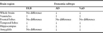
Barber et al.Reference Barber, Ballard, McKeith, Gholkar and O'Brien50 looked at differences between the brains of 27 patients with DLB, 25 with AD, 24 with VaD (matched for age, sex, education and duration of illness) and 26 controls, by carrying out MRI-based volume measurements of the whole brain, ventricles, frontal lobe, temporal lobe, hippocampus and amygdala (Table 1). An important finding in this study was that in the brains of subjects with DLB there was a relative preservation of temporal lobe structures compared with subjects with AD. Hippocampal and amygdala volumes were also found to be significantly larger in subjects with DLB compared with subjects with AD.Reference Barber, Ballard, McKeith, Gholkar and O'Brien50 The study may be criticised on the basis that diagnosis of dementia subtype was made according to clinical, rather than pathological features in all but seven cases, but the findings have since been replicated in numerous other studies.Reference Harvey, Hughes, McKeith, Briel, Ballard, Gholkar, Scheltens, Perry, Ince and O'Brien51–Reference Whitwell, Weigand, Shiung, Boeve, Ferman, Smith, Knopman, Petersen, Benarroch, Josephs and Jack53 A previous study on the same sample of patients had used a visual rating scale to assess MRI scans, rather than volumetric measures. Whilst medial temporal lobe atrophy was more common in all dementia patients compared with control subjects, DLB patients had ‘significantly less atrophy than those with AD’.Reference Barber, Gholkar, Scheltens, Ballard, McKeith and O'Brien54 The results of these two studies are therefore consistent and imply that a simple visual rating scale can be used reliably in clinical practice to assess MRI scans of patients.
MRI-based volume measurements of different brain regions in DLB, AD and VaD compared with controlsReference Barber, Ballard, McKeith, Gholkar and O'Brien50

↑, volume of the brain region significantly larger compared with controls. ↓, volume of the brain region significantly smaller compared with controls. No difference, volume of the brain region not significantly different compared with controls.
Another study of 27 DLB patients, 27 AD patients and 27 control subjects, examined using an MRI-based volumetric technique, confirmed that the volume of the hippocampus was smaller in both dementia groups compared with the controls, but more preserved in DLB than AD patients.Reference Hashimoto, Kitagaki, Imamura, Hirono, Shimomura, Kazui, Tanimukai, Hanihara and Mori55 However, in this study, no difference was found in the atrophy of the amygdala between the two dementia groups.
Studies examining the brains of DLB patients at autopsy have indicated that the relatively preserved whole brain volume found in DLB may be due to ‘relative preservation of synapse integrity and neuronal counts’.Reference Burn, Perry, O'Brien, Charney and Nestler6 The different patterns of temporal lobe atrophy between AD and DLB, particularly greater volume loss of the hippocampus in AD, is likely to play a part in explaining why memory impairment is a much more prevalent clinical feature in AD compared with DLB.Reference Barber, McKeith, Ballard, Gholkar and O'Brien56
These studies highlight the potentially useful role of imaging techniques in the differentiation of AD and DLB. However, it may be that these findings are only present after the dementia has progressed for a number of years, and so are not contributory to differential diagnoses at the very start of the illness.
An additional imaging modality, single photon emission computerized tomography (SPECT), is being used increasingly in the differential diagnosis of dementia, due to its ability to detect specific subclinical changes occurring in DLB, namely nigrostriatal degeneration and profound reductions in the striatal dopamine transporter,Reference Piggott, Marshall, Thomas, Lloyd, Court, Jaros, Burn, Johnson, Perry, McKeith, Ballard and Perry57, Reference Walker and Walker58 by using ligands such as iodine-123-FP-CIT that bind to the dopamine transporter molecule in the membranes of the nigrostriatal nerve terminals.Reference Walker and Walker58, Reference Booij, Habraken, Bergmans, Tissingh, Winogrodzka, Wolters, Janssen, Stoof and van Royen59 Studies have demonstrated high levels of diagnostic accuracy, in terms of both sensitivity and specificity, when using 123I-FP-CIT SPECT to differentiate DLB from other dementia subtypes.Reference McKeith, O'Brien, Walker, Tatsch, Booij, Darcourt, Padovani, Giubbini, Bonuccelli, Volterrani, Holmes, Kemp, Tabet, Meyer and Reininger60–Reference O'Brien, McKeith, Walker, Tatsch, Booij, Darcourt, Marquardt and Reininger63 There is a potential role for this mode of imaging as a confirmatory investigation in cases where the diagnosis of DLB is probable on the basis of clinical features, but not entirely certain, rather than as a routine investigation in all patients.Reference Walker, Costa, Walker, Shaw, Gacinovic, Stevens, Livingston, Ince, McKeith and Katona62, Reference O'Brien, McKeith, Walker, Tatsch, Booij, Darcourt, Marquardt and Reininger63
Conclusion
The differences between DLB and AD may not be immediately obvious when a patient initially presents, emphasizing the need for detailed assessment. Although there are distinguishing features, such as attentional deficits and extrapyramidal symptoms in DLB and early working memory deficits in AD, it is still questionable whether DLB and AD are totally separate entities. It may just be that these more specific features lie at the very extremes of the clinical spectrum of an overall general neurodegenerative disease, with features such as senile plaque pathology lying towards the middle and Lewy bodies and neurofibrilliary tangles towards opposite ends. Research is needed to greater define the distinctions between these two conditions, especially at an early stage before hallmark symptoms appear. Even if the long-term prognosis of an older person with a chronic dementia cannot be changed, providing a specific diagnosis enables an efficient long-term management plan to be formulated and individualized for each patient.
Conflicts of interest
The author declared no conflicts of interest.