Introduction
Lymphatic filariasis (LF) is a mosquito-transmitted disease, caused by the nematode parasites Wuchereria bancrofti, Brugia malayi and B. timori. These parasites are endemic in over 100 countries and place more than 1.1 billion people (20% of the world's population) at risk of infection (Melrose & Rahmah, Reference Melrose and Rahmah2006). Approximately 130 million people living in the tropics and sub-tropics are infected with these parasites, of whom 115 million are infected with W. bancrofti and 13 million with B. malayi (Michael & Bundy, Reference Michael and Bundy1997). Present strategies to eliminate LF are reliant on just a few drugs, diethylcarbamazine (DEC), ivermectin and albendazole (Ottesen, Reference Ottesen2006). Clinical trials with a combination of these look promising (Campbell, Reference Campbell1991; Ismail et al., Reference Ismail, Jayakody, Weil, Fernando, de Silva, de Silva and Balasooriya2001); however, microfilaraemia reappears after a period of drug withdrawal (Ottesen et al., Reference Ottesen, Duke, Karam and Behbehani1997).
Vector control and administration of antifilarials has reduced infection and disease rates significantly. However, limitations of these programmes, including selective activity of these drugs on microfilariae (mf), the need for 10–15 years of annual treatments, logistical obstacles and the potential emergence of drug-resistant strains (Bourguinat et al., Reference Bourguinat, Pion, Kamgno, Gardon, Duke, Boussinesq and Prichard2007; Lustigman & McCarter, Reference Lustigman and McCarter2007) demand alternative strategies. The life cycle of filarial parasites involves various developmental stages and each stage has a complex antigen make-up. A vaccine that targets the infective larval stage (L3) of B. malayi could provide an additional tool to guarantee successful elimination of infection with this parasite. Thus, an essential step in the development of immunoprophylactic measures is to identify host immune responses toward antigens of B. malayi L3.
The development of vaccines against a multicellular parasite, which is well adapted to divert the host's immune system to ensure its long-term survival, is not an easy task. However, it is feasible; examples exist of successful helminth vaccines for livestock (Benthony et al., Reference Benthony, Loukas, Hotez and Knox2006) and a vaccine against hookworm disease is progressing toward human trials (Hotez et al., Reference Hotez, Zhan, Bethony, Loukas, Williamson, Goud, Hawdon, Dobardzic, Dobardzic, Ghosh, Bottazzi, Mendez, Zook, Wang, Liu, Essiet-Gibson, Chung-Debose, Xiao, Knox, Meagher, Inan, Correa-Oliveira, Vilk, Shepherd, Brandt and Russell2003). Attempts at immunoprophylaxis against filariasis have been made in several experimental models employing different species and life stages of filarial parasites (Kazura et al., Reference Kazura, Cicirello and McCall1986; Chatterjee et al., Reference Chatterjee, Singh and Misra1992; Pearlman et al., Reference Pearlman, Kroeze, Hazlett, Chen, Mawhorter, Boom and Kazura1993). Significant levels of protection against homologous challenge infection have been observed with the L3 attenuated by irradiation (Oothuman et al., Reference Oothuman, Denham, McGreevy, Nelson and Rogers1979; Bancroft & Devaney, Reference Bancroft and Devaney1993; Bleiss et al., Reference Bleiss, Oberländer, Hartmann, Adam, Marko, Schönemeyer and Lucius2002; Lustigman et al., Reference Lustigman, MacDonald and Abraham2003). Crude or partially purified somatic and secretory antigens have been shown to provide partial protection against parasitic helminth infection (Schallig & Van Leeuwen, Reference Schallig and Van Leeuwen1997; Ferreira et al., Reference Ferreira, Santoro and Goes1998; Abdel Rahman et al., Reference Abdel Rahman, Abouel Ezz and Abdel Megeed2005). Protection studies using partially purified antigens of animal filariids revealed marginally better efficacy using various methods of purification (Storey & Mettias, Reference Storey and Mettias1980; Sumati et al., Reference Sumati, Saxena, Tandon, Saxena, Singh, Misra and Chatterjee1990; Dikshit & Misra, Reference Dikshit and Misra1994; Dixit et al., Reference Dixit, Gaur, Sahoo, Joseph, Murthy and Murthy2006). However, soluble antigen does not include many of the particulate or insoluble components of the parasite which might possess protective characteristics. Differential centrifugation is a technique traditionally used to isolate subcellular organelles on the basis of sedimentation coefficient (De Duve & Beaufay, Reference De Duve and Beaufay1981). Attempts have been made to separate subcellular organelles, to add to our knowledge of the cell biology of the parasites (Fry & Jenkins, Reference Fry and Jenkins1983; Schroter et al., Reference Schroter, Braun, Englert, Beck, Schmid and Kalbacher1999; Hunter & Korzick, Reference Hunter and Korzick2005) and for exploration of their prophylactic efficacy (Parker et al., Reference Parker, Field, Berry and Manclark1978; Ruiz et al., Reference Ruiz, Esteva, Cabeza Meckert, Laguens and Segura1985; Blasco et al., Reference Blasco, Gamazo, Winter, Jiménez de Bagüés, Marín, Barberán, Moriyón, Alonso-Urmeneta and Díaz1993).
In the present study, we carried out fractionation of a crude somatic extract of a subperiodic strain of the human lymphatic filariid, B. malayi, by differential centrifugation, in order to determine the protective efficacy of subcellular fractions. To better define the immunological basis for the protective effect exerted by the vaccine candidate, we also examined several immunological parameters in both immunized and non-immunized rodent hosts. Specifically, antigen-specific serum antibody titre and isotypes were determined in addition to lymphocyte proliferation, measurement of Th1/Th2 cytokines and nitric oxide release by peritoneal macrophages following stimulation with parasite antigens.
Materials and methods
Experimental animals and infection
Healthy, 6-week-old male Mastomys coucha, (GRA ‘Giessen strain’) and 8-week-old, male Meriones unguiculatus (jird), were used in the study. The animals were housed under standard conditions of temperature (23 ± 1°C), relative humidity (55 ± 10%) and 12 h/12 h light/dark cycles at the National Laboratory Animal Centre (NLAC), CDRI, Lucknow, and were fed with a standard pellet diet and tap water ad libitum. All the animals and the various animal handling protocols employed in the present study were duly approved by the Institutional Animal Ethics Committee.
Antigen preparation and fractionation by differential centrifugation
Adult B. malayi were collected from the peritoneal cavity of infected jirds 90–180 days after larval inoculation (McCall et al., Reference McCall, Malove, Ah and Thompson1973). The mixed population of adult worms was homogenized in an Elvejhm tissue grinder using 10% (w/v) cold 150 mm potassium chloride (KCl). The homogenate was filtered through a muslin cloth to remove debris. Fractionation was carried out as described earlier (Sumati et al., Reference Sumati, Saxena, Tandon, Saxena, Singh, Misra and Chatterjee1990). The homogenate was centrifuged sequentially as follows: 1000 g for 15 min, 10,000 g for 30 min, 100,000 g for 60 min. Each pellet thus obtained was resuspended in 150 mm KCl and defined as the ‘nucleus rich’ fraction (NR), ‘mitochondria rich’ fraction (MT), and ‘microsome rich’ fraction (MC), respectively. The supernatant obtained after the final centrifugation was defined as the soluble fraction (CYT). Protein concentrations were determined by the method of Bradford (Reference Bradford1976) and fractions stored at − 20°C until further use. The fractions were analysed for enzyme markers and protein to determine the amount recovered, which was not less than 80% each time the process of fractionation was carried out.
Immunization schedule and larval challenge
Mastomys were divided into six groups (ten animals per group). One group of animals served as non-immunized control (UNIMM). Another group, defined as ADJ, received adjuvant to serve as an age-matched immunization control. Animals of groups NR, MT, MC and CYT received subcutaneous (sc) injections of the respective fractions (10 μg protein/animal). Booster doses (75 μg protein/animal) were given 15 and 23 days after the first immunization. The first injection was given in Freund's complete adjuvant (FCA; Sigma, St Louis, Missouri, USA) and the rest in Freund's incomplete adjuvant (FIA). Seven to eight days after the final booster, five animals from each group were euthanized for immune profiling and the other five animals were challenged through subcutaneous (sc) route with 100 L3 of B. malayi recovered from Aedes aegypti mosquitoes (Ash & Riley, Reference Ash and Riley1970; Misra-Bhattacharya et al., Reference Misra-Bhattacharya, Katiyar, Bajpai, Tripathi and Saxena2004) for protection studies.
Monitoring of microfilaraemia
Ninety days after larval challenge, 10 μl blood was taken from the tail vein of each animal between 12.00 and 13.00 hours (Singh et al., Reference Singh, Misra, Murthy, Katiyar, Agarwal and Sircar1997) to prepare a thick blood smear on a glass slide which was air-dried and stained with Leishman stain to assess the microfilarial density. Microfilarial examination was continued monthly until the end of experiment (7 months after L3 challenge).
Effect on adult worm recovery
Seven months after challenge, the animals were euthanized for the recovery of adult worms. Various tissues, namely heart, lung, lymph glands and testes, were isolated in phosphate-buffered saline (PBS, pH 7.2) and teased to recover adult worms. The male and female worms were counted and the intra-uterine contents of the female worm were examined under a microscope to assess the effect of vaccination on female reproductive potential (Misra-Bhattacharya et al., Reference Misra-Bhattacharya, Katiyar, Bajpai, Tripathi and Saxena2004). Any change in percentage worm recovery was calculated by comparison with the worms recovered from non-immunized control animals challenged under identical conditions.
IgG antibody level assessment by enzyme-linked immunosorbent assay
Blood was collected from the retro-orbital plexus of animals for isolation of serum just before L3 challenge and at monthly interval thereafter. Serum antibody titre was assessed by enzyme-linked immunosorbent assay (ELISA) as described earlier (Singh et al., Reference Singh, Misra, Murthy, Katiyar, Agarwal and Sircar1997). In brief, ELISA plates (Nunc, Denmark) were coated with adult B. malayi somatic antigen (BmAg; 1 μg ml− 1) in carbonate buffer, pH 9.6, overnight at 4°C and thereafter blocked with 1% gelatin in PBS-T (0.05% Tween-20 in PBS) for 120 min at 37°C. Primary antibody (mastomys serum) was added at serial twofold dilutions and strips were reincubated with a 1:5000 dilution of secondary antibody (rabbit anti-mouse–IgG–horseradish peroxidase (HRP); Sigma) at 37°C for 90 min. Absorbance was read at 492 nm by ELISA reader (Infinite M200, Tecan, Switzerland) after adding the substrate, orthophenylenediamine (OPD, Sigma), and the reading at optimum dilution was represented as mean ± SE.
Antibody isotyping
The concentrations of various antibody isotypes (IgG1, IgG2a, IgG2b, IgG3, IgA, IgM) were determined by antigen-mediated ELISA method in the serum of immunized animals following the manufacturer's guidelines (Sigma, USA). Appropriately diluted BmAg (1 μg ml− 1) was used to coat the microtitre plate and was incubated overnight at 4°C. The wells were washed three times with PBS-T. Serum (100 μl of 1:200 dilution for IgG1, IgG2a, IgM and 1:50 dilution for IgG2b, IgG3, IgA) was added to the coated wells. After 2 h of incubation at room temperature, the plate was washed and goat anti-mouse isotype specific monoclonal antibody (1:1000 diluted in PBS) was added and kept for 30 min at room temperature. After washing, 100 μl of diluted (1:5000 in PBS-T) secondary antibody (rabbit anti-goat–IgG–HRP conjugated) was added to each well and incubated for 15 min. Freshly prepared substrate (100 μl of the following: 20 mg of OPD dissolved in 25 ml of citrate buffer, pH 5.0, and 20 μl of H2O2) was added to each well. The reaction was stopped after 5 min by adding 50 μl of H2SO4 (2.5n) to each well. Readings were taken at 492 nm on an ELISA plate reader to determine the antibody isotype level.
Lymphocyte proliferation assay
The lymphoproliferative response of the animals of different groups was determined as described earlier (Misra et al., Reference Misra, Mukherjee, Dikshit and Chatterjee1998). The spleen was removed and a single cell suspension was prepared aseptically in incomplete RPMI 1640 medium (containing 1% antibiotic antimycotic cocktail; Sigma). Red blood cells were lysed with chilled ammonium chloride solution (0.83%) and splenocytes were suspended at a concentration of 5 × 10⁶ cells ml− 1 in complete RPMI (media fortified with 10% fetal calf serum). Then 100 μl of cell suspension was plated in 96-well flat-bottom NUNC culture plates and stimulated with 100 μl/well of Concanavalin A (Con A; 5 μg ml− 1), 100 μl of lipopolysaccharide (LPS; 5 μg ml− 1) and 100 μl of antigen fraction (Ag; 10.0 μg ml− 1), while the unstimulated lymphocytes served as the control. The cells were cultured for 3 days in the case of the mitogens, and for 5 days in the case of the antigen at 37°C in 5% CO2. Sixteen hours prior to termination of culture, the cells were pulsed with 0.5 μCi [3H]thymidine (BARC, India) and harvested on a NUNC harvester. Incorporated [3H]thymidine was measured in a liquid scintillation β-counter (LKB Wallac LS 1800; Beckman Instruments, Fullerton, California, USA). Results were expressed as stimulation indices, which denote the ratio of mean counts per minute (cpm) of stimulated cultures to mean cpm of unstimulated controls.
Cytokine profiling
The splenocytes were isolated and plated as mentioned for lymphocyte proliferation assay. Cells were stimulated with antigen fraction and Con A and incubated for 2 days at 37°C in 5% CO2. The supernatant was collected and Th1 (interferon-γ (IFN- γ), tumour necrosis factor-α (TNF-α), interleukin-1β (IL-1β)) and Th2 cytokines (interleukin-4 (IL-4), interleukin-6 (IL-6) and interleukin-10 (IL-10)) were quantified using Duo set ELISA kits (R & D Systems, Minneapolis, Minnesota, USA) according to the manufacturer's protocol. The ELISA plates were coated with the specified dilution of capture antibody and incubated overnight at 4°C. The plates were washed with PBS-T and 200 μl blocking buffer (PBS, 1% bovine serum albumin (BSA), 0.05% sodium azide (NaN3)) was added. After 1 h incubation, wells were washed with PBS-T, 100 μl of pooled culture supernatant was added and the plates were kept for 2 h at 37°C. Then 100 μl of detection antibody (100 ng ml− 1) was added and plates were incubated for 2 h at 37°C. After washing, 100 μl of streptavidin–HRP conjugate diluted (1:200 dilution) in reagent diluent (Tris-buffered saline; 0.1% BSA, 0.05% Tween-20) was added and incubated for 20 min. Tetramethyl benzidine (TMB; 100 μl/well) was added after washing the plates, and they were incubated for 20 min. The reaction was stopped by adding 50 μl of 2.5m H2SO4. The OD540nm and OD450nm were read using an ELISA plate reader.
Macrophage function assay
Nitric oxide production was estimated by measuring the nitrite concentration in cell culture supernatants using the Griess reagent, as described earlier (Green et al., Reference Green, Wagner, Glogowski, Skipper, Wishnok and Tannenbaum1982; Gupta et al., Reference Gupta, Bajpai, Tripathi, Srivastava, Jain and Misra-Bhattacharya2004). On the day of autopsy, the peritoneal macrophages were isolated in cold incomplete RPMI medium. The cells were washed, counted and plated (3 × 10⁵ cells/well) into flat-bottom 96-well NUNC culture plates and incubated for 2 h at 37°C in 5% CO2 in air to allow adherence of macrophages to the plastic bottom. Non-adherent cells were removed by rinsing the wells once with incomplete RPMI, to finally obtain a macrophage monolayer in each well. Complete RPMI (100 μl) was added to the wells, which served as unstimulated blanks. LPS (1.0 μg/ml) and antigen fraction (5 μg ml− 1) were added in triplicate wells and the plate was further kept at 37°C for 24 h. Culture supernatant was collected and NO production was estimated by Griess reagent (0.1% NCl–naphthyl ethylene diamine hydrochloride in distilled water, 1% sulphanilamide in 5% phosphoric acid, added in equal volume prior to use). The absorbance was read at 540 nm using an ELISA plate reader. Results were expressed as μmoles NO/106 cells and a standard curve was plotted using NaNO2 to quantitate the test results.
Analysis of T- and B-lymphocyte populations by flow cytometry
The expression of specific molecules on the membranes of T and B lymphocytes were determined by direct immunofluorescence staining as per manufacturer's instruction (AbD Serotec, Kidlington, Oxford, UK). A single cell suspension of splenocytes was prepared (as mentioned for the lymphocyte proliferation assay, above). The RPMI medium was replaced by PBS and flow cytometric analysis of the lymphocyte subsets and activation markers was carried out: CD19 (B cells), CD4 (T-helper cells), CD8a (suppressor/cytotoxic T cells), CD30 (activated T and B cells) and CD45 (role in antigen-receptor activation of T and B cells), using fluorescein isothiocyanate (FITC)-conjugated anti-mouse monoclonal antibodies (Serotec, UK). The appropriate monoclonal antibody was added to cells (l × 106 splenocytes/tube), samples vortexed and then incubated in a cold rack for 15 min. The cells were washed three times with dilution buffer (PBS, 1% BSA, 0.1% NaN3) and fixed in 500 μl of 2.0% paraformaldehyde. A minimum of 10,000 gated cells were included in the analysis. The percentage of positive cells was measured with a fluorescence-activated cell sorter FACS Calibur (Becton Dickinson, Franklin Lakes, New Jersey, USA) and data were analysed by CellQuest Software (Becton Dickinson).
Statistical analysis
Results have been presented as mean ± SE of ten animals in two experiments, and the data were analysed statistically using Newman–Keuls multiple comparison test. The criteria for statistical significance between the results of immunized and non-immunized control groups were as follows: P < 0.05 was considered as significant, P < 0.01 to P < 0.001 as highly significant.
Results
Parasitological parameters
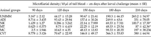
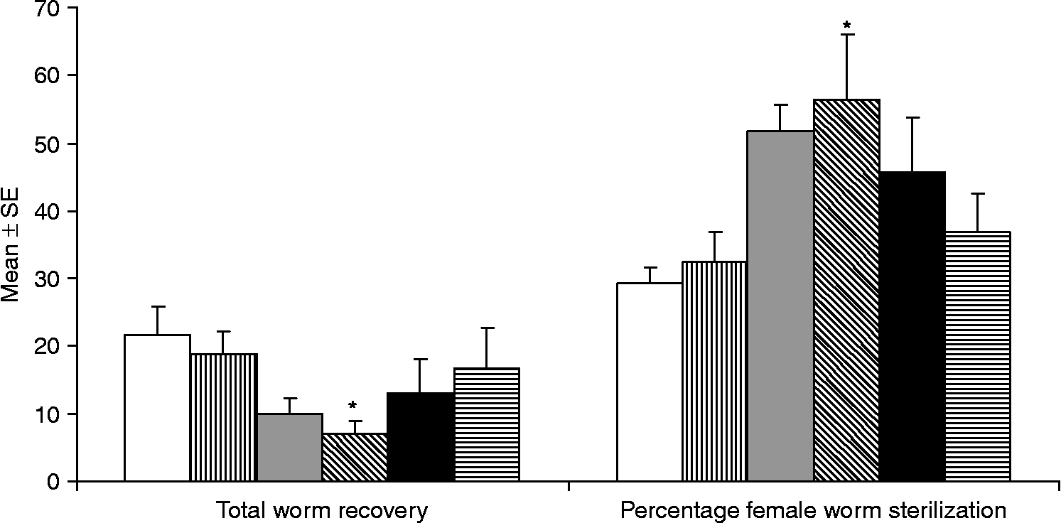
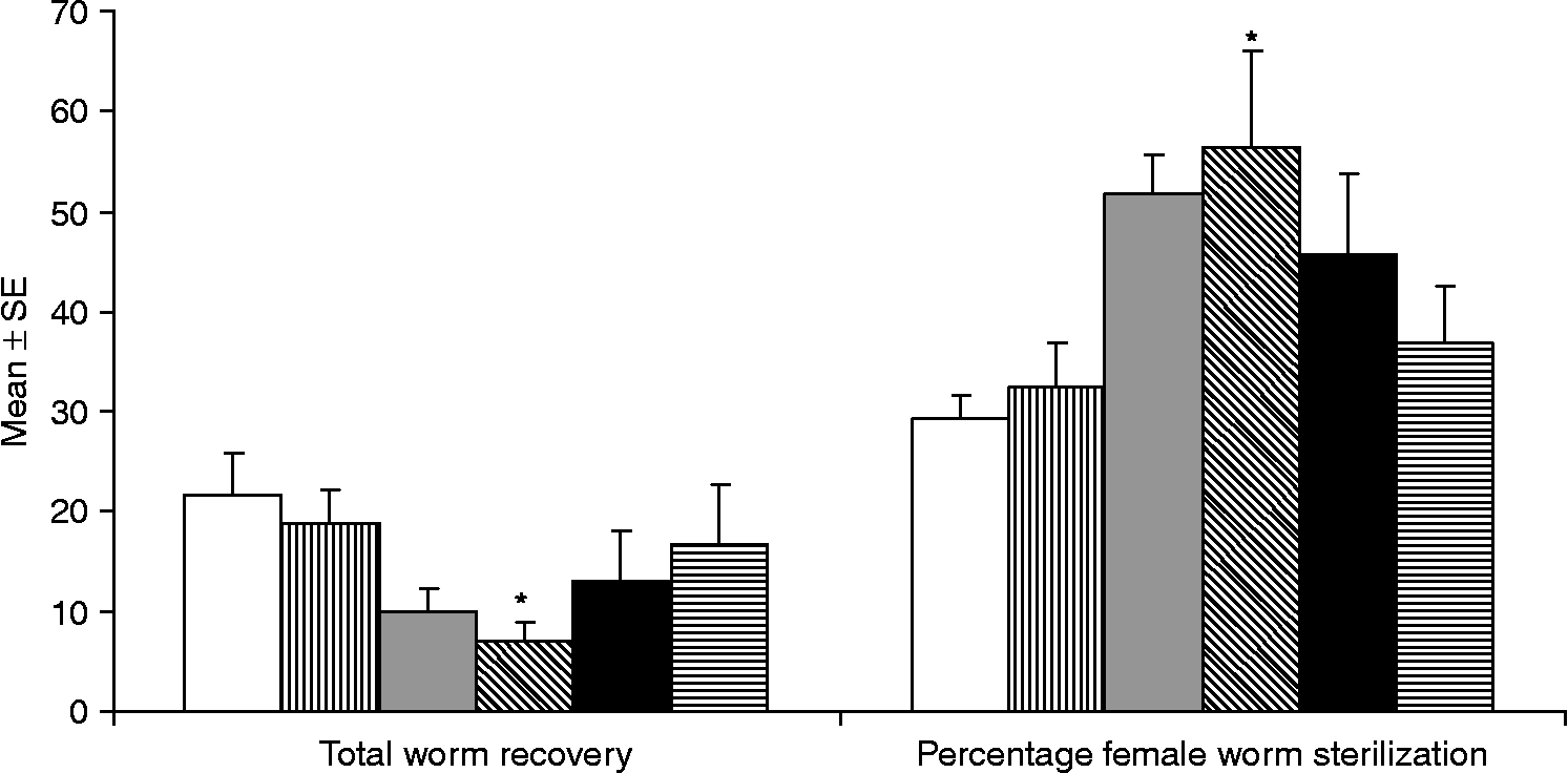
The rise in microfilaraemia in animals of the three groups (NR, MT and MC) was not as progressive as in other groups (table 1); however, at the end of observation period, a significant difference in mf density was observed in NR and MT groups, where the percentage mf count was significantly (P < 0.05) reduced by 62 and 67.2%, respectively, over adjuvant immunized control groups. The microfilaraemia in the CYT group was highest amongst all the groups and was higher than that observed in the control groups. There was a considerable reduction in adult worm recovery from NR (52.3%) and MT (65.9%, P < 0.05) groups as compared to the non-immunized animals (fig. 1). There was a mild reduction in adult worm recovery in MC and CYT groups also (36.7 and 19.22%, respectively). The percentage of female worms with an embryostatic effect in the MT immunized group (56.46%) was significantly higher (P < 0.05) than in the non-immunized (29.23%) and adjuvant immunized control groups (32.49%).
Microfilaraemia in 10 μl of tail blood at various time periods after larval challenge (starting from day 90) in M. coucha immunized with ‘nucleus rich’ (NR), ‘mitochondria rich’ (MT), ‘microsome rich’ (MC) or soluble (CYT) fractions, along with adjuvant (ADJ) and non-immunized (UNIMM) control groups. The values are expressed as mean±SE from a total of ten animals in two independent experiments.
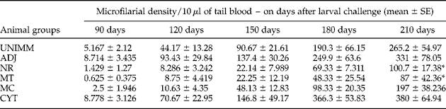
*P < 0.05.
Worm recovery and percentage of female worms sterilized on day 210 after subcutaneous challenge with B. malayi L3 in M. coucha immunized with ‘nucleus rich’ (grey bar), ‘mitochondria rich’ (bar with diagonal lines), ‘microsome rich’ (black bar) and soluble (bar with horizontal lines) fractions, along with adjuvant (bar with vertical lines) and non-immunized (white bar) control groups. Values are means ± SE of data from a total of ten animals in two independent experiments. *P < 0.05.
Antibody titre and isotypes
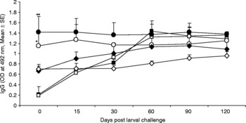
The elevated IgG levels against filarial antigen were observed in all the immunized groups just before larval challenge. NR showed a significant increase in IgG level (OD 1.416 ± 0.26; P < 0.01) followed by MT (1.1475 ± 0.027; P < 0.05), in comparison with non-immunized (0.2067 ± 0.028) and adjuvant immunized (0.1913 ± 0.004) controls (fig. 2).
Antigen-specific IgG antibody titre in sera of mastomys immunized with different subcellular fractions of B. malayi at various time points by ELISA. Groups of mastomys were immunized with ‘nucleus rich’ (●), ‘mitochondria rich’ (○), ‘microsome rich’ (♦) and soluble (⋄) fractions. Adjuvant (□) and non-immunized (■) animals served as controls. All immunized animals showed elevated antibody level on the day of larval challenge (day 0) while the adjuvant and non-immunized controls showed a low titre. After larval challenge, the IgG levels in control groups started to rise up to day 60 and remained stable thereafter; whereas all the immunized groups maintained a high antibody titre throughout the observation period. Each point represents the mean ± SE of OD492nm of sera of ten animals at 1:800 dilution. *P < 0.05, **P < 0.01.

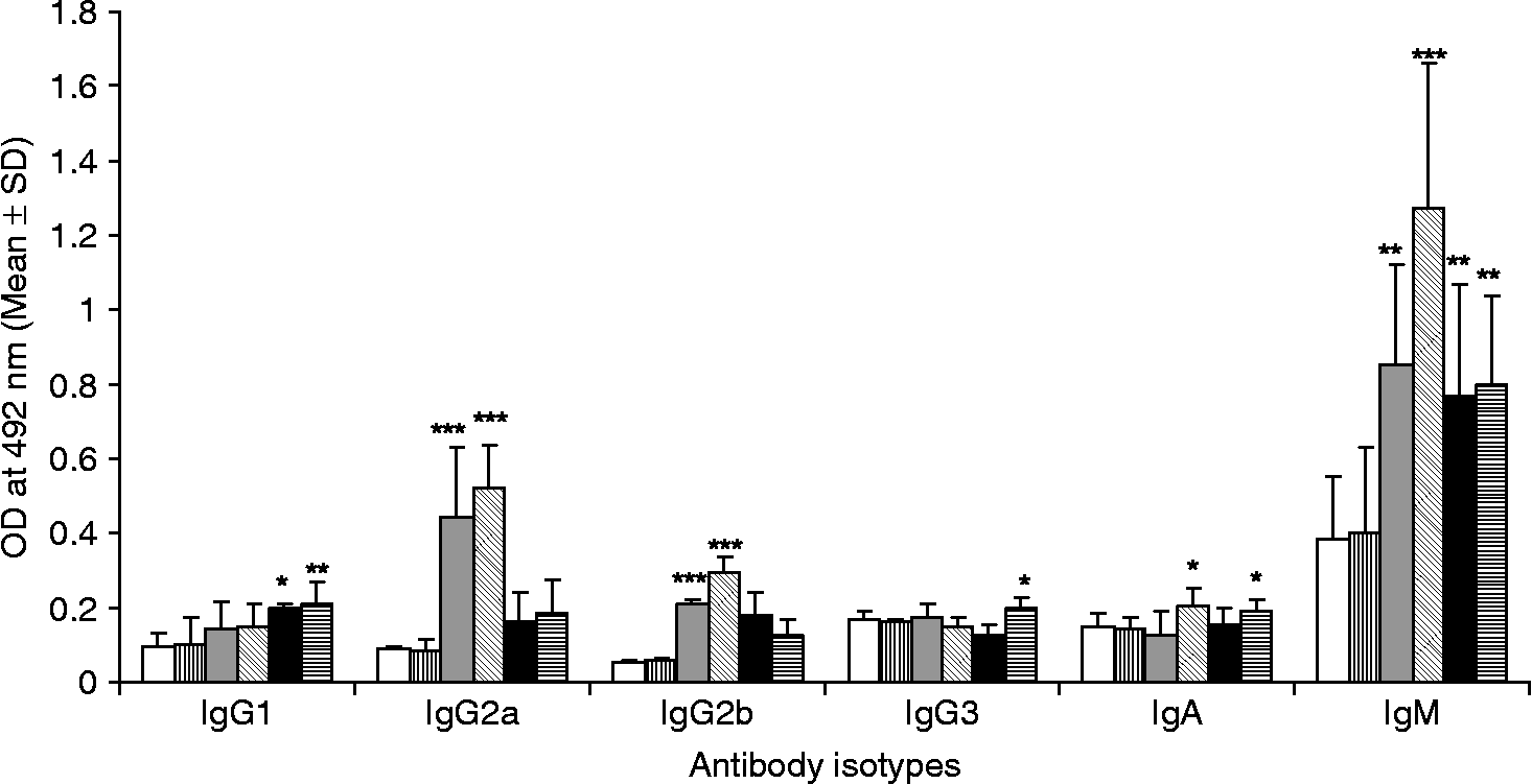
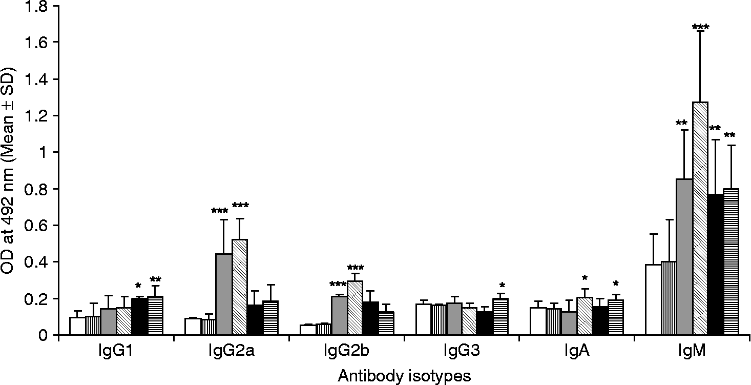
Investigations were carried out to correlate protection with immunoglobulin isotype generated following immunization with various fractions. The relationship between Th1 and Th2 responses could be determined following various immunization regimens by calculating the IgG1/IgG2a ratio. Immunization with NR and MT elicited highly significant differences (P < 0.001) in the levels of IgG2a, IgG2b and IgM (fig. 3), with marginal difference in levels of IgG1, IgG3 and IgA as compared to non-immunized and adjuvant control animals. These two groups showed a low IgG1/IgG2a ratio, suggesting a Th1-biased response. Higher levels of IgG1 were observed in MC and CYT groups, increasing their IgG1/IgG2a ratio.
Antibody isotype level in the sera of animals immunized with ‘nucleus rich’ (grey bar), ‘mitochondria rich’ (bar with diagonal lines), ‘microsome rich’ (black bar), and soluble (bar with horizontal lines) fractions along with adjuvant (bar with vertical lines) and non-immunized (white bar) control groups. IgG1, IgG2a, IgG2b, IgG3, IgA and IgM antibodies were checked by ELISA for specific antibodies against the respective fractions. The bars represent means ± SD of OD492nm. *P < 0.05, **P < 0.01, ***P < 0.001.
Lymphoproliferation
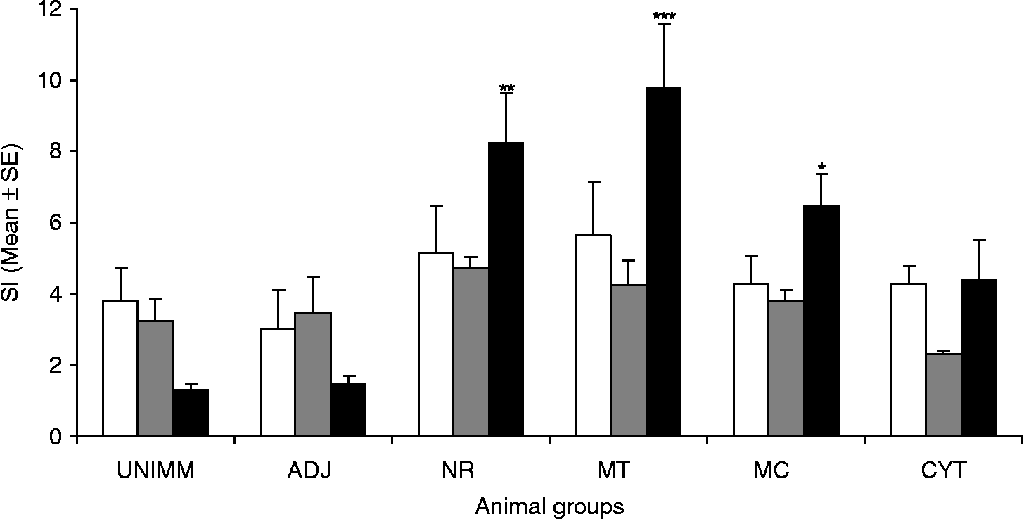
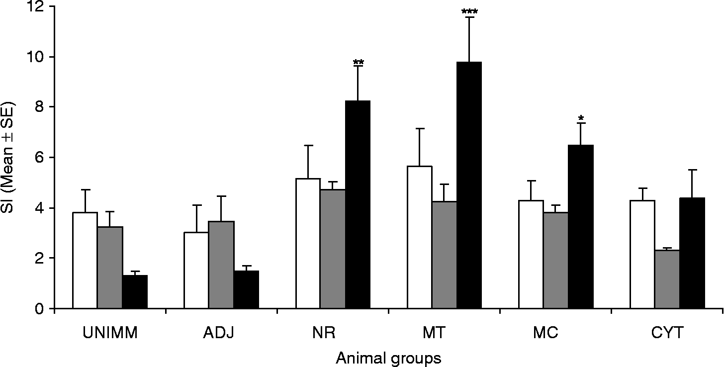
The ability of T and B cells from immunized animals to recognize and respond to parasite antigens and mitogens was investigated by stimulating splenocytes in vitro. The antigen-specific T-cell proliferation was found to be significantly higher in NR (P < 0.01) and MT groups (P < 0.001) as compared to the non-immunized group (fig. 4). Likewise, immunization with both these fractions also demonstrated substantial B-cell stimulation. The MC and CYT groups showed an increased antigen-specific proliferative response but low B-cell stimulation.
Lymphoproliferation of splenocytes isolated from animals of different groups. The splenocytes were stimulated in vitro with Concanavalin A (white bar), lipopolysaccharide (grey bar) and antigenic fractions (black bar). Animals were immunized with: ADJ, adjuvant; and NR, ‘nucleus rich’; MT, ‘mitochondria rich’; MC, ‘microsome rich’ and CYT, soluble fractions of B. malayi. UNIMM, non-immunized animals. The immunized groups showed a significant increase in antigen-specific proliferation. The results are expressed as mean stimulation index (SI) ± SE. *P < 0.05, **P < 0.01, ***P < 0.001.
Th1/Th2 cytokine level
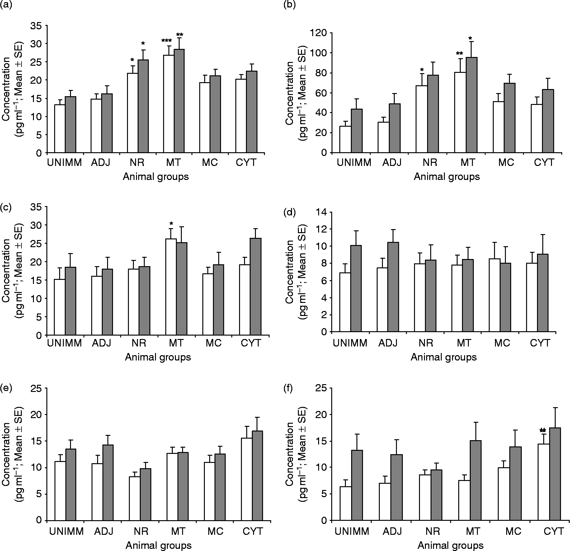
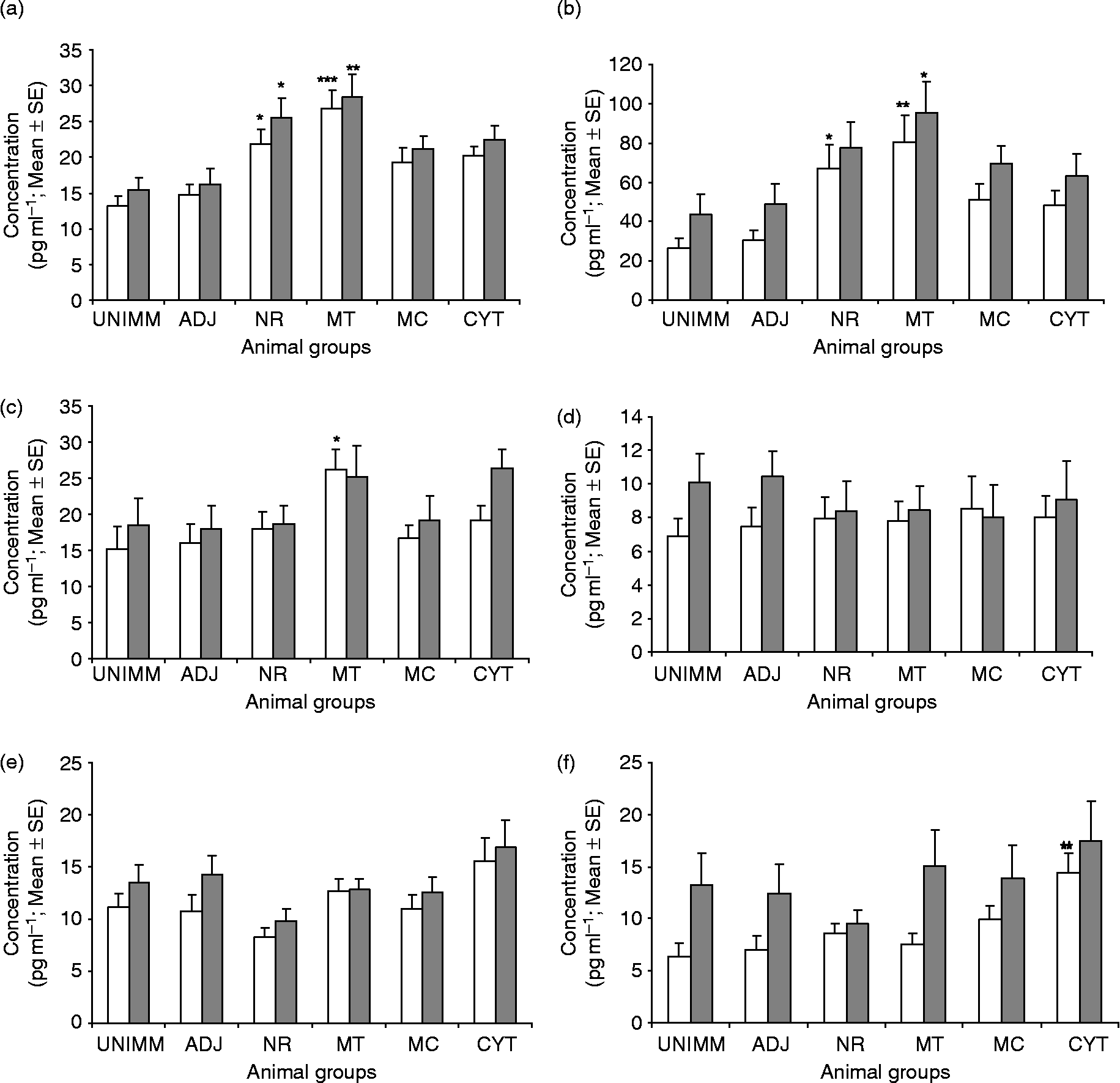
If cell-mediated immunity is involved in the protective immune response, then the observation that all immunized groups generate positive T-cell responses clearly suggests that the responses induced by these antigen fractions may differ. The T-helper response in animals immunized with various fractions was investigated by measuring the release of the cytokines IFN-γ, TNF-α, IL-1β, IL-4, IL-6 and IL-10 by capture ELISA in the culture supernatants of T cells primed in vitro with respective antigen fractions and the mitogen Con A. A significant increase in levels of antigen-specific TNF-α was observed in the case of MT (P < 0.001) and NR (P < 0.05) immunized animals. The level of TNF-α in response to mitogen was also significantly higher in both MT (P < 0.01) and NR (P < 0.05) immunized groups (fig. 5a). In addition, there was increased release of antigen-specific IFN-γ in MT (P < 0.01) and NR (P < 0.05) immunized animals as compared to unimmunized controls. However, in response to mitogen, only MT immunized animals showed a significant (P < 0.05) increase (fig. 5b). The other cytokine IL-1β also revealed enhanced release (P < 0.05) in the case of MT fraction after priming with a homologous fraction (fig. 5c). These animals, however, did not show any noticeable effect on IL-4, IL-6 and IL-10 levels. MC and CYT immunized animals revealed marginally higher antigen-specific IL-4 and IL-6 levels, respectively, as compared to other groups; however, the differences were statistically not significant (fig. 5d and e). Nevertheless, CYT induced significantly high levels of immunomodulatory cytokine IL-10 (P < 0.01) (fig. 5f).
Levels of (a) TNF-α, (b) IFN-γ, (c) IL-1β, (d) IL-4, (e) IL-6 and (f) IL-10 in the culture supernatants of lymphocytes of animals immunized by various fractions were quantified by antibody capture ELISA. The cells were stimulated in presence of antigen fraction (white bar) and mitogen, Concanavalin A (grey bar). Animals were immunized with: ADJ, adjuvant; and NR, ‘nucleus rich’; MT, ‘mitochondria rich’; MC, ‘microsome rich’ and CYT, soluble fractions of B. malayi. UNIMM, non-immunized animals. The values represent means ± SE of the data from ten animals. *P < 0.05, **P < 0.01, ***P < 0.001.
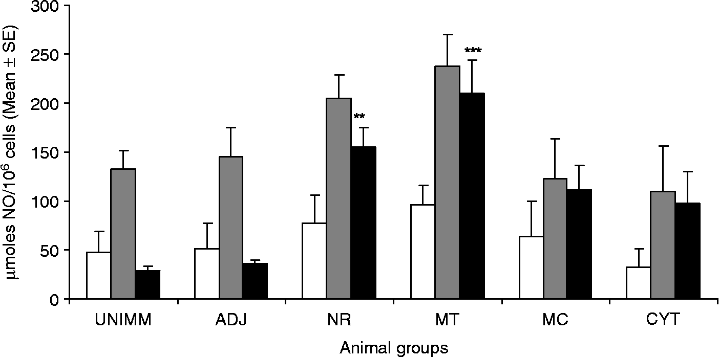
NO production
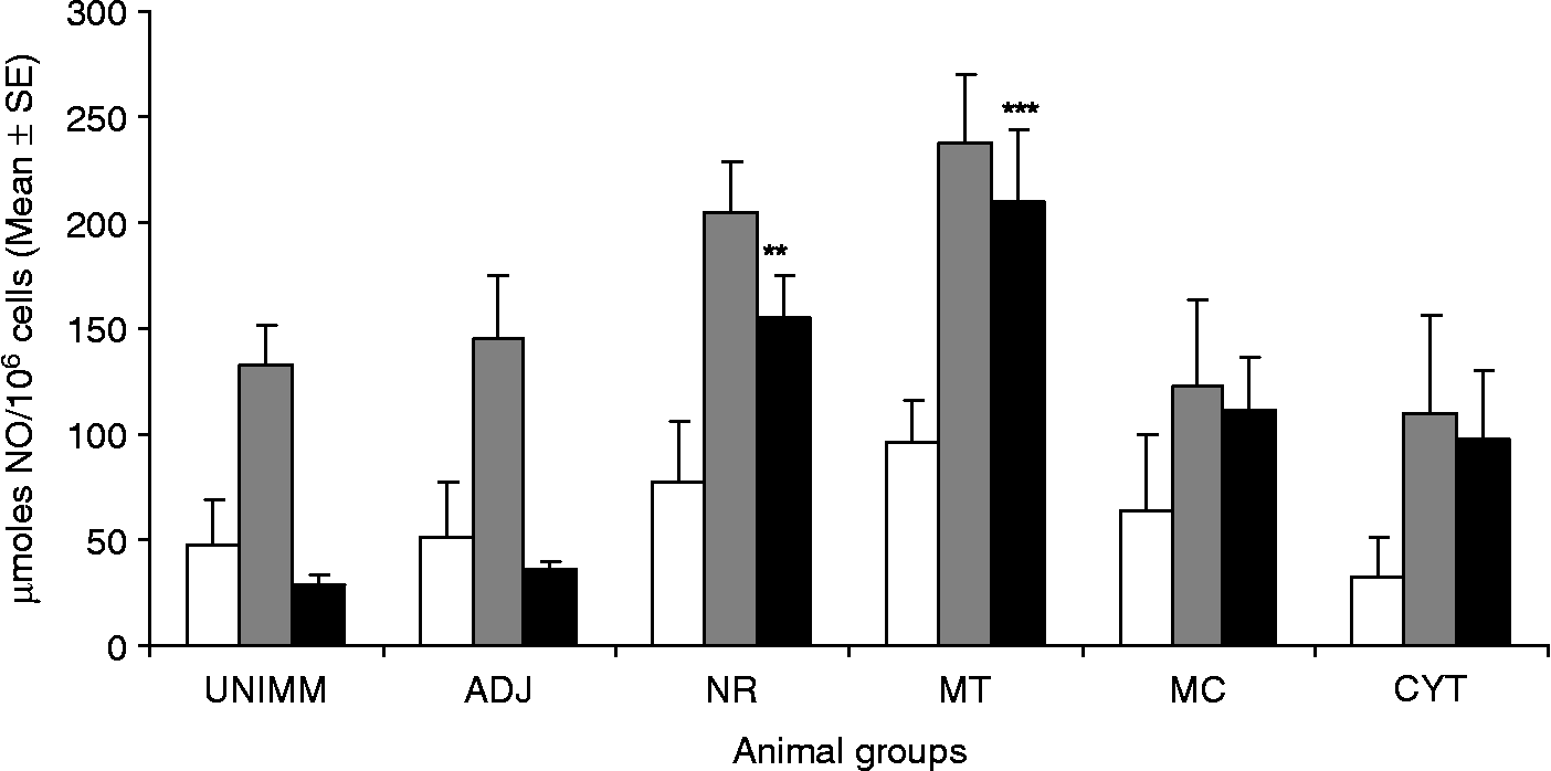
Induction of release of nitric oxide by the macrophages of immunized and non-immunized animals was estimated by measuring the nitrite level in the cell culture supernatant just before larval challenge, as shown in fig. 6. The nitrite level was also measured after LPS stimulation of cells which served as a positive control. Stimulation of macrophages with antigen led to a significant increase in the levels of NO in NR (P < 0.01) and MT (P < 0.001) immunized animals. The other two immunized groups, MC and CYT showed low levels of nitric oxide.
Nitric oxide production by peritoneal macrophages of immunized and control groups of mastomys post vaccination (day 7 of final booster) after in vitro stimulation with lipopolysaccharide (grey bar) and antigenic fraction (black bar). Animals were immunized with: ADJ, adjuvant; and NR, ‘nucleus rich’; MT, ‘mitochondria rich’; MC, ‘microsome rich’ and CYT, soluble fractions of B. malayi. UNIMM, non-immunized animals. White bars indicate the production of nitric oxide by unstimulated cell culture. Each bar represents the mean ± SE of ten animals. **P < 0.01, ***P < 0.001.
T- and B-cell populations
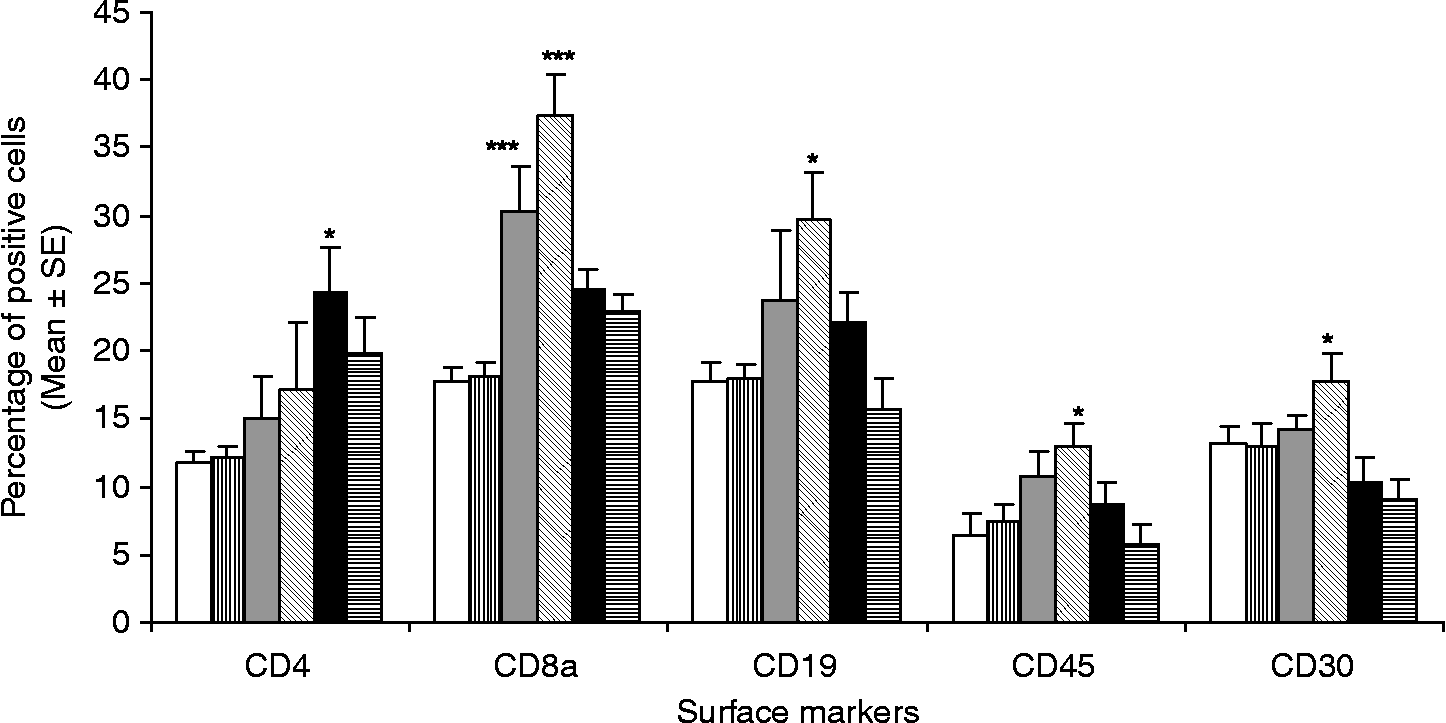
Increased expression of the B-cell marker CD19 in NR, MT and MC immunized animals was observed, with MT showing a significantly higher value of 29.75 ± 5.05% (P < 0.05) as compared to that of the non-immunized (17.7 ± 1.5%) and adjuvant control groups (18.02 ± 0.967%). The increased CD19 cell population was positively correlated with high antibody titre in the immunized animals. CD4+T cells are associated with a helper T-cell response and there was a significant increase in the population of CD4+T cells in all the immunized groups as compared to controls (11.67–12.13%) with the MC group demonstrating the highest values (24.3 ± 3.42%; P < 0.05). CD8a cells are associated with a cytotoxic response and there was highly significant increase in the population of CD8a cells (fig. 7) in MT (37.36 ± 3.12%; P < 0.001) and NR immunized groups (30.34 ± 3.4%; P < 0.001) followed by the MC group (24.58 ± 1.22%).
T- and B-lymphocyte populations determined by flow cytometry. Population of CD4+, CD8a+T lymphocytes and CD19 B lymphocytes, along with CD30 and CD45 surface markers, compared among different animal groups using fluorescein isothiocyanate-conjugated CD marker specific goat anti-mouse monoclonal antibodies. The bars in the graph indicate animals immunized with ‘nucleus rich’ (grey bar), ‘mitochondria rich’ (bar with diagonal lines), ‘microsome rich’ (black bar), and soluble (bar with horizontal lines) fractions along with adjuvant (bar with vertical lines) and non-immunized (white bar) control groups. Results were analysed by CellQuest Software and represented means ± SE of percentage of positive lymphocytes. *P < 0.05, **P < 0.01, ***P < 0.001.
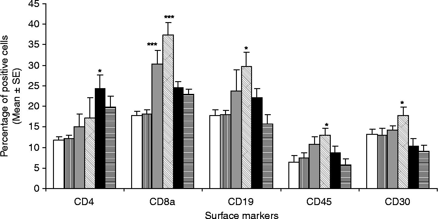
CD30 is a member of TNF-receptor superfamily and is expressed by activated T and B cells. This receptor is associated with negative selection of T cells in thymus and T-cell receptor (TCR)-mediated cell death and is also a known Th2 cell-surface marker. There was a significant increase in the number of CD30 cells in the MT immunized group (17.77 ± 1.97%) followed by the NR group (14.18 ± 1.1%) as compared to non-immunized (11.265 ± 1.2%) and adjuvant (13.02 ± 1.6%) control groups. CD45 is a member of the protein tyrosine phosphatase (PTP) family which has been shown to be an essential regulator of T- and B-cell antigen receptor signalling. There was significant difference observed in the case of the MT immunized group (P < 0.05), whereas NR and MC immunized groups demonstrated only a slight increase in the number of CD45 cells.
Discussion
One of the main objectives of this study was to investigate the protectivity of subcellular fractions of B. malayi on the basis of their ability to prevent establishment of infection in immunized animals after L3 challenge. The second objective was to identify immune responses uniquely associated with the high degree of protection by comparing them with non-protective and control groups.
Adult filarial worms live for a prolonged period inside the host and strongly polarize the immune system towards a predominant Th2 (or perhaps an impaired Th1) response (Ravindran, Reference Ravindran2001) with concomitant inhibition/suppression of Ag-specific proliferation and IFN-γ production (King, Reference King2001; Babu & Nutman, Reference Babu and Nutman2003; Maizels & Yazdanbakhsh, Reference Maizels and Yazdanbakhsh2003; Semnani et al., Reference Semnani, Law, Kubofcik and Nutman2004). Protective immunity is shown to be associated with Th1 polarization, as the absence of infection (in endemic normals; EN) correlates with elevated production of IL-2 and IFN-γ (Elson et al., Reference Elson, Calvopina, Paredes, Araujo, Bradley, Guderian and Nutman1995; Dimock et al., Reference Dimock, Eberhard and Lammie1996; Saeftel et al., Reference Saeftel, Volkmann, Korten, Brattig, Al-Qaoud, Fleischer and Hoerauf2001) along with a stronger proliferative response by T cells to filarial antigens (Ottesen & Ramachandran, Reference Ottesen and Ramachandran1995). Many studies have been carried out to assess the protective immunity of various vaccine candidates against nematode parasites (Babayan et al., Reference Babayan, Attout, Harris, Taylor, Le Goff, Vuong, Rénia, Allen and Bain2006; Pokharel et al., Reference Pokharel, Rai, Nandakumar Kodumudi, Reddy and Rathaur2006; Wang et al., Reference Wang, Cui, Wei, Han, Zhang and Li2006), but only a low or moderate degree of protection has been achieved. The inefficacy of vaccines tested against helminths might have resulted, in part, from too narrow a focus on single antigens and their ability to induce effective antibody responses. Ultimately, the most effective vaccines will probably elicit broad T-helper responses, including potent cell-mediated and humoral components, and should prepare the host body such that it could combat the incoming larvae. In the absence of a satisfactory adulticidal drug, there remains a need for exploration of alternative measures for prevention and protection against filarial infection. As the L3 of filarial parasites is the critical stage for infection and a potential target of protective immunity (Devaney & Osborne, Reference Devaney and Osborne2000), we examined the response of immunized animals before and after challenge with live L3. It was observed that two subcellular fractions of B. malayi gave a considerable amount of protection against infective larval challenge. Suppressed microfilaraemia, low adult worm recovery and significant sterilization of the female worms were observed in the case of animals immunized with NR and MT fractions. This indicates that these fractions led to a marked reduction in the establishment of L3 and also had an adverse effect on the reproductive potential of the established worms.
Several studies have indicated that antibody titre and isotype are important in protection against nematode infection (Weil et al., Reference Weil, Li, Liftis and Chandrashekar1992; Hussain et al., 1987; Blackwell & Else, Reference Blackwell and Else2001; Herbert et al., Reference Herbert, Nolan, Schad and Abraham2002; Bungiro et al., Reference Bungiro, Sun, Harrison, Shoemaker and Cappello2008). The antigen-specific serum IgG titre in all the immunized groups was comparatively higher than non-immunized and adjuvant controls, just before the larval challenge. After which, the titre in control groups started to rise up to day 60 and remained stable thereafter; whereas all the immunized groups maintained a high antibody titre throughout the observation period. The elevated population of CD19 cells in NR, MT and MC immunized groups correlated with serum IgG levels. Although the level of IgG in all the immunized animals was higher than the controls, only two fractions (NR and MT) rendered significant protection against infection. Therefore, it becomes obvious that the elevated level of circulating antibody is not the sole factor responsible for protection.
Serum antibody isotypes were assessed in order to determine the magnitude and nature of the immune response to vaccination. The antibody isotype offers a good surrogate marker of Th1 and Th2 immune responses (Rizzo et al., Reference Rizzo, DeKruff and Umetsu1992). In the murine system, Th1 response is associated with the production of IgG2a antibody isotype, whereas Th2 response is associated with IgG1 isotype (Pulendran et al., Reference Pulendran, Smith, Caspary, Brasel, Pettit, Maraskovsky and Maliszewski1999). By correlating the protection data with isotype levels, it could be observed that the amount of IgG2a, IgG2b and IgM were highest in the case of the MT and NR groups, indicating their collective role in eliciting a protective immune response. On the contrary, the IgG1 level was found to be lower in both the protected groups in comparison to other immunized groups. Thus, a Th1-biased immune response (low IgG1/IgG2a ratio) was observed in the case of protective fractions (NR and MT).
Both T and B cells play an important part in development of resistance to infection in mice, as indicated by mutant strains that lack T lymphocytes alone or in combination with B cells, which fail to reject worms and permit them to persist to maturity (Babu et al., Reference Babu, Shultz, Klei and Rajan1999; Martin et al., Reference Martin, Saeftel, Vuong, Babayan, Fischer, Bain and Hoerauf2001; Spencer et al., Reference Spencer, Shultz and Rajan2003). Complete clearance of B. malayi is delayed in the absence of both Th1 and Th2 pathways and the early immune response to filarial parasites involves both type 1 and type 2 cytokine secretion (Goff et al., Reference Goff, Martin, Oswald, Vuong, Petit, Ungeheuer and Bain2000; Taubert & Zahner, Reference Taubert and Zahner2001; Babu et al., Reference Babu, Blauvelt and Nutman2007). The protective response elicited by immunization with cell fractions may be more closely associated with a cell-mediated than with a humoral immune response. To explore this possibility, we assessed the proliferative responses of lymphocytes and the Th1/Th2 cytokine levels. We found that the NR and MT fractions were capable of producing a significantly high antigen-specific T-cell response while other groups showed moderate T-cell proliferation. The MT group, which showed the highest level of protection, exhibited the highest levels of IFN-γ, TNF-α and IL-1β, cytokines that are characteristic of a Th1 response. T cells from the non-protected group produced antigen-specific IL-4 and IL-10, indicating a Th2-type response, which was low in the protected groups. Thus the non-protected groups show a mixed Th1/Th2 response, and the protected groups primarily exhibit a Th1-type response. These observations correlate with antibody isotype levels, as IL-4 primes the lymphocytes for switching to IgG1 while IFN-γ induces IgG2a production.
Murine models of leishmaniasis and schistosomiasis show a correlation of in vivo NO synthesis with protective immunity against infection (Oswald et al., Reference Oswald, Wynn, Sher and James1994). NO has been shown to be an important component of the innate defence mechanism against filarial parasites, both in vivo and in vitro (Rajan et al., Reference Rajan, Porte, Yates, Keefer and Shultz1996; Taylor et al., Reference Taylor, Cross, Mohammed, Trees and Bianco1996; Thomas et al., Reference Thomas, McCrossan and Selkirk1997; Pfaff et al., Reference Pfaff, Schulz-Key, Soboslay, Geiger and Hoffmann2000; Gupta et al., Reference Gupta, Bajpai, Tripathi, Srivastava, Jain and Misra-Bhattacharya2004). NO production by macrophages is positively controlled by several up-regulatory stimuli (including IFN-γ, TNF-α and IL-1β) and negatively controlled by others (principally IL-10, IL-4). There was significantly higher antigen-stimulated nitric oxide production in the NR and MT immunized groups, which correlated with the levels of different cytokines.
The first host cells to encounter the L3 deposited on the skin are Langerhans' cells (LC) which are essential for the initiation and propagation of immune responses; TNF-α and IL-1β are the two independent cytokine signals required for migration of LC (Cumberbatch et al., Reference Cumberbatch, Griffiths, Tucker, Dearman and Kimber1999). Several studies using animal models have addressed the interaction of LC with parasites and the consequences of these interactions (Sato & Kamiya, Reference Sato and Kamiya1995; Flohe et al., Reference Flohe, Bauer, Flohe and Moll1998). As IgM is required for leucocyte adherence to L3 (Rajan et al., Reference Rajan, Ramalingam and Rajan2005), its elevated levels, along with increased production of TNF-α and IL-1β by antigen-stimulated lymphocytes in the protected group, MT, suggests their collective role in adversely affecting the establishment of L3.
It seems likely that while a Th1-type response might be associated with a partial degree of protection, it is not sufficient to ensure complete protection, and other immune mechanisms must also be activated. Although we measured immune responses in immunized animals as readout for the success of our vaccination protocol, we do not know which component of the immune response mediated the observed positive effect on worm recovery. In summary, immunization of mice with ‘mitochondria rich’ and ‘nucleus rich’ fractions induces an immune response that provides significant protection against challenge with B. malayi. The nature of the immune response appears to be cell mediated, with specific antibodies playing a considerable role in the protective response. The protective response is more closely associated with a Th1 than with a Th2 cytokine profile.
Acknowledgements
The authors thank Mr A.K. Roy and Mr R.N. Lal for their technical assistance in experimental maintenance of B. malayi infection. Financial assistance in the form of the Senior Research Fellowship from the Council of Scientific and Industrial Research, New Delhi, to S.S., is gratefully acknowledged.