The indications and methods of treatment for muscular ventricular septal defects are hard to define with diverse ideas on best management within our specialty.Reference Cantinotti, Assanta, Murzi and Lopez 1
The St Jude® muscular ventricular septal defect device (St Jude Medical, St Paul, Minnesota, United States of America) has been the standard device for the treatment of muscular defects, based on the Amplatzer platform.Reference Carminatti, Buttera and Chessa 2 There has been no clamour from clinicians for a different device design, but Occlutech’s delivery system and device construct may offer some advantages over the St Jude® device. This is the first reported use of the new Occlutech® muscular ventricular septal defect device (Occlutech GmbH, Jena, Germany) in the literature.
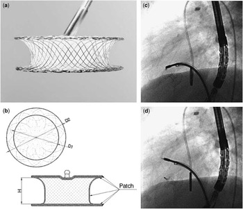
The “Occlutech® mVSD Occluder” (Fig 1) is a double-disc, braided Nitinol device. The diameter of the device waist defines its nominal size. The discs are symmetrical and larger than the waist by between 6 and 8 mm, depending on device size. The height of the waist is 7 mm. Polyethylene terephthalate is sewn into the device. The device has one hub onto which all the Nitinol strands attach. This forms the attachment for the typical Occlutech® “ball and socket” controlled release mechanism. The device is available in sizes ranging from 4 to 20 mm in 2-mm increments, deliverable in sheath sizes of 6–11 Fr. The preparation, loading, and deployment are the same as in the “Flex ” systems for Occlutech’s atrial septal devices.Reference Haas, Happel and Soetemann 3 The instructions for use recommend similar device sizing to St Jude’s muscular device.
( a ) Occlutech device’s potential angulated attachment to its delivery cable without tension or distortion. ( b ) Device dimensions described in the text. ( c and d ) Minimal change of orientation and position before and immediately after release.

Case report
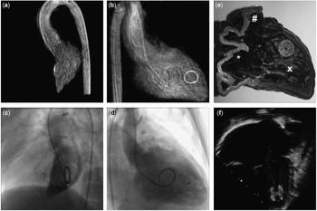
A 10-kg patient with multiple muscular ventricular septal defects had initial palliation at 2 months with a pulmonary artery band. By 18 months, she had become progressively desaturated with poor weight gain. Transoesophageal echocardiography and an MRI scan delineated the anatomy and allowed us to create a three-dimensional printed model of the ventricular septum (Fig 2). Most of the defects had spontaneously closed, leaving one significant anterior muscular defect and a smaller more apical one. We used this model to pre-select and narrow down a range of device types and sizes before committing to cardiac catheterisation.Reference Dankowski, Baszko and Sutherland 4
( a and b ) Left anterior oblique and right anterior oblique views of a three-dimensional (3D) reconstructed left ventricle angiogram; the circle represents the “target” area where the ventricular septal defect is positioned. ( c and d ) Angiography in the same views with the Occlutech device in situ. ( e ) Right ventricle aspect of a 3D printed model used for procedural planning with a device in place. “*” marks the inlet portion; “#” marks the outflow tract; “X” marks the trabecular apex. ( f ) Echocardiographic view of the device in situ.

During cardiac catheterisation, the right ventricular function had decreased significantly. After successfully dilating the pulmonary band, we performed rotational angiography in the left ventricle. This defined the plane of the ventricular septum and allowed us to use an overlay tool to highlight the area on the leftward aspect of the septum where the defect lay (Fig 2). We crossed the defect from the left ventricle and created an arteriovenous circuit in the usual manner.Reference Kenny, Morgan and Bajwa 5
Transoesophageal echocardiography was used to accurately size the ventricular septal defect (4–5 mm). We selected an 8-mm Occlutech® muscular ventricular septal defect device, slightly oversized to account for the significantly hypertrophied ventricular septum. A 7-Fr, 45-cm Mullins sheath was advanced through the right heart across the septum and positioned in the ascending aorta. Using echocardiographic and three-dimensional image overlay, the device was deployed by forming the left-sided disc in the outflow tract and pulling it onto the septum. The waist and right ventricular disc were then quickly deployed to minimise haemodynamic instability (Fig 2).
After complete deployment, the haemodynamic instability resolved, allowing us to carefully assess the device position and haemodynamic effect.
Echocardiographic and angiographic imaging looked satisfactory with good device conformation and no residual leak. There was no alteration in device position after release. The patient recovered quickly, and 1 month later the device remains in good position without any evidence of residual leak, despite a decrease in the degree of septal and right ventricular hypertrophy.
Discussion
Currently available devices perform well in a range of anatomical and physiological circumstances. Difficulties in right ventricular disc conformation or interference with tricuspid valve tissue may be overcome by using single disc devices such as the St Jude ADO I (St Jude Medical). Proximity of the target lesion to the free walls of either ventricle have caused theoretical concerns regarding erosion into the pericardial space. Thrombus formation on either disc rarely occurs due to the high blood flow velocity along both aspects of the ventricular septum, but rapid endothelisation remains at least a theoretical advantage.
The Occlutech® device has features that may prove clinically beneficial. The wire structure and weave cause the Occlutech® device to be softer overall. This may confer a lower risk of damage to related anatomy such as the ventricular free walls. The absence of a left ventricular hub may decrease the time taken to complete endothelisation of the left ventricle aspect, but this featureless left disc may prove problematic when attempting to retrieve an embolised device.Reference Pedra, Pedra and Costa 6
A key characteristic is the junction between the delivery system and the device. The ball and socket design allows a wide range of angulation between the device and the delivery cable (Fig 1). This minimises the change in configuration that occurs after device release allowing a more confident pre-release assessment. Its utility in the ventricle also decreases haemodynamic instability, by allowing a more neutral orientation and minimising distortion of the local anatomy, particularly important in smaller hearts and in patients with poor baseline haemodynamics.Reference Zartner, Christians, Stelter, Hraska and Schneider 7
Conclusion
The first reported case using this device illustrates the need for continued device development, even where an excellent option already exists. The equivalent St Jude® device is the major market player and has been successfully implanted in thousands of patients with excellent results. As we continue to push anatomical and physiological boundaries in our practice, subtle differences in device design may determine success or failure.Reference Shibbani, Kenny, McElhnney, Hijazi and Moran 8
Acknowledgements
The author thanks Miss Karolina Bilska for her transoesophageal echo guidance during the procedure and Dr Gur Mainzer for assisting with the procedure.
Financial Support
This research received no specific grant from any funding agency, commercial, or not-for-profit sectors.
Conflicts of Interest
None.