Hypoplastic left heart syndrome still remains one of the most challenging congenital cardiac diseases, either because of its complex surgical and postoperative management, or its long term prognosis. After Norwood and colleaguesReference Norwood, Lang and Hansen1 reported the possibility of palliation for this condition in the early 1980s, a modification for shunting to the pulmonary arteries was introduced by Kishimoto and colleagues,Reference Kishimoto, Kawahira, Kawata, Miura, Iwai and Mori2 and popularized over the last few years by Sano and associates.Reference Sano, Ishino and Kawada3 The modification consists in restoring the flow of blood to the lungs by placing a conduit from the right ventricle to the pulmonary arteries, rather than constructing a modified Blalock-Taussig shunt. It is argued that the ventriculo-pulmonary shunt permits better coronary arterial perfusion, avoiding diastolic run-off into the pulmonary circulation,Reference Pizarro and Norwood4–Reference Maher, Pizarro and Gidding6 and thus improving the postoperative course. Clinical outcomes attest to improved early results in most centres.Reference Sano, Ishino and Kado7 As yet, however, it is unclear whether the long-term outcome, with the need for a right ventricular incision, will be comparable to that obtained using the classic approach with an arterio-pulmonary shunt. In the hope of shedding more light on this issue, we obtained the hearts from neonates and children who did not survive after Norwood procedure, reviewing the pathological findings to establish whether significant differences existed in the impact of the two techniques.
Materials and methods
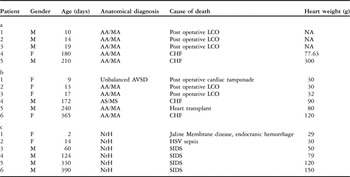
We reviewed 11 consecutive heart specimens obtained from patients having the diagnosis of hypoplastic left heart syndrome who had been submitted to a Norwood procedure. The specimens came from the anatomical collection of Congenital Heart Diseases, Institute of Pathological Anatomy, at the University of Padua. We divided the specimens in 2 groups according to whether the flow of blood to the lungs had been provided through a modified Blalock-Taussig shunt, or via a conduit placed from the right ventricle to pulmonary arteries. The hearts were obtained from 8 neonates who died after Norwood procedure prior to a cavo-pulmonary connection, with half having undergone a classic shunt and the other half the ventriculo-pulmonary modification. The remaining 3 hearts came from infants who died, or underwent cardiac transplantation, after a bi-directional cavo-pulmonary connection, with 2 of these having a classic arterio-pulmonary shunt, and the other the ventriculo-pulmonary modification. The detailed diagnoses, and causes of death, of these patients are listed in Table 1a and 1b. We compared the findings in these 11 specimens with findings from 6 normal hearts, matched for age and weight, belonging to patients who had died from noncardiac causes as neonates or infants (Table 1c). We performed macroscopic and microscopic analysis for each specimen, taking 2 full thickness samples of the myocardial walls from the right ventricular inflow and outflow tracts, far away from the area of scar tissue surrounding the site of placement of the conduit in those undergoing the ventricular modification, evaluating the impact of the ventriculotomy on the overall right ventricular myocardium. All samples were stored in formalin 10% in 0.1 M (pH = 7.4) phosphate buffer, dehydrated in a crescent series of ethanol, and then embedded in paraffin. Sections of 7 μm thickness were stained with Hematoxylin and eosin, the Heidenheim modified Azan Mallory stain, and Sirius red. Other sections were incubated with Monoclonal Mouse Anti-human CD31 (Dakocytomation, Glostrup, Denmark). Morphometric analysis and measurements were done following methods first introduced by Olivetti et al.Reference Olivetti, Melissari, Balbi, Quaini, Sonnenblick and Anversa8 This method is now validated and currently applied in our lab through a computerized image analyzer system consisting in an optic microscope Olympus BH2, connected to a computer via a video-camera (JVC 3-CCD, Japan), and a software for image analysis (Image PRO-Plus 4.0, Media Cybernetics, Silver Spring, MA, USA).
Anatomic and pathogical findings in our 3 groups of specimens: a, classic Norwood with Blalock Taussig Shunt; b, Norwood with ventriculo-pulmonary shunt, and c, Controls.

Legend: AA: aortic atresia; CHF: congestive heart failure; HSV: herpes simplex virus; LCO: low cardiac output syndrome; MA: mitral atresia; MS: mitral stenosis; NA: not applicable (heart lung block specimen); NrH: normal heart; SIDS: sudden infant death.
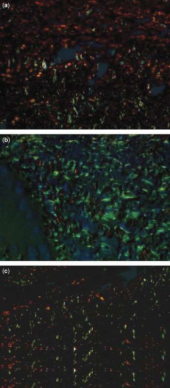
We analyzed the diameters of the myocytes in micrometers, the proportional areas of myocardial fibrosis, and the density of capillaries, calculating the number of capillary vessels per 0.045 millimetres squared, for both samples taken from each specimen. We measured 3 fields for the outer, or epicardial, the middle, and the inner, or endocardial, layers at 20 times magnification. In addition, by means of analysis of sections stained with Sirius red staining under a polarized light, we differentiated Collagen I fibres (orange-red, ubiquitary, parallel between them, structured, with low remodelling capacity) and Collagen III fibres (thin yellow-green, with reticular and less organized structure, with higher remodelling capacity). We evaluated 5 fields, using 40 times magnification, for each sample area and expressed the findings as a percentage.
Statistical analysis
For each parameter, we calculated the means and standard deviations, using Student’s t test to evaluate statistically significant differences among the 3 groups, the different areas of sampling, and the different types of collagen. Data were considered statistically significant when the value for p was less than 0.05.
Results
The groups were homogeneous for distributions of age and weight (Table 1). Among the 3 different groups, the ratios of males to females were 4 to 1, 2 to 4, and 4 to 2 for those undergoing the classic arterial shunt, the ventricular modification, and the control hearts, respectively. Preoperative diagnosis in those undergoing shunting was hypoplastic left heart syndrome in all, associated with an unbalanced atrioventricular septal defect in 1 patient who underwent a ventricular-to-pulmonary arterial shunt. Aortic atresia was present in 82% of the surgical cases. All the patients died either from cardiac failure or low cardiac output. There was no evidence of obstruction in the aortic arch, or shunt-related problems, detected at autopsy. Cardiac transplantation had been performed in 1 patient after a ventricular-to-pulmonary shunt because of congestive cardiac failure occurring after a bi-directional cavopulmonary anastomosis. In the control group, all heart specimens were normal at macroscopic evaluation.
Pathological findings
Diameter of the myocytes
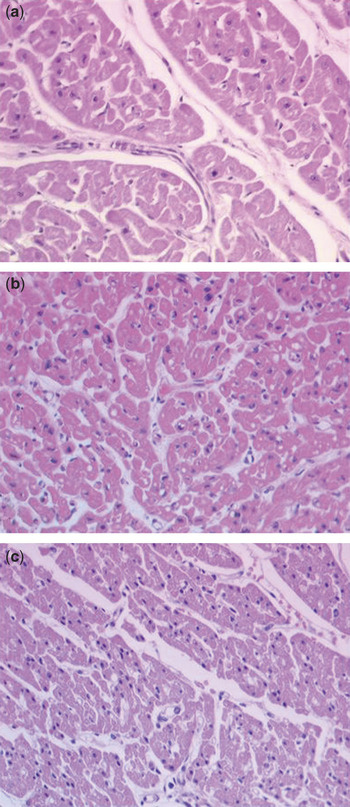
Hypertrophy of the cardiomyocytes, expressed as mean myocytic diameter, was increased in both groups of patients when compared to their controls, being 10.74 ± 0.66 μm in those undergoing the classic arterial shunt; 10.23 ± 0.71 μm in those undergoing the ventricular modification, and 5.77 ± 0.92 μm in the control hearts, these differences being statistically significant between each pathological group and the controls (p equal to 0.05). No significant difference was observed between the 2 groups of shunted patients (Fig. 1). The measurements of the diameters of the myocytes in the blocks obtained from the inflow and outflow tracts was similar, without difference between the two regions, suggesting that the remodelling is not directly related to the ventriculotomy.
Histological section showing (a) the middle layer of the myocardium of the ventricular walls in patient in whom we constructed a classic systemic-to-pulmonary arterial shunt and (b) in a patients in whom the shunt was placed from the right ventricle to the pulmonary arteries. Note than in both sections the diameter of the myocytes is greater than in the controls (c). Section stained with haematoxylin and eosin, and magnified 20 times.

• Inflow: The myocytes had a diameter of 10.46 ± 2.15 in those undergoing the classic arterial shunt (p = 0.008), and 9.73 ± 1.99 in those undergoing the ventricular modification (p = 0.004), as opposed to 5.72 ± 0.24 in controls.
• Outflow: The myocytic diameter was 11.03 ± 1.83 in those having a classic arterial shunt (p = 0.003), and 10.72 ± 2.22 in those having ventriculo-pulmonary shunts (p = 0.002), as opposed to 5.82 ± 0.23 in controls.
Myocardial fibrosis
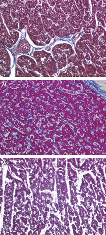
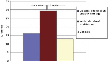
The extent of fibrosis was increased in those with ventriculo-pulmonary shunts when compared to those having a classic arterial shunt and controls (Fig. 2). The proportion of fibrous tissue was 29.40 ± 15.80% in those with ventriculo-pulmonary shunts, 16.15 ± 17.07% in those with classic arterial shunts, and 13.05 ± 8.49% in the controls (Fig. 3). The fibrosis was both interstitial and perivascular in those who had had ventriculo-pulmonary shunts, but was mainly perivascular following classic arterial shunting. Analysis of the inflow and the outflow ventricular tracts revealed similar patterns of fibrosis, confirming that the extent of fibrosis was not related to the ventriculotomy.
Histological section showing the endocardial layer of the myocardium in (a) a representative patient with an arterio-pulmonary, and (b) a patient with a ventriculo-pulmonary shunt compared to (c) a control. The extent of fibrosis is more marked in the patient undergoing the ventriculo-pulmonary shunt. Section stained with the Heidenhain modification of the Mallory trichrome technique, with 20 times original magnification.

Fibrosis expressed as mean values area in the three groups of patients, those with a classic arterial shunt, those with a ventricular modification, and the control hearts.

• Inflow: The proportion was 29.55 ± 6.12 after a ventriculo-pulmonary shunt, as opposed to 14.01 ± 8.3 in controls (p = 0.004), and 16.94 ± 5.83 in those with a classic arterial shunt (p = 0.006).
• Outflow: The proportion was 29.25 ± 7.57 after a ventriculo-pulmonary shunt, as opposed to 12.09 ± 8.41 in controls (p = 0.004), and 15.36 ± 4.60 after a classic arterial shunt (p = 0.005).
Types of collagen
The ratio between collagen I and collagen III was 0.94 for those having a classic arterial shunt and controls, but was 0.61 for those having a ventriculo-pulmonary shunt. The ratios were similar for analysis at the inflow or the outflow tracts. The proportion of Collagen I was higher in the inner-endocardial layers of those undergoing a classic arterial shunt when compared to those having the ventricular modification (60.10 ± 27.99% versus 44.98 ± 26.87%, p = 0.035), while the proportion of Collagen III was higher the mid-myocardial and outer-epicardial layers of those having a ventriculo-pulmonary shunt when compared to the controls (62.48 ± 25.21% versus 47.61 ± 58.97%, p = 0.04, Fig. 4).
Histologic section showing the epicardial layer of the myocardium stained with Sirius Red and visualised using polarized light. Note the higher content of collagen type III, stained green-yellowish, compared with collagen I, stained orange-red, in the patient with an arterio-pulmonary shunt (b) compared to the patient with a ventriculo-pulmonary shunt (a) and a control (c). The slide is magnified 40 times.

Density of capillaries
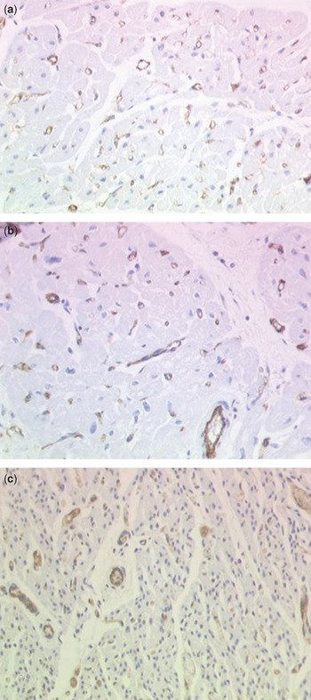
When we calculated the density of capillaries as the number of vessels found per 0.045 mm2, the result proved lower in those who had undergone a classic arterial shunt, at 65.23 ± 20.33, and lower in patients who had been submitted to the ventricular modification, at 82.70 ± 6.41, than in the control subjects, who had values of 118.14 ± 12.19 (Fig. 5). Similar differences were found when analyzing the blocks removed from the ventricular inflow and the outflow tracts:
Immunoistochemical evidence of capillary vessels in the middle layer of the myocardium in (a) a representative patient with an arterio-pulmonary shunt, (b) a patient with a ventriculo-pulmonary shunt and (c) a control heart. The density of capillaries is increased in the shunted patients in comparison with the normal myocardium. Mouse anti human anti-CD31, monoclonal antibody (Dakocytomation, Glostrup, Denmark), magnified 20 times from original magnification.

• Inflow: 57.77 ± 27.92 in those with a classic shunt, versus 127.01 ± 16.54 in controls (p = 0.002), and 86.70 ± 6.94 in those undergoing a ventricular modification versus the control values (p = 0.0009).
• Outflow: 72.68 ± 12.75 in those with a classic arterial shunt versus 109.29 ± 7.84 in the controls (p = 0.0014), and 78.70 ± 5.88 after the ventricular shunt, with a p value of 0.0001 compared to the control values.
Discussion
The Norwood sequence of operations for hypoplasia of the left heart remains the most challenging surgical repair for the paediatric cardiac surgeon. After the initial efforts by Norwood and his associates in the early 1980s,Reference Norwood, Lang and Hansen1 this complex malformation has stimulated the scientific community to work for the best treatment to offer these children and their parents. Nowadays, despite a excellent outcome at the first stage, as reported, for example, by Bove and his colleagues,Reference Bove, Ohye and Devaney9 the hypoplastic left heart syndrome remains a demanding cardiac disease in most centres. Despite the excellence of reconstructive surgery, most deaths occur in the first 24 to 48 hours after surgery due to haemodynamic instability caused by unpredictable rapid variations in pulmonary resistances. A recent multicentric North American study revealed that overall survival is no more than 72% at 1 month, 60% at 1 year, and 54% at 5 years.Reference Ashburn, McCrindle and Tchervenkov10
Over the years, efforts to achieve a balanced circulation during the early postoperative period have focused on limiting the flow of blood to the lungs, and improving systemic blood flow, such as by reducing the size of the systemic-to-pulmonary arterial shunt,Reference Bove, Ohye and Devaney9 management of ventilation on the basis of serial analysis of blood gases,Reference Hoffman, Ghanayem and Kampine11 focusing on retention of carbon dioxide,Reference Jobes, Nicolson, Steven, Miller, Jacobs and Norwood12 using hypoxic admixtures,Reference Bradley, Simsic and Atz13 and systemic vasodilators such as phenoxybenzamine.Reference Tweddell, Hoffman and Fedderly14 There have also been several modifications proposed for the surgical conduct of the Norwood sequence, with the hope of improving early results.Reference Fraser and Mee15–17 Recently, Sano and associatesReference Sano, Ishino and Kawada3 have popularized a modification that was considered by Norwood himself in the earlier days of his own experience [personal communication], and was reproposed by Kishimoto and colleagues in 1999.Reference Kishimoto, Kawahira, Kawata, Miura, Iwai and Mori2 This modification consists in the creation of a conduit from the right ventricle to the bifurcation of the pulmonary trunk. This is thought to eliminate aortic diastolic run-off into the pulmonary circulation, which is characteristic of the modified Blalock Taussig shunt. The reduction in diastolic run-off leads to increased diastolic pressure,Reference Pizarro and Norwood4–Reference Maher, Pizarro and Gidding6 as nicely demonstrated by Ohye et al,Reference Ohye, Ludomirsly, Devaney and Bove18 and facilitates better perfusion of the coronary arteries and other end-organs, thus improving myocardial perfusion and ventricular function. Simplified clinical postoperative courses and improved hospital survival have been reported elsewhere.Reference Sano, Ishino and Kado7, Reference Pizarro, Malec and Maher19 As yet, however, it is not known if this modification results in improved right ventricular performance over the long term when compared to the traditional classical approach, which does not involve a ventricular incision.
The theoretical advantages of the modified Blalock Taussig shunt are improved growth of the pulmonary arteries owing to the anterograde flow across the shunt during the entire cardiac cycle. The lower diastolic pressure with modified Blalock Taussig shunt is believed to compromise coronary arterial flow, with potential impact on cardiac function, especially in the immediate postoperative course. A conduit from the right ventricle to the pulmonary arteries provides anterograde flow to the pulmonary arteries during systole. There are some concerns. First, it produces diastolic reversal of flow, causing ventricular volume overload prior to the second stage of palliation, with a potential impact in the long term on ventricular performance. Second, it may be associated with reduced growth of the pulmonary arteries prior to second stage, with potential elevation of pressures in the Fontan baffle, and hypoxaemia and an increased risk of protein losing enteropathy. Third, the ventricular incision required for insertion of the conduit carries an unknown risk for late arrhythmias and diminished ventricular function.
Recently, Hughes et al.Reference Hughes, Shekerdemian, Brizard and Penny20 have reported an echocardiographic study on 9 infants who survived to Norwood procedure, with either a classic arterial or a ventricular shunt, showing that strain Doppler echocardiography provides a good noninvasive means of quantifying myocardial deformation, and that the right ventricular longitudinal systolic contractility is improved in patients who have undergone the ventricular modification as compared to a classic shunt. These patients, however, were studied in the early period of follow-up, between 27 and 50 days after the operation, at which time ventricular remodelling is incomplete. Tanoue and associatesReference Tanoue, Kado, Shiokawa, Fusazaki and Ishikawa21 reported an elegant study of ventricular function after the ventricular modification, again making comparisons with a classic shunt, based on calculation of contractility, afterload, and ventricular efficiency using pressure and volume data of cardiac catheterization, and Simpson’s rule to derive right ventricular functional data. Despite some intrinsic limits of this study, which is a small retrospective non-randomized investigation, and uses a method validated only in an animal model, they reported some interesting findings. Although ventricular efficiency was not different in the 2 groups, contractility following a bi-directional cavopulmonary shunt and the Fontan procedure in patients undergoing the classic shunt was superior to that measured in patients who had undergone the ventricular modification. As far as we are aware, these are the first investigators to attempt quantitative comparison for ventricular function in the 2 groups. Tabbutt et al.Reference Tabbutt, Dominguez and Ravishankar22 have also reported the experience from a single centre using the 2 different techniques. They found no differences in either survival or hospital course after the first stage procedure. Overall survival at 3 years was similar in the 2 groups. Mortality between the first and second stages was lower using the ventricular modification, but a trend of increasing mortality and late poor outcome was shown in these patients after the second stage of Norwood palliation.
To the best of our knowledge, ours is the first pathological evaluation of the state of the ventricular myocardium in patients subsequent to palliation of hypoplasia of the left heart comparing the consequence of a classic systemic-to-pulmonary arterial with a shunt placed from the right ventricle to the pulmonary arteries. Despite the evident limits of a pathological investigation, we surmise that our analysis has underlined some significant points. Our findings show that ventricular myocardial remodelling after the Norwood procedure is characterized by an increase in fibrosis, and myocytic diameters, independent of the surgical technique. In particular, as shown in Figures 2 and 3, cardiomyocytic hypertrophy is similar in the groups of patients undergoing different forms of arterial shunting, but the degree of fibrosis is greater in those with a shunt created from the right ventricle to the pulmonary arteries, where it is both interstitial and perivascular, compared to those with a classic systemic-to-pulmonary arterial shunt, where it is essentially perivascular. Moreover, the content of collagen I is proportionally higher in those with a classic arterial shunt, especially in the endocardial myocardial layers, which are subjected to a higher mural stress. The content of collagen III is proportionally higher in those who have had the shunt placed from the right ventricle top the pulmonary arteries, especially in the mid-myocardial and epicardial layers of the walls. The higher content of fibrous tissues in those undergoing a Norwood procedure involving a shunt placed from the ventricle to the pulmonary arteries is probably due to the wider area occupied by the loosely disposed thin fibrils, in contrast with collagen I, which is arranged in the form of bundles, with thick closely-packed fibrils. On the basis of features of the types of collagen, we conclude that those undergoing the ventricular modification have much more potential for myocardial remodelling. This could produce a deterioration of ventricular function over the long term, potentially associated with earlier haemodynamic and electrical instability. All the morphological substrates investigated were found in both the inflow and outflow tracts of the right ventricle, despite the presence of a right ventriculotomy in those undergoing the ventricular modification. Thus, it is not the scarring produced by the ventriculotomy which is the culprit for the changes observed, but rather the dynamic volume overload, which may alter permanently and globally the viable myocardium. This is in agreement with the common concept that cardiac function after the Norwood procedure is characterized by volume overload, with higher than normal diastolic pressures that may jeopardize ventricular myocardial perfusion. Our findings suggest that this change is more significant in those undergoing the ventricular modification of the shunting procedure. Whether this is attributable to the reversal of diastolic flow in the non-valved conduit placed from the right ventricle to the pulmonary arteries, or to the ventriculotomy itself, is not clear. The consequences of a ventriculotomy on the onset of late arrhythmias can be demonstrated only with long-term follow-up. Our pathological study, nonetheless, has revealed the existence of an abnormal substrate potentially responsible for tachyarrhythmias, as already reported for a ventricular incision.Reference Hanna, Colan, Bridges, Mayer and Castaneda23
In conclusion, we have shown greater remodelling of the ventricular myocardial extracellular matrix in patients having a shunt from the right ventricle to the pulmonary arteries when compared to those having a classic arterio-pulmonary shunt, with this remodelling progressing even after the neonatal period. This may influence a later suboptimal ventricular performance. Thus, we should not underestimate the importance of long-term follow-up of patients who have undergone the ventricular modification of the Norwood procedure. Comparative morbidity over the long term is potentially more important than overall survival over the early and mid-terms, keeping in mind that our target is achievement of best quality of life for our patients, and not just the improved statistics relating to operative survival for our records.