Inflammation is an adaptive response to damage of vascularised tissues, which develops according to a stereotyped sequence of events controlled by local production of a number of compounds known as inflammatory mediators. There is plenty of evidence to suggest that the plasminogen activation system is implicated in the regulation of all phases of the inflammatory response, but it plays a crucial role in leucocyte recruitment to the site of the inflammation (Del Rosso et al. Reference Del Rosso, Fibbi, Pucci, Margheri and Serrati2008, Reference Del Rosso, Margheri, Serrati, Chilla, Laurenzana and Fibbi2011). The urokinase plasminogen activator (u-PA) and its cellular receptor (u-PAR), expressed on the surface of leucocytes, provide a functional unit regulating the interaction of leucocytes with the extracellular matrix. This interaction is crucial for the migration of leucocytes and their movement in the damaged tissue (Reichel et al. Reference Reichel, Kanse and Krombach2012).
To breach the mechanical barrier imposed by the basement membrane, leucocytes use limited proteolytic activity at regions of cell contact. Typically, u-PAR clusters are observed at the leading edges of migrating cells (Sitrin et al. Reference Sitrin, Pan, Blackwood, Huang and Petty2001). u-PA bound to u-PAR remains proteolytically active and thus, can convert the inactive proenzyme plasminogen to active plasmin. Plasmin, in turn, is capable of degrading certain matrix components, in addition to activating other matrix degrading enzymes such as metalloproteinases (Politis et al. Reference Politis, Zavizjon, Cheli and Baldi2002). Thus, u-PA and u-PAR act synergistically in a pro-inflammatory fashion. Plasminogen activator inhibitor 1 (PAI-1) blocks the activation of plasminogen because it binds to u-PA bound to u-PAR causing internalisation of the u-PA/u-PAR complex within the leucocyte. The internalised complex is subsequently degraded. Thus, internalisation of the complex after binding of the PAI-1 inhibits cell migration (Degryse et al. Reference Degryse, Sier, Resnati, Conese and Blasi2001). Our group investigated the role of PAI-1 and the enigmatic plasminogen activator inhibitor 2 (PAI-2) during mastitis and suggested that PAI-1 and PAI-2 may play a role related to the successful localisation of the infection possibly by preventing a shift from acute to chronic inflammation (Theodorou et al. Reference Theodorou, Daskalopoulou, Chronopoulou, Baldi, Dell'orto and Politis2010).
Proteolytic breakdown of milk proteins generates a number of peptides endowed with various biological properties (antimicrobial, antihypertensive, antithrombotic and immunomodulatory activities) (Baldi et al. Reference Baldi, Politis, Pecorini, Fusi, Chronopoulou and Dell'Orto2005; Korhonen & Pihlanto, Reference Korhonen and Pihlanto2007). The ability of milk-born peptides to modulate the function of monocytes and neutrophils, two cell populations key components of the non-specific (innate) immunity has been previously demonstrated (Baldi et al. Reference Baldi, Politis, Pecorini, Fusi, Chronopoulou and Dell'Orto2005; Politis & Chronopoulou, Reference Politis and Chronopoulou2008). Whether yoghurt contains peptides capable of modulating the function of monocytes and neutrophils is not known. In yoghurt, there are two main sources of peptides. The first source is the outcome of the deliberate proteolysis caused by lactic acid bacteria cultures. The proteolytic machinery of lactic acid bacteria includes a cell wall bound proteinase and several intracellular peptidases (Law & Haandrikman, Reference Law and Haandrikman1997). The transport system of these bacteria enables them to internalise oligopeptides up to 18 amino acids in length. Longer oligopeptides are a major source of bioactive peptides in yoghurt when they are further degraded by gastrointestinal enzymes or by intracellular peptidases of lysed bacteria following yoghurt consumption (Law & Haandrikman, Reference Law and Haandrikman1997). The second source is from the generation of peptides by inadvertent proteolysis of proteins in milk prior to processing caused by native proteolytic systems (i.e. plasmin) during storage. Lastly, inadvertent proteolysis can be caused by the proteolytic enzyme machinery of the natural milk microbes present in milk. Pasteurisation minimises the ability of these microorganisms to grow but their proteolytic enzymes remain within milk and the resulting yoghurt and thus, heat-resistant proteolytically active enzymes can cause proteolysis.
To the best of our knowledge, there are no studies investigating the effect of yoghurt peptides on the expression of u-PA related genes in ovine monocytes and neutrophils. Thus, the objective of the present study was to investigate the ability of water soluble extracts (WSE) obtained from yoghurts made from bovine and ovine milk to modulate the expression of u-PA, u-PAR, PAI-1 and PAI-2 in ovine monocytes and neutrophils. Furthermore, the effect of WSE on total cell-associated, membrane-bound u-PA activity and free u-PA binding sites on monocytes and neutrophils was examined.
Material and methods
Unless otherwise stated all reagents were purchased from Sigma Chemical (St. Louis, MO).
Yoghurt samples and Water soluble extracts
A total of eight traditional type commercial Greek yoghurt brands were selected to participate in the present experiment. Four yoghurts were made using ovine and another four yoghurts were made using bovine milk. All brands selected had comparable composition of main nutrients (Fat 4,5%, Protein 5,6% and Total solids 15,8%). The protein composition of the four yoghurts made from bovine milk was elevated to the level of 5,6% by protein fortification. Yoghurt samples were stored for up to 25 days at 4 °C.
Water soluble extracts (WSE) were obtained from all 8 yoghurts at day 3 and 25 of storage using the method proposed by Kuchroo and Fox (Reference Kuchroo and Fox1982). The determination of WSE protein concentration was performed using the Kjeldahl method (N × 6·38) (AOAC, 1990).
Immunocompetent cells isolation and treatment with WSEs
Monocytes and neutrophils were isolated using methods previously described by us (Politis et al. Reference Politis, Theodorou, Lampidonis, Chronopoulou and Baldi2012). The purity of monocytes was 92% (88–97%). Cell viability was assessed by the trypan blue dye exclusion methodology (typically >95% of total cells obtained were viable). All reagents and water used were endotoxin free to avoid monocyte activation. Following isolation, cells were cultured in cell culture medium (RPMI 1640 containing 2 mM L-glutamine, 1·5 g/l HCO3 and 10 mM HEPES supplemented with 5·5 mM glucose, 1 mM sodium pyruvate, 50 mg/ml penicillin and streptomycin and 10% foetal bovine serum) in the presence or absence of crude WSE (0·3 mg/ml). Preliminary experiments indicated that this was the optimal concentration. Furthermore, there was no effect of yoghurt storage time on WSE effectiveness. Thus, WSE from yoghurts stored for 25 days were used throughout the experiment. All treatments were performed in duplicate while six samples without addition of WSE were used as a control group. After 3 h of incubation, cell culture medium was removed, cells were washed 3 times with PBS and then phorbol myristate acetate (PMA, 81 nM) was added to activate the cells. After incubation for 30 min, cells were washed again 3 times with PBS and then processed either for determination of u-PA activity or for RNA isolation.
Determination of total cell-associated and membrane-bound u-PA activity and u-PA free binding sites
Total cell-associated u-PA activity (intracellular + membrane-bound) was measured in activated monocytes and neutrophils. Cells were lysed by addition of 500 µl of 1 mm sodium bicarbonate, centrifuged at 12 000 × g for 3 min, aliquoted and stored at −80 °C. Activity of u-PA was determined following the procedure described by Politis et al. (Reference Politis, Hidiroglou, White, Gilmore, Williams, Scherf and Frigg1996). For membrane-bound u-PA activity determination, cells were resuspended at various cell concentrations in 250 µl of 100 mm Tris buffer (pH 8·0). Membrane-bound u-PA was determined following the procedure described in detail by Politis et al. (Reference Politis, Hidiroglou, Cheli and Baldi2001). The free, unoccupied u-PA binding sites present on the cell membrane can be fully saturated following incubation with purified u-PA. Thus, the difference of membrane-bound u-PA before and after incubation with u-PA reflects the presence of free u-PA binding sites on the cell membrane. Following activation, cells were resuspended in 500 µl of HBSS + 20 mm HEPES containing purified u-PA (Sigma; 10 U/ml). After incubation for another 30 min at 37 °C, neutrophils were washed three times with HBSS and then resuspended at various cell concentrations in 250 µl of 100 mm Tris buffer (pH 8·0) for the determination of the membrane-bound u-PA.
RNA extraction and gene expression
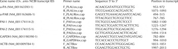
Total RNA was extracted from approximately 5 × 10⁶ cells using TriReagent (Sigma-Aldrich Co., USA) according to the manufacturer's instructions. Extracted RNA was treated with DNase I using a commercially available kit (New England Biolabs). The quality and quantity of the RNA extracted were confirmed by spectrophotometry as well as gel electrophoresis. Relative levels of mRNA for target genes were quantified with real-time, quantitative RT-PCR using SYBR Green chemistry. The amount of sample RNA was normalised by using glyceraldehyde 3-phosphate dehydrogenase (GAPDH) and β-actin (ACTB) as housekeeping genes. A pair of primers for each of the genes used in this study was constructed using PERLprimer software (Marshall, Reference Marshall2004). All primers were designed to be specific for Capra hircus and Ovis aries except for ACTB because it was not possible (Table 1). Total RNA (100–500 ng) was reverse transcribed with the PrimeScript First Strand cDNA Synthesis Kit (Takara), according to the manufacturer's instructions using a mix of random hexamers and oligo-dT primers. Real time PCR was performed in the 7500 Real Time system (Applied Biosystems) using the SYBR® Select Master Mix (Life Technologies) according to the manufacturer's protocol. Each reaction (10 µl) contained 5 ng RNA equivalents as well as 300 nM of forward and reverse primers for each gene. The reactions were incubated at 95 °C for 2 min followed by 40 cycles of 15 s at 95 °C and 60 s at 60 °C. This was followed by a melt curve analysis to determine the reaction specificity. Each sample was measured in duplicates. A modified version of the Pfaffl (Reference Pfaffl2001) normalization method was used for normalization against the two reference genes (Hellemans et al. Reference Hellemans, Mortier, De Paepe, Speleman and Vandesompele2007).
Sequences and relative positions of forward (F) and reverse (R) primers for Urokinase plasminogen activator (u-PA), urokinase plasminogen activator receptor (u-PAR), plasminogen activator inhibitor type 1 (PAI-1), plasminogen activator inhibitor type 2 (PAI-2), glyceraldehyde 3-phosphate dehydrogenase (GAPDH) and β-actin (ACTB) used in Real Time PCR

Statistical analysis
Data were analysed using the SPSS statistical package (version 17·0). For all relative expression analyses Control group was set to 1 and the expression of the treatments with bovine and ovine WSE is presented as x-fold expression to the Control group. The normality of the distribution for each variable was tested using the Kolmogorov-Smirnov (KS) test, while the homogeneity of variance using the Levene's test. All comparisons between groups were conducted by one-way ANOVA using Tukey's HSD post-hoc test and data are given as means ± sem. Significance for all cases was set at 0·05.
Results
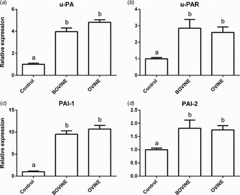
The ability of WSE obtained from yoghurts made from bovine or ovine milk to modulate the expression of u-PA, u-PAR, PAI-1 and PAI-2 by ovine monocytes was examined and the results are presented in Fig. 1. Data indicated that treatment with WSE increased the expression of u-PA, u-PAR and PAI-1 by approximately 4-fold, 2·7-fold and 10-fold, respectively. WSE increased the expression of PAI-2 by approximately 70%. There were no differences in the effectiveness of WSE related to milk origin (bovine versus ovine).
Effect of treatment of ovine monocytes with WSE obtained from ovine and bovine yoghurt on expression of (a) Urokinase Plasminogen Activator (u-PA), (b) Urokinase Plasminogen Activator Receptor (u-PAR), (c) Plasminogen Activator Inhibitor type 1 (PAI-1) and (d) Plasminogen Activator Inhibitor type 2 (PAI-2). Values are means ± sem. Differences between columns labelled with different letters are statistically significant at P < 0·05.

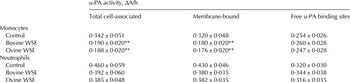
Changes in total cell-associated, membrane-bound u-PA activity and u-PA binding sites of monocytes treated with and without WSE were determined and the results are presented in Table 2. Treatment of monocytes with WSE resulted in decreases in total cell-associated and membrane-bound u-PA. Both WSE (bovine and ovine) were equally effective. WSE had no effect on free u-PA binding sites on ovine monocytes.
Effect of treatment of ovine monocytes and neutrophils with WSE obtained from ovine and bovine yoghurt on total cell associated, membrane-bound urokinase plasminogen activator (u-PA) activity and u-PA binding sites

Values are means ± sd for 10 independent samples
** Values are significantly different from those of the control (P < 0·01)
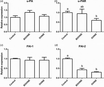
The ability of WSE obtained from yoghurts made from bovine or ovine milk to modulate the expression of u-PA, u-PAR, PAI-1 and PAI-2 by neutrophils was examined and the results are presented in Fig. 2. Data indicate that treatment with WSE decreased by 50–60% the expression of PAI-2. Only the ovine WSE decreased by 40% the expression of u-PAR. There was no effect of WSE on expression of u-PA and PAI-1 by neutrophils.
Effect of treatment of ovine neutrophils with WSE obtained from ovine and bovine yoghurt on expression of (a) Urokinase Plasminogen Activator (u-PA), (b) Urokinase Plasminogen Activator Receptor (u-PAR), (c) Plasminogen Activator Inhibitor type 1 (PAI-1) and (d) Plasminogen Activator Inhibitor type 2 (PAI-2). Values are means ± sem. Differences between columns labelled with different letters are statistically significant at P < 0·05.

Changes in total cell-associated, membrane-bound u-PA activity and u-PA binding sites of neutrophils treated with and without WSE were determined and the results are presented in Table 2. Treatment of neutrophils with WSE had no effect on total cell-associated and membrane-bound u-PA activity and free u-PA binding sites on ovine neutrophils.
Discussion
The first finding emerging from the present study is that yoghurt peptides modulated the expression of u-PA related genes and overall the u-PA system at the protein level in ovine leucocytes. However, the u-PA system was affected in a differential manner in the two cellular populations (monocytes and neutrophils) investigated. In monocytes the key observation is that all four genes (u-PA, u-PAR, PAI-1 and PAI-2) were up-regulated following treatment with yoghurt WSE. At first glance, this is a puzzling observation. The up-regulation of u-PA and u-PAR favours plasminogen activation and, thus, monocyte migration. The zymogen pro(u-PA) is bound to u-PAR on the leucocyte plasma membrane. Minute amounts of plasmin present in the monocyte cell vicinity convert the zymogen pro(u-PA) to active u-PA enzyme, which in turn, converts plasma-associated plasminogen to plasmin. This process is known as ‘reciprocal zymogen activation’ (Theodorou et al. Reference Theodorou, Daskalopoulou, Chronopoulou, Baldi, Dell'orto and Politis2010). This efficient machinery generates proteolytic activity that is restricted to the plasma membrane. This is very important because when plasmin diffuses away from the cell membrane it is rapidly inactivated by circulating plasma inhibitors (Plesner et al. Reference Plesner, Behrendt and Ploug1997). The up regulation of u-PA and u-PAR favours monocyte chemotaxis because the generated proteolytic activity will enable monocytes to transverse the endothelial barrier and migrate to the site of the inflammation. What makes the situation intriguing and perhaps surprising is that WSE up-regulated the expression of PAI-1 and PAI-2, as well. This obviously does not favour monocyte migration. While both u-PA and PAI-1 are thought to act as chemoattractants, binding of PAI-1 to the complex u-PA/u-PAR causes internalisation of the u-PA and blocks u-PA-induced chemotaxis (Degryse et al. Reference Degryse, Sier, Resnati, Conese and Blasi2001). A different explanation for the paradoxical up-regulation of both activators and inhibitors of the system has been offered by Politis (Reference Politis1996) who suggested that upregulation of PAI-1 may be blocking the u-PA system but essentially it might be a mechanism that continuously changes (renewing) the proteolytic activity localised on monocyte cell membrane. Closer examination of our results reveals that the up-regulation of PAI-1, which is 20–100 times more efficient than PAI-2 (Kucharewicz et al. Reference Kucharewicz, Kowal, Buczko and Bodzenta-Lukaszyk2003), is much higher (10-fold) than that of u-PA and u-PAR (3–4 fold) (Fig. 1). This creates an imbalance shifting the u-PA system towards inhibition. This conclusion is further supported by our findings at the protein level where treatment of monocytes with WSE resulted in reduced total cell-associated and membrane-bound u-PA activity in ovine monocytes. Even though an alternative explanation has been offered above (renewal of proteolytic activity) the majority of evidence is that up- regulation of the inhibitors blocks monocyte chemotaxis. Our results at the molecular and protein levels, taken together, suggest that yoghurt WSE promotes anti-inflammatory activities in ovine monocytes.
The overall picture of how WSE affected the u-PA system in ovine neutrophils is very different from that in monocytes. First, WSE had no effect on expression of two key genes (u-PA and PAI-1) in neutrophils. Second, WSE caused small decreases (50–60%) in the expression of PAI-2 and only WSE extracts from yoghurts made from ovine milk decreased the expression of u-PAR by 40% in neutrophils. Our view is that these competing small changes in expression essentially cancel each other and they are not sufficient to shift the system towards inhibition or activation. In line with this suggestion, our results show that WSE had no effect on total cell-associated and membrane-bound u-PA activity in ovine neutrophils. The reason why yoghurt peptides affected the u-PA system in monocytes and not in neutrophils is not known and anything said will be pure speculation. Monocytes have common properties with neutrophils, but they have distinctive morphological and functional properties, depending upon the state of differentiation. The most important difference between these two cellular populations is that monocytes following activation to macrophages have a greater ability for cytokine and overall protein production than the neutrophils (Dale et al. Reference Dale, Ruhlman and Baxter2015). We speculate, based on our findings, that cytokine and protein production is not only lower in neutrophils but it may not be easily modulated.
The second finding emerging from the present study is that WSE from yoghurts made from ovine or bovine milk were essentially equally effective towards monocytes and neutrophils. The only small difference was that WSE from yoghurt made from ovine milk but not that made from bovine milk down-regulated the expression of u-PAR in neutrophils.
The main question is what is the physiological significance of yoghurt peptides. One could argue that these peptides could play a role in the in vivo inflammatory response at the level of the mammary gland, since a portion of these peptides is generated very early during the naturally occurring milk proteolysis prior to processing. However, the small amount of peptides generated this way, taken together with the the fact that immunocompetent cells come into contact with milk peptides only when the former are present in milk (at the end of the migratory process), makes the possibility that the peptides may play a role in vivo highly unlikely. Thus, the emphasis of our research is directed towards the possible effects of the consumption of bioactive peptides present in yoghurt.
In conclusion, yoghurt made from ovine or bovine milk contains peptides that affect mainly the expression of u-PA related genes in ovine monocytes and to a lesser extent in ovine neutrophils. The main finding in monocytes is that the upregulation of the inhibitors is higher than that of the activators of plasminogen, shifting the system towards inhibition. Thus, yoghurt peptides possess anti-inflammatory activity.
Future work will focus in identifying individual peptides derived from yoghurt affecting the plasminogen activation system.
The present study was supported by the General Secretariat for Research and Technology – Greek Ministry of Education and Religious Affairs (Cooperation 2011-Project 1265).