

Introduction
The domestic cat is a valuable model for developing assisted reproductive techniques in endangered felids (Pope, Reference Pope2014). The Felidae family has 38 species of which 18 have been classified as vulnerable or endangered according to the International Union for Conservation of Nature and Natural Resources (2021). At present, several species of wild felids have been born after the transfer of embryos generated by in vitro fertilization (IVF) (Veraguas et al., Reference Veraguas, Echeverry, Castro, Rodriguez-Alvarez, Shrivastav and Singh2017; Pope, Reference Pope2019). Furthermore, interspecific somatic cell nuclear transfer (iSCNT) has allowed live offspring from the African Wildcat and Sandcat (Gómez et al., Reference Gómez, Pope, Giraldo, Lyons, Harris, King, Cole, Godke and Dresser2004, Reference Gómez, Pope, Kutner, Ricks, Lyons, Ruhe, Dumas, Lyons, López, Dresser and Reiser2008). This demonstrates that the domestic cat oocyte is capable of reprogramming somatic cells from other felid species, allowing the production of embryos and live offspring. The advantage of iSCNT is that embryos from endangered species can be generated without the necessity to collect gametes from them. However, the efficiency of this technique is low. A reduced proportion of cloned embryos reached the blastocyst stage and only two species of endangered felids have been born using this technique (Gómez et al., Reference Gómez, Pope, Ricks, Lyons, Dumas and Dresser2009, Reference Gómez, Pope, Biancardi, Dumas, Galiguis, Morris, Wang and Dresser2011, Reference Gómez, Biancardi, Jenkins, Dumas, Galiguis, Wang and Earle Pope2012).
The embryo aggregation method enhances the developmental competences of embryos generated by SCNT (Buemo et al., Reference Buemo, Gambini, Moro, Hiriart, Fernández-Martín, Collas and Salamone2016). This method consists of removing the zona pellucida and then culturing together two or more cloned embryos that are genetically identical but with different epigenetic problems, these embryos develop into a single blastocyst (Boiani et al., Reference Boiani, Eckardt, Leu, Schöler and McLaughlin2003). It has been proposed that embryo aggregation increases the blastocyst rate by reducing the epigenetic problems associated with incomplete nuclear reprogramming (Boiani et al., Reference Boiani, Eckardt, Leu, Schöler and McLaughlin2003). It has been described that embryo aggregation increases the developmental competence of domestic cat cloned embryos (Moro et al., Reference Moro, Hiriart, Buemo, Jarazo, Sestelo, Veraguas, Rodriguez-Alvarez and Salamone2015b). Furthermore, embryo aggregation enhances the developmental competence of embryos from Cheetah, Tiger and Bengal cat generated by iSCNT (Moro et al., Reference Moro, Jarazo, Buemo, Hiriart, Sestelo and Salamone2015a, Reference Moro, Hiriart, Buemo, Jarazo, Sestelo, Veraguas, Rodriguez-Alvarez and Salamone2015b). Recently, our research group reported that embryo aggregation allows the production of blastocysts from kodkod by iSCNT (Veraguas et al., Reference Veraguas, Aguilera, Echeverry, Saez-Ruiz, Castro and Rodriguez-Alvarez2020a). These reports demonstrate that embryo aggregation can improve the competence of felid cloned embryos, being a useful method for the genetic preservation of endangered felids. However, the in vivo developmental competence of these zona-free cloned embryos has not been reported. In addition, no live births have been achieved after the embryo transfer (ET) of domestic cat zona-free blastocysts generated by IVF (Veraguas-Davila et al., Reference Veraguas-Davila, Cordero, Saez, Saez-Ruiz, Gonzalez, Saravia, Castro and Rodriguez-Alvarez2021).
The lack of an intact zona pellucida might be associated with several problems in the in vivo development of domestic cat embryos. During early development, the zona pellucida allows contact among blastomeres, protects the embryo against external factors and regulates embryo–maternal communication (Cohen et al., Reference Cohen, Malter, Elsner, Kort, Massey and Mayer1990; Wassarman et al., Reference Wassarman, Chen, Cohen, Litscher, Liu, Qi and Williams1999; Van Soom et al., 2009). It has been reported that domestic cat embryos in a hatching blastocyst stage are not able to implant, proposing the idea that an intact zona pellucida might be needed to achieve in vivo embryo survival and implantation (Kanda et al., Reference Kanda, Oikawa, Nakao and Tsutsui1995; Pope, Reference Pope2014). However, the specific reasons that cause this implantation failure are still unknown.
Recently, our research group reported that domestic cat embryos cultured without zona pellucida were not capable of implanting after ET (Veraguas-Davila et al., Reference Veraguas-Davila, Cordero, Saez, Saez-Ruiz, Gonzalez, Saravia, Castro and Rodriguez-Alvarez2021). In that study, the domestic cat blastocysts cultured without the zona pellucida had a reduced expression of the pluripotency markers SOX2 and NANOG and an increased expression of BAX compared with embryos with an intact zona pellucida (Veraguas-Davila et al., Reference Veraguas-Davila, Cordero, Saez, Saez-Ruiz, Gonzalez, Saravia, Castro and Rodriguez-Alvarez2021). This alteration in the gene expression pattern could be associated with the low implantation potential observed in the zona-free blastocysts. In mice, SOX2 regulated the expression of trophectoderm markers TEAD4, YAP1, EOMES and CDX2 (Keramari et al., Reference Keramari, Razavi, Ingman, Patsch, Edenhofer, Ward and Kimber2010). It could be possible that a low expression of SOX2 might alter the expression of trophectoderm markers in the zona-free blastocysts from domestic cat. An altered expression of trophectoderm markers might affect the formation of trophectoderm linage and the subsequent implantation (Marikawa & Alarcon, Reference Marikawa and Alarcon2012). For these reason, we hypothesized that domestic cat blastocysts cultured without a zona pellucida might have altered expression of the trophectoderm markers, which might be related to their reduced implantation potential. To prove this, the objective of this research was to evaluate the expression pattern of trophectoderm markers in domestic cat blastocysts cultured without a zona pellucida or with an intact zona pellucida.
Materials and methods
All chemical reagents were purchased from Sigma Aldrich Chemicals Company (St. Louis, MO, USA), except for those otherwise indicated.
Ethics statement
All procedures that involved animal manipulation (ovariohysterectomy and orchiectomy) were approved by the Ethics Committee (Comité de Bioética de la Facultad de Ciencias Veterinarias de la Universidad de Concepción). The procedures were authorized by the animal owners through signed consent, according to the guidelines of the Ethics Committee (Certificate of approval no. CBE-08–2020).
Animals
Healthy female and male domestic cats aged between 6 months and 5 years were selected as oocyte and sperm donors for in vitro embryo production.
Experimental design
Two experimental groups were selected: (1) domestic cat embryos generated by IVF and cultured in vitro (zona intact, ZI group); and (2) domestic cat embryos generated by IVF and cultured in vitro without a zona pellucida (zona free, ZF group). The ovaries from domestic cat were collected by ovariohysterectomy. The immature cumulus–oocyte complexes (COCs) were collected from these ovaries by slicing and then were subjected to in vitro maturation (IVM). Subsequently, the in vitro matured COCs were subjected to IVF using epididymal sperm. After IVF, the cumulus cells of the presumptive zygotes were removed. Only in the ZF group, the zona pellucida of the presumptive zygotes was removed and this were cultured in vitro using the well of the well (WOW) system. In the ZF and ZI groups, embryos were cultured in vitro for 7 days. The cleavage, morula and blastocyst rates were estimated. The total cell number and the diameter of the blastocysts were assessed. Finally, the relative expression of trophectoderm markers TEAD4, YAP1, CDX2 and EOMES, the cell adhesion marker E-cadherin and the apoptosis marker CASP3 were evaluated by RT-qPCR in the blastocysts.
Ovariohysterectomy and cumulus–oocyte complexes (COCs) collection
The ovaries of female domestic cats were collected by ovariohysterectomy. For ovariohysterectomy, anaesthesia was induced using an (i.m.) dose of 0.5 mg/kg acepromazine (Ingor; Centrovet, Chile) and 5 mg/kg ketamine (Ketamine 100; Chemie Chile) and maintained by administering acepromazine/ketamine (i.v.) at the same dose. Ovaries were transported to the laboratory in a sterile solution containing 0.9% NaCl and 0.1% gentamycin (Gentamicin 10%, Veterquimica, Chile) at 38.5°C. COCs were recovered from the ovaries by slicing in a 100-mm Petri dish containing 10 ml of TCM-199 with Earle’s salts supplemented with 0.18 mM HEPES, 5% FBS and 50 μg/ml gentamycin (He199) at 38.5°C. Only grades I and II immature COCs were subjected to IVM.
In vitro maturation
IVM of immature COCs was performed in four-well dishes containing 500 µl of TCM-199 with Earle’s salts supplemented with 0.3% fraction-V BSA, 0.1 IU/ml FSH-LH (Pluset, Serono, Italy), 1 μg/ml 17β-estradiol, 0.36 mM sodium pyruvate, 2 mM glutamine, 2.2 mM calcium lactate, 20 ng/ml EGF, 10 µl/ml ITS (insulin, transferrin, selenium) and 50 μg/ml gentamycin (IVM-199) in a humidified gas atmosphere with 5.0% CO2, at 38.5°C for 26 h.
Sperm collection
Only male domestic cats older than 10 months of age were used as sperm donors for the generation of embryos by IVF. The anaesthesia procedure was the same as that described for ovariohysterectomy. Additionally, lidocaine (Lidocalm, 2% lidocaine chlorhydrate, Dragpharma, Chile) was administered in the genital area as local anaesthesia. Testes were transported to the laboratory in sterile 0.9% NaCl solution with 0.1% gentamycin at room temperature. Epididymal sperm were collected as previously described by our research group (Veraguas et al., Reference Veraguas, Saez, Aguilera, Echeverry, Gallegos, Saez-Ruiz, Castro and Rodriguez-Alvarez2020b). The caudal portions of the epididymis were cut into small pieces of ∼1 mm in a 100 mm Petri dish containing 10 ml of He199 supplemented with 0.3% Fraction-V BSA instead of FBS (He199-BSA) at 38.5°C. The medium with the sperm was passed through a sterile nylon filter (40 µm) into a 50-ml sterile tube. The filtrate was centrifuged at 1500 rpm for 5 min, and the pellet was resuspended in 5 ml of 20% Andromed medium (diluted in sterile distilled water according to the manufacturer’s instructions) (Minitub, Tiefenbach, Germany) and refrigerated at 4°C for 24 h (Veraguas et al., Reference Veraguas, Saez, Aguilera, Echeverry, Gallegos, Saez-Ruiz, Castro and Rodriguez-Alvarez2020b).
In vitro fertilization
Once IVM was finished, COCs were washed in TALP medium supplemented with 6 mg/ml BSA, 0.36 mM sodium pyruvate, 1 mM glutamine, 2.2 mM calcium lactate, 1% MEM nonessential, amino acids (NEAA), 0.01 mg/ml heparin sodium salt and 50 μg/ml gentamycin (TALP-IVF). The refrigerated sperm were allowed to swim up for 30 min in He199-BSA at 38.5°C (Veraguas et al., Reference Veraguas, Saez, Aguilera, Echeverry, Gallegos, Saez-Ruiz, Castro and Rodriguez-Alvarez2020b). The supernatant was collected and then centrifuged at 1500 rpm for 5 min. The pellet was collected and resuspended in TALP-IVF. For IVF, 20–30 COCs were placed in four-well dishes containing 500 µl of TALP-IVF and co-incubated with 1.5 to 2.5 × 10⁶ spermatozoa/ml in a humidified atmosphere of 5.0% CO2 in air at 38.5°C for 24 h. Subsequently, cumulus cells were removed from the presumptive zygotes using a 0.5 mg/ml hyaluronidase solution and vortexed for 6 min.
In vitro embryo culture
In the ZI group, presumptive zygotes were cultured in four-well dishes containing 500 µl of SOF medium supplemented with 0.37 mM trisodium citrate, 2.77 mM myo-inositol, essential and nonessential amino acids (final concentration 1×), 50 µg/ml gentamycin, 3 mg/ml essentially fatty acid-free BSA, 10 µl/ml ITS and 10 ng/ml EGF (S-SOF). Next, 10–20 embryos were placed into each well.
In the ZF group, the zona pellucida from presumptive zygotes was removed before embryo culture. The zona pellucida of presumptive zygotes was removed by incubation in a 2 mg/ml pronase solution for 4 min. Subsequently, the presumptive zygotes were washed three times in He199 supplemented with 30% FBS (He199–30) to remove pronase. ZF embryos were cultured in four-well dishes using the WOW system (Vajta et al., Reference Vajta, Korösi, Du, Nakata, Ieda, Kuwayama and Nagy2008). The WOW system consists of microwells in a culture dish in which ZF embryos can be placed, and the microwells prevent the disaggregation of the blastomeres, allowing in vitro development (Vajta et al., Reference Vajta, Korösi, Du, Nakata, Ieda, Kuwayama and Nagy2008). The microwells were created using an aggregation needle (BLS Ltd, Aggregation needles DN-09N). For this, each well of the four-well dish was covered with 300 µl of SOF, the aggregation needle was pressed into the plastic, and circular movements were made to create one microwell (Veraguas-Davila et al., Reference Veraguas-Davila, Cordero, Saez, Saez-Ruiz, Gonzalez, Saravia, Castro and Rodriguez-Alvarez2021). Eighty microwells were made in a four-well dish (20 in each well). Next, the four-well dish was washed at least three times with SOF and then covered with 500 µl of S-SOF. Each ZF embryo was cultured individually in one microwell, and 15–20 ZF embryos were cultured per well.
In both experimental groups, the culture dishes were incubated in a humidified atmosphere of 5.0% CO2, 5.0% O2 and 90% N2 at 38.5°C and cultured for 7 days. At day-5 of culture, the culture medium was replaced by S-SOF supplemented with 10% FBS instead of BSA and ITS concentration was reduced to 1 µl/ml cleavage, morula and blastocyst rates were estimated at days 2, 5 and 7, respectively.
Morphological evaluation of blastocysts
Total cell counting
The blastocysts were fixed in a 3% glutaraldehyde solution for 72 h at 4°C. Fixed blastocysts were stained with 5 μg/ml Hoechst 33342 for 20 min. The stained embryos were placed on a slide and covered with a coverslip. Visualization was achieved using the EVOS FL Auto Cell Imaging System (Thermo Fisher Scientific).
Diameter measurement
The blastocysts from the ZI and ZF groups were imaged using a camera mounted to a stereomicroscope (Micrometrics CMOS Digital Camera), and the diameter of the blastocysts was measured from the captured images using Micrometrics SE Premium Software.
Gene expression analysis
RNA extraction and cDNA synthesis
For gene expression analysis. ZI and ZF day 7 blastocysts were pooled within their respective groups. 10 pools were made in both groups, and all the pools had five day-7 blastocysts. The pooled samples were stored at −80°C until RNA extraction and cDNA synthesis.
RNA extraction was performed using the Ambion® Cells-to-cDNA™ II Kit (Thermo Fisher Scientific, Austin, TX, USA) as previously described by our research group (Veraguas et al., Reference Veraguas, Cuevas, Gallegos, Saez-Ruiz, Castro and Rodriguez-Alvarez2018). For total RNA extraction, the samples were washed in cold PBS to eliminate the remaining culture medium. Next, 60 µl of lysis buffer was added, and the samples were incubated at 75°C for 10 min. Subsequently, the samples were treated with DNase I (0.04 U/µl) for gDNA digestion. The cDNA synthesis was done using the SuperScript™ IV First-Strand Synthesis System (Invitrogen, Thermo Fisher Scientific). Here, 10 µl of total RNA was used in a 20-µl final reaction containing first, 50 ng/µl random primers hexamers and 10 mM each dNTP were added and the RNA–primer mixture was incubated to 65°C for 5 min and was kept to 4°C for at least 1 min. Then, the reaction was completed by adding 4µl 5× SSIV buffer, 1 µl 100 mM DTT, 1 µl ribonuclease inhibitor and 1 µl SuperScript™ IV Reverse Transcriptase (200 U/μl). The mix was centrifuged and then incubated to 23°C for 10 min, 55°C for 10 min and finally to 80°C for 10 min. The cDNA samples were kept at −20°C until qPCR was performed.
Real-time quantitative polymerase chain reaction (qPCR)
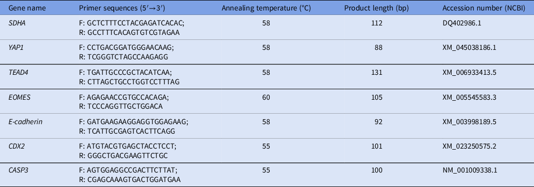
Gene expression analysis was performed by real-time qPCR using the standard curve method as we previously described Veraguas et al., Reference Veraguas, Cuevas, Gallegos, Saez-Ruiz, Castro and Rodriguez-Alvarez2018. Standard curves were generated using 2 µl of PCR products for each specific gene. The PCR products were previously purified using the E.Z.N.A gel extraction kit (Omega, BioTek, Santiago, Chile) and quantified using a spectrophotometer (Epoch, BioTek Instruments, Inc., Winooski, VT, USA). The standard curves were generated using eight 10-fold dilutions from the PCR products previously quantified. For qPCR analysis, each sample was loaded in duplicate (technical replicates). PCR was performed using 2 µl of cDNA from the samples, 1 µl of primers (10 pmol each, forward and reverse), and 5 µl of the KiCqStart SYBR Green ReadyMix, Low ROX (Sigma Aldrich) in a final volume of 10 µl. The reaction was run on an MX3000P Real-Time PCR device (Agilent, Santa Clara, CA, USA). Only experiments with an efficiency (including standard curves and samples) within the range 90–110% and a correlation coefficient of at least 0.9 were used for analysis. For the analysis, the expression of each gene was normalized using the housekeeping gene SDHA. The primers used and PCR conditions for each gene are shown in Table 1.
Primer sequences and RT-qPCR conditions for the gene expression analysis

Statistical analysis
The Wilcoxon non-parametric test was used to evaluate the in vitro development and gene expression patterns between the ZI and ZF groups. The t-test was used to evaluate the diameter and the total cell number in the blastocysts between groups. The statistical software Infostat (2014 Version; University of Cordoba, Argentina) was used for these analyses.
Results
In vitro development of domestic cat embryos cultured with and without zona pellucida
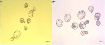
In both groups, the presumptive zygotes were in vitro cultured for seven days. In the ZF group, the presumptive zygotes were cultured into microwells to prevent the disaggregation of the blastomeres (Figure 1). No differences were observed in the cleavage, morula and blastocyst rates between the ZI and ZF groups (P > 0.05). This indicates that the microwell culture system used in this research prevents the disaggregation of blastomeres allowing the in vitro development of domestic cat ZF embryos (Table 2).
In vitro development of domestic cat embryos. (A) Domestic cat blastocysts generated by IVF and in vitro cultured for 7 days (40×). (B) Domestic cat blastocysts generated by IVF and in vitro cultured without zona pellucida for 7 days (50×).

In vitro development of domestic cat embryos cultured with (ZI) and without (ZF) zona pellucida for 7 days

Morphological evaluation of domestic cat blastocysts cultured with and without zona pellucida
Total cell counting
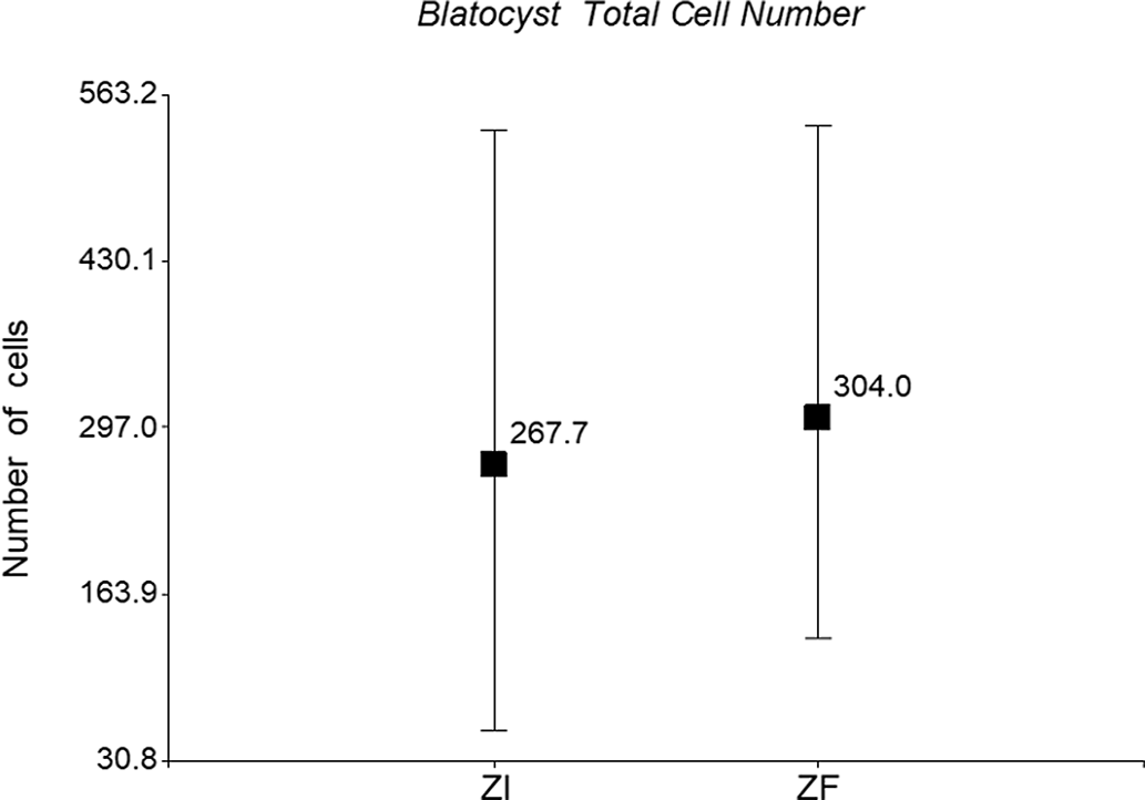
For total cell counting, 10 ZF blastocysts and 10 ZI blastocysts at day 7 of IVC were evaluated. No differences were observed in the total cell number [mean ± standard deviation (SD)] of blastocysts from the ZF (304.0 ± 142.9) and ZI groups (267.7 ± 166.2) at day 7 of culture (P > 0.05) (Figure 2).
Total cell counting. Total cell number (mean; min/max) of blastocysts after 7 days of IVC. ZI (267.7; 55.0/535.0) and ZF group (304.0; 129.0/539.0). *Significant differences between groups (P < 0.05).

Blastocyst diameter
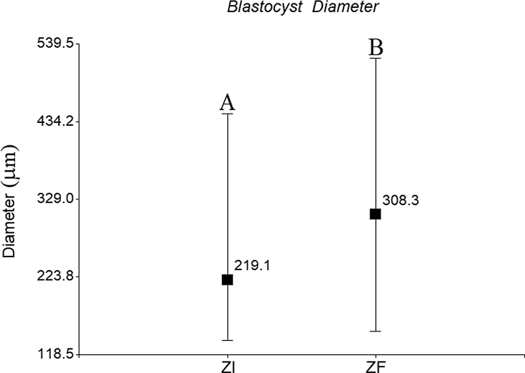
For diameter measurement, 50 blastocysts were evaluated in each group (ZI and ZF). The diameter (mean ± SD) of the blastocysts generated in the ZF group (308.3 ± 96.3 µm) was significantly higher than that of their counterparts from the ZI group (219.1 ± 67.8 µm) (P < 0.05) (Figure 3). This increase in blastocyst size observed in the ZF group might be caused by the absence of the zona pellucida, which was not related to an increase in total cell number.
Diameter measurement (mean; min/max) of day-7 blastocysts from the ZI (219.1; 137.7/444.8) and ZF group (308.3; 149.8/520.3). A,BDifferent superscripts indicate significant differences between groups (P < 0.05).

Gene expression analysis of domestic cat blastocysts cultured with and without zona pellucida
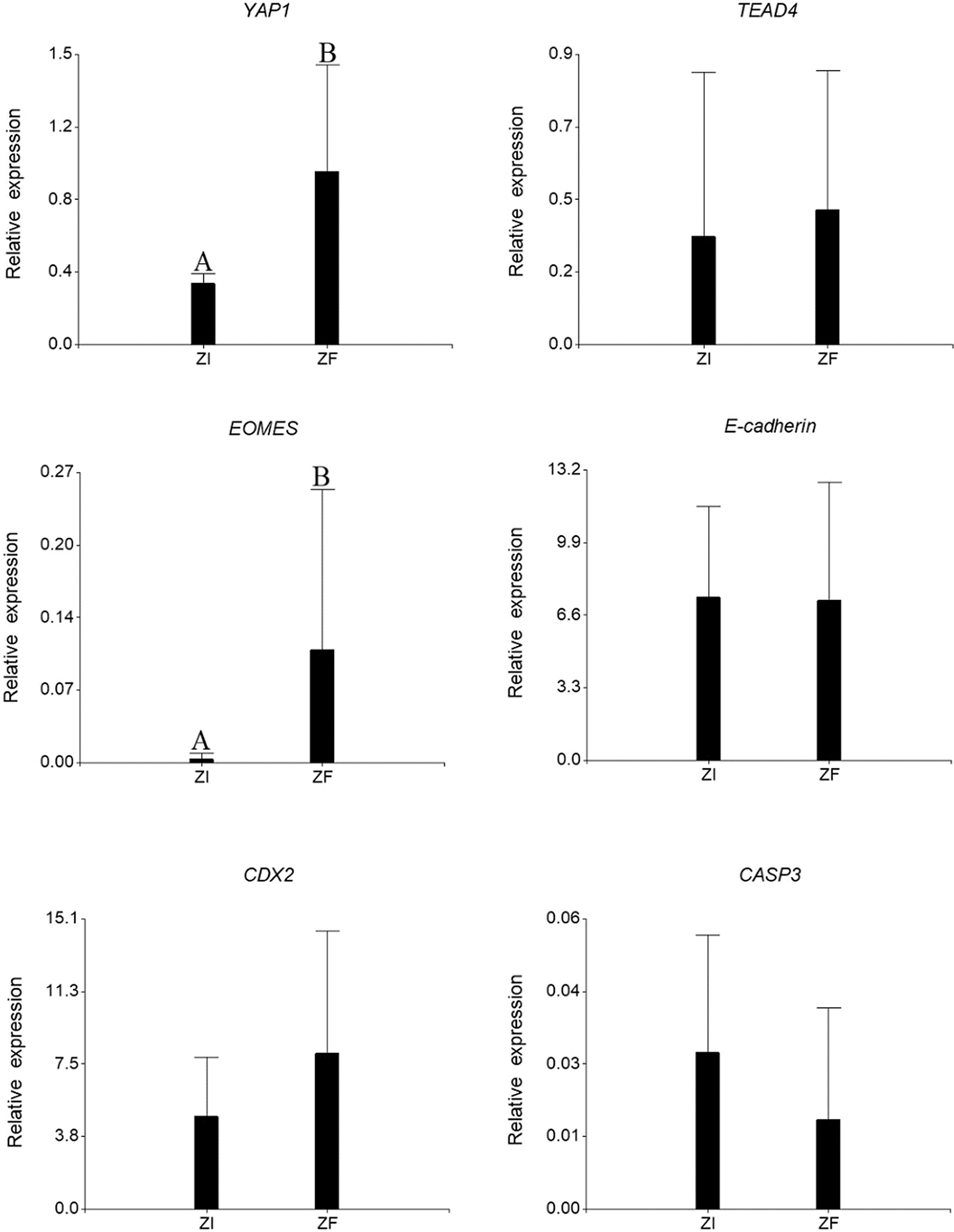
The gene expression pattern of day 7 blastocysts from the ZF and ZI groups was evaluated. Regarding the expression of trophectoderm markers. No statistical differences were observed in the relative expression of TEAD4 and CDX2 between the blastocysts from the ZF and ZI groups (P > 0.05). However, the relative expression of YAP1 and EOMES was significantly higher in the ZF blastocysts than in the ZI blastocysts (P < 0.05). No differences were found in the relative expression of the cell adhesion marker E-cadherin between both groups (P > 0.05). In addition, no statistical differences were observed in the relative expression of the pro-apoptotic marker CASP3 between ZF and ZI blastocysts (P > 0.05) (Figure 4).
Relative expression analysis (mean ± SD) of trophectoderm (YAP1, TEAD4, EOMES and CDX2), cell adhesion (E-cadherin) and apoptosis genes (CASP3) in day-7 blastocysts from the ZI and ZF groups. The housekeeping gene SDHA was used as an internal control. A,BDifferent superscripts indicate significant differences between groups (P < 0.05).

Discussion
In this study, the removal of the zona pellucida did not affect the in vitro development of domestic cat embryos. In embryos from bovine, equine and porcine the removal of the zona pellucida did not affect the in vitro development when a culture system that prevents the disaggregation of blastomeres is used (Lee et al., Reference Lee, Park, Choi, Kim, Ka and Lee2007; Rodríguez-Alvarez et al., Reference Rodríguez-Alvarez, Sharbati, Sharbati, Cox, Einspanier and Castro2010; Gambini et al., Reference Gambini, Jarazo, Olivera and Salamone2012). In the WOW culture system, embryos without zona pellucida are cultured into microwells, this ensures a stretch contact among the blastomeres allowing in vitro development (Vajta et al., Reference Vajta, Peura, Holm, Páldi, Greve, Trounson and Callesen2000). The WOW culture system has allowed the production of embryos by handmade cloning (HMC) and embryo aggregation, which has enhanced the efficiency of SCNT in different species (Vajta et al., Reference Vajta, Lewis, Trounson, Purup, Maddox-Hyttel, Schmidt, Pedersen, Greve and Callesen2003; Gambini et al., Reference Gambini, De Stefano, Bevacqua, Karlanian and Salamone2014; Moro et al., Reference Moro, Hiriart, Buemo, Jarazo, Sestelo, Veraguas, Rodriguez-Alvarez and Salamone2015b). Live births of bovine, equine and porcine have been achieved after the transfer of ZF embryos generated by SCNT or IVF (Oback et al., Reference Oback, Wiersema, Gaynor, Laible, Tucker, Oliver, Miller, Troskie, Wilson, Forsyth, Berg, Cockrem, McMillan, Tervit and Wells2003; Lagutina et al., Reference Lagutina, Lazzari and Galli2006; Gambini et al., Reference Gambini, Jarazo, Olivera and Salamone2012). However, when domestic cat embryos generated without zona pellucida have been transferred, the live births of kittens have not been achieved (Veraguas-Davila et al., Reference Veraguas-Davila, Cordero, Saez, Saez-Ruiz, Gonzalez, Saravia, Castro and Rodriguez-Alvarez2021).
We previously reported that domestic cat blastocysts cultured without a zona pellucida had a decreased relative expression of SOX2 and NANOG (Veraguas-Davila et al., Reference Veraguas-Davila, Cordero, Saez, Saez-Ruiz, Gonzalez, Saravia, Castro and Rodriguez-Alvarez2021). However, more studies are needed to prove if this downregulation of SOX2 and NANOG affects the in vivo development and implantation of domestic cat ZF blastocysts. In SOX2 knockdown embryos, the expression levels of TE associated markers TEAD4, YAP, CDX2, EOMES, FGFR2 and FGF4 were downregulated and an increase in apoptosis was observed (Keramari et al., Reference Keramari, Razavi, Ingman, Patsch, Edenhofer, Ward and Kimber2010). This demonstrated that the expression of SOX2 might be related to the establishment of the TE linage. In the present study, it was demonstrated that domestic cat blastocysts cultured without a zona pellucida had an increased expression of YAP1 and EOMES in ZF blastocysts.
It has been described that members of the Hippo pathway such as TEAD4, YAP and LATS allow the differentiation of the TE from the ICM by suppressing the expression of pluripotency genes (Nishioka et al., Reference Nishioka, Inoue, Adachi, Kiyonari, Ota, Ralston, Yabuta, Hirahara, Stephenson, Ogonuki, Makita, Kurihara, Morin-Kensicki, Nojima, Rossant, Nakao, Niwa and Sasaki2009; Wicklow et al., Reference Wicklow, Blij, Frum, Hirate, Lang, Sasaki and Ralston2014; Frum et al., Reference Frum, Watts and Ralston2019; Emura et al., Reference Emura, Saito, Miura and Sawai2020). YAP1 is a transcriptional co-activator of the Hippo pathway and, during embryo development, participates in the segregation of the ICM and TE (Wicklow et al., Reference Wicklow, Blij, Frum, Hirate, Lang, Sasaki and Ralston2014). During embryo development, the expression of SOX2 is suppressed by YAP1, WWTR1 and TEAD4 (Frum et al., Reference Frum, Watts and Ralston2019). These genes prevent a premature expression of SOX2 and restrict the expression of SOX2 to the ICM in mouse embryos (Wicklow et al., Reference Wicklow, Blij, Frum, Hirate, Lang, Sasaki and Ralston2014; Frum et al., Reference Frum, Watts and Ralston2019). In porcine embryos, the downregulation of YAP1 and LATS2 alters the expression of the pluripotency markers OCT4 and SOX2 (Emura et al., Reference Emura, Saito, Miura and Sawai2020). In embryonic stem cells (ES), the depletion of YAP1 inhibits differentiation, meanwhile its overexpression stimulates differentiation (Chung et al., Reference Chung, Lee, Uprety, Shen, Lee and Kim2016; LeBlanc et al., Reference LeBlanc, Lee, Yu, Kim, Kambhampati, Dupont, Seruggia, Ryu, Orkin and Kim2018). This demonstrates that YAP1 has a crucial role in the regulation of differentiation during the embryo development.
The downregulation of SOX2 that we previously observed in domestic cat ZF blastocysts might be related to the overexpression of YAP1 found in this research. The removal of the zona pellucida and the subsequent embryo culture in microwells might be causing an earlier differentiation of the ZF blastocysts. We have previously reported an increased diameter in domestic cat ZF blastocysts compared with ZI blastocysts (Veraguas et al., Reference Veraguas, Aguilera, Echeverry, Saez-Ruiz, Castro and Rodriguez-Alvarez2020a; Veraguas-Davila et al., Reference Veraguas-Davila, Cordero, Saez, Saez-Ruiz, Gonzalez, Saravia, Castro and Rodriguez-Alvarez2021), which was consistent with this study. This indicates a rapid growth of the domestic cat ZF-blastocyst due to the lack of restriction by the absence of zona pellucida. The WOW allows the development of domestic cat ZF embryos until the blastocyst stage, however the zona pellucida is a tridimensional structure and the microwells might not be enough to regulate the embryo development. We might theorize that the culture of domestic cat ZF embryos in microwells leads to a faster growth and differentiation in the blastocyst stage. For this reason, the overexpression of YAP1 and downregulation of SOX2 might have indicated an increase in the differentiation process in the domestic cat ZF blastocysts.
Additionally, YAP1 has a regulatory role in apoptosis during cell differentiation (LeBlanc et al., Reference LeBlanc, Lee, Yu, Kim, Kambhampati, Dupont, Seruggia, Ryu, Orkin and Kim2018). The loss of YAP1 generates an increase in apoptosis and CASP3 expression in ESCs during differentiation (LeBlanc et al., Reference LeBlanc, Lee, Yu, Kim, Kambhampati, Dupont, Seruggia, Ryu, Orkin and Kim2018). In domestic cat embryos, the in vitro culture without zona pellucida generates an increase in the relative expression of BAX at the blastocyst stage (Veraguas-Davila et al., Reference Veraguas-Davila, Cordero, Saez, Saez-Ruiz, Gonzalez, Saravia, Castro and Rodriguez-Alvarez2021). In these same ZF blastocysts, the relative expression of the anti-apoptotic gene BCL2 was not altered (Veraguas-Davila et al., Reference Veraguas-Davila, Cordero, Saez, Saez-Ruiz, Gonzalez, Saravia, Castro and Rodriguez-Alvarez2021). Despite that, in the present study, the expression of CASP3 was not affected in domestic cat ZF blastocysts. This could be related to the overexpression of YAP1 that might be regulating the expression of CASP3. According to these results, the removal of the zona pellucida does not increase the relative expression of the pro-apoptotic gene CASP3 during the in vitro culture.
Finally, in this study, the relative expression of EOMES was increased in the ZF blastocysts. The T-box gene Eomesodermin or EOMES, is essential for trophoblast development and mesoderm formation in mouse embryos (Russ et al., Reference Russ, Wattler, Colledge, Aparicio, Carlton, Pearce, Barton, Surani, Ryan, Nehls, Wilson and Evans2000). Embryos lacking EOMES arrest at the blastocyst stage and their trophectoderm cells do not differentiate into trophoblasts (Russ et al., Reference Russ, Wattler, Colledge, Aparicio, Carlton, Pearce, Barton, Surani, Ryan, Nehls, Wilson and Evans2000). EOMES expression is promoted by members of the Hippo pathway and by CDX2, and ensures trophoblast differentiation (Niwa et al., Reference Niwa, Toyooka, Shimosato, Strumpf, Takahashi, Yagi and Rossant2005; Sasaki, Reference Sasaki2010; Wu et al., Reference Wu, Gentile, Fuchikami, Sutter, Psathaki, Esteves, Araúzo-Bravo, Ortmeier, Verberk, Abe and Schöler2010). In CDX2 knockdown embryos, the expression of EOMES is downregulated (Wu et al., Reference Wu, Gentile, Fuchikami, Sutter, Psathaki, Esteves, Araúzo-Bravo, Ortmeier, Verberk, Abe and Schöler2010). In contrast, SOX2 knockdown in human ESCs results in the overexpression of EOMES (Adachi et al.,Reference Adachi, Suemori, Yasuda, Nakatsuji and Kawase2010). In the present study, the overexpression of EOMES observed in the ZF blastocysts might be caused by the reduced expression of SOX2 previously observed in these embryos (Veraguas-Davila et al., Reference Veraguas-Davila, Cordero, Saez, Saez-Ruiz, Gonzalez, Saravia, Castro and Rodriguez-Alvarez2021). Furthermore, the overexpression of EOMES could generate an accelerated differentiation of the trophectoderm cells in the domestic cat ZF blastocysts.
In conclusion, the in vitro culture of domestic cat embryos without the zona pellucida affects the gene expression of the resulting blastocysts. An overexpression of the trophectoderm markers YAP1 and EOMES was observed in the ZF blastocysts and might be related to the reduced expression of SOX2 previously reported in these embryos. More studies are needed to elucidate if this overexpression of YAP1 and EOMES is due to an accelerated differentiation process or if it is a compensatory mechanism due to the lack of zona pellucida.
Acknowledgements
We express our appreciation to the veterinary medical team Darling Ruíz Saéz, María Consuelo Álvarez and Yazmin Vasquez Araya for their help in the surgical procedures. In memoriam: The first author of this article wants to thank to Daniel Veraguas Bordoli for his help and support during this research and the past investigations. Thanks for always believing in me.
Financial support
This work was supported by ANID FONDECYT Postdoctorado 3200352, de la Agencia Nacional de Ciencia y Tecnología (ANID) del Ministerio de Ciencias de Chile.
Declaration of interest
The authors declare that there are no conflicts of interest to report.
Ethical standards
All the procedures used in this research were in accordance with the ethical standards of animal welfare and were approved by the Comité de Bioética de la Facultad de Ciencias Veterinarias de la Universidad de Concepción.