Introduction
First described in a case report by English otologist Joseph Toynbee in 1850, keratosis obturans is a clinical condition where patients classically present with acute conductive hearing loss, severe otalgia, widening of the external auditory canal and a thickened tympanic membrane.Reference Toynbee1–Reference Persaud, Hajioff, Thevasagayam, Wareing and Wright2 Currently, both the aetiology and pathogenesis of keratosis obturans is incompletely understood; clinical conditions attributed to keratosis obturans include eczema, seborrheic dermatitis, furunculosis and bronchiectasis.Reference Bhide, Kale, Pai, Tepan, Pandit and Raleraskar3–Reference Bharadwaj, Walling, Rees and Novotny5 This results in an abnormal migratory pathway, where a complete lack of epithelial migration or an abnormally slow movement of the epithelium in the external auditory canal leads to the build-up of squamous debris, resulting in keratosis obturans.Reference Michaels and Soucek6–Reference Corbridge, Michaels and Wright7 As a chronic disease process, keratosis obturans requires life-long surveillance and serial debridement to improve external auditory canal patency and provide serviceable hearing. As a result, the aim of this systematic review was to present clinical data relating to the management principles of keratosis obturans. Although the level of evidence in the included studies is low, it is important to recognise that this collection of research articles represents the current body of available otolaryngology literature regarding this important otological condition.
Materials and methods
Study protocol
This study was designed and performed using the Preferred Reporting Items for Systematic Reviews and Meta-Analyses protocol and is exempt from institutional review board approval.Reference Moher, Shamseer, Clarke, Ghersi, Liberati and Petticrew8
Search strategy
Independent searches of the PubMed (Medline), Scopus, Embase, Web of Science and Cochrane databases were performed on 7 July 2018, by the first and second authors, to identify all studies which described keratosis obturans using the Boolean method and relevant search term combinations with no language restrictions. Search components were ‘keratosis’ and ‘obturans’ and their combination.
Review process
After the medical literature was extracted, article titles, abstracts and full-text manuscripts were independently reviewed by two authors (V A Patel and J A Harounian) to identify studies for inclusion or exclusion.
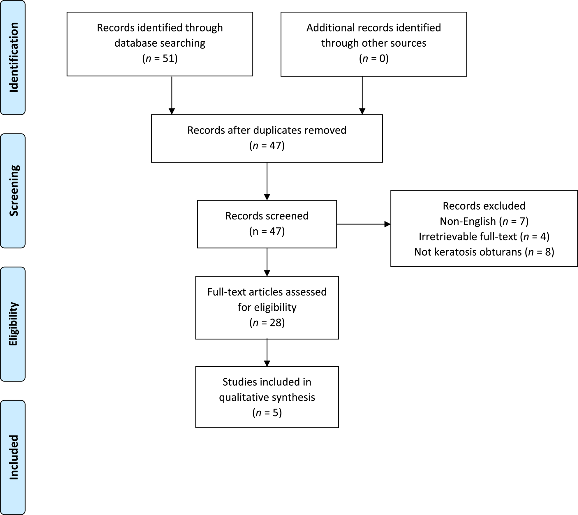
Inclusion criteria included full-length studies in English or those able to be translated into English that primarily focused on the condition of keratosis obturans. Studies that did not meet inclusion criteria were excluded. After initial review, any discrepancies between the two authors were submitted to a third author (H Isildak) for a final decision on inclusion or exclusion. Finally, full-length original articles were assessed for data extraction eligibility. Due to the heterogeneity of the study methods and measures, a meta-analysis was not deemed to be possible. A qualitative synthesis of the studies was employed to integrate study results. Similarly, data imputation was not performed. The utilised search strategy is summarised in Figure 1.
Search strategy protocol.

Results
Study characteristics
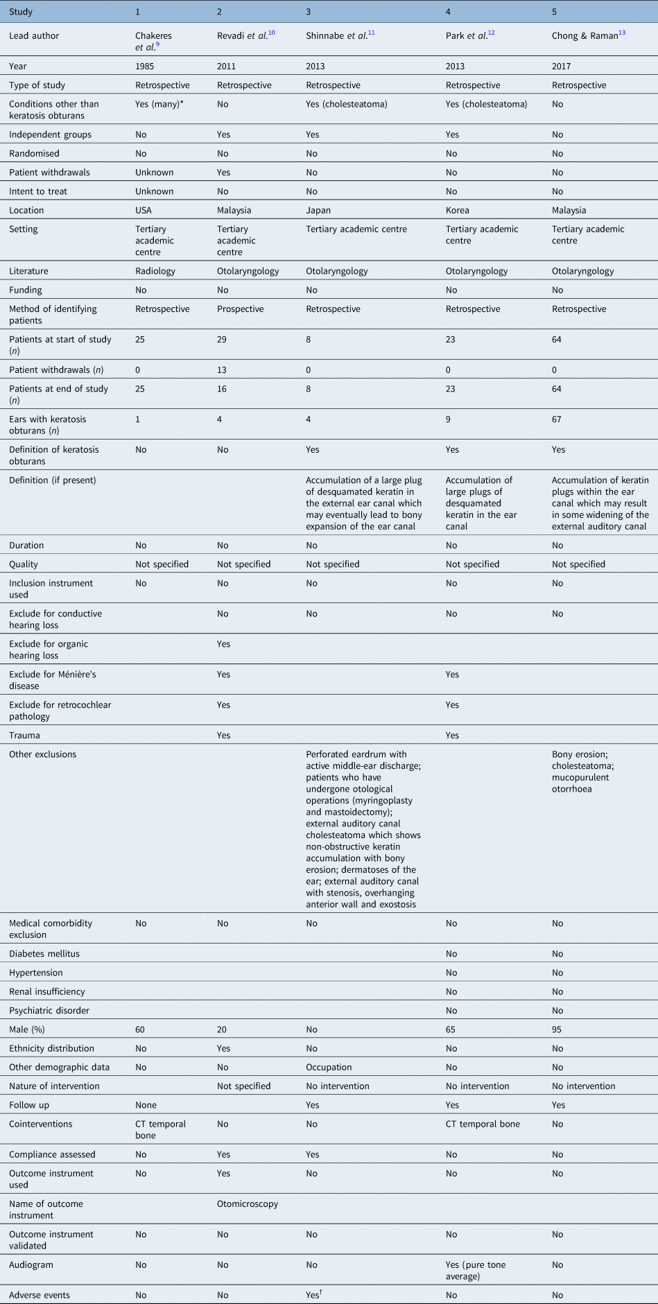
The database search yielded a total of 51 studies, 47 of which remained after duplicates were removed. Seven studies were excluded due to being written in a non-English language, four articles had full text that was irretrievable and eight articles discussed other medical conditions unrelated to keratosis obturans. Dual investigator screening of the remaining 28 abstracts and full-text articles gave 5 studies that were included in the final analysis (Figure 1). Characteristics of the collected studies are summarised in Table 1. With respect to the five studies, one was from the USA, two were from Korea, one was from Japan and one was from Malaysia.Reference Chakeres, Kapila and Lamasters9–Reference Chong and Raman13
Study data available for qualitative analysis

*Seventy-five per cent from the keratosis obturans group had also tympanic membrane perforation with middle-ear cholesteatoma; †facial nerve palsy from inflammation (n = 1). CT = computed tomography
Risk of bias
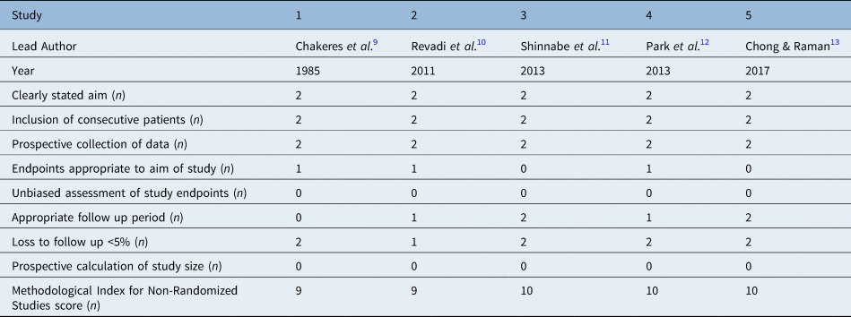
The methodological quality of each study was evaluated using the Methodological Index for Non-Randomized Studies criteria (Table 2).Reference Slim, Nini, Forestier, Kwiatkowski, Panis and Chipponi14 The Methodological Index for Non-Randomized Studies scores ranged from 9 to 10, with 16 being a perfect score. The mean and median Methodological Index for Non-Randomized Studies scores were 9.5 and 9.5, respectively. Studies with scores more than 11 were considered to have a low risk of bias. As a result, in this analysis, all included studies are known to have a risk of bias. Most studies were deficient in categories of prospective power calculation, blinded evaluations, randomisation and an overall unbiased assessment of study endpoints.
Keratosis obturans: Methodological Index for Non-Randomized Studies criteria

Patient sample
Study sample sizes ranged from 8 to 64 patients. All studies encountered in this analysis were retrospective in nature. The range of follow up periods varied greatly from 3 weeks to 3.5 years. Notably, three studies had statements of reported follow up but did not specify an exact timeline for follow up. No study included a calculated mean and standard deviation with regards to follow up periods. All studies included patients afflicted with either unilateral or bilateral keratosis obturans. Three studies included patients with conditions other than keratosis obturans in their sample size; the most common other condition was cholesteatoma. Only two studies excluded patients who were not afflicted with keratosis obturans and clearly stated so. Most studies maintained their original sample size, with only one study losing more than 5 per cent of their sample size at follow up. Finally, all studies had clearly stated aims and correct inclusion sampling. It is important to note that despite the retrospective nature of the included studies, all studies involved a prospective collection of clinical data.
Description of keratosis obturans
The definition of keratosis obturans was only present in 60 per cent of studies; when present, 3 included studies classified keratosis obturans as the accumulation of desquamated keratin plus cholesteatoma within the external auditory canal. Two of the three studies included sequelae of external auditory canal keratosis obturans in the definition, referring to widening or expansion of the bony external auditory canal. One of the studies made it a point to contrast the definition of keratosis obturans with external auditory canal cholesteatoma. The most commonly accepted distinguishing features of keratosis obturans from external auditory canal cholesteatoma may be the presence of overlying epithelium and the absence of osteonecrosis. Furthermore, Chong and Raman developed a grading system for keratosis obturans, which was otherwise unprecedented in the otolaryngology literature.Reference Chong and Raman13 Finally, three studies mentioned the use of temporal bone computed tomography to either confirm the presence or assess the extent of keratosis obturans.
Outcome reporting
Two studies (40 per cent) reported an outcome instrument to evaluate corresponding endpoints. Revadi et al. used a malleable wire or marked stillette, along with binocular microscopy and a 30-degree otoendoscope, to investigate lateral displacement of desquamated epithelium within the external auditory canal.Reference Revadi, Prepageran, Raman and Sharizal10 One study reported the use of audiometry with specific reference to pure tone audiometry thresholds to demonstrate the effect of keratosis obturans on hearing; however, the degree of hearing loss was not quantified. Rather, only qualitative statements such as ‘mild conductive hearing loss’ was cited.Reference Park, Jung and Oh12 One study noted ‘conductive hearing loss of mild to moderate severity in pure tone audiometry (10–30 dB air–bone gap)’.Reference Chong and Raman13 No studies commented on the validation of the outcome instrument utilised for evaluation of their corresponding endpoints.
Discussion
This study was the first in the otolaryngology literature to comprehensively examine the medical literature regarding treatment and rehabilitative outcomes associated with keratosis obturans and has several notable strengths.
This systematic review presents research studies according to the Preferred Reporting Items for Systematic Reviews and Meta-Analyses protocol and was conducted via well-established methodology with an assessment of eligibility and data extraction and comparison performed by two independent reviewers for each analysed study. Furthermore, a validated scale for evaluating methodological quality and risk of bias was included, which is imperative in assessing both the generalisability and reliability of the existing clinical data.
The Methodological Index for Non-Randomized Studies tool is another validated instrument that assesses the methodological quality of studies, whether comparative or non-comparative. Our study employed this instrument to determine the level of bias present in the included research studies. All studies had scores close to but lower than 11, indicating the presence of a high risk of bias. This high level of bias in all studies analysed raises a significant concern about the internal validity of the studies because most studies were deficient in categories of prospective power calculation, randomisation and standardised assessment of study endpoints. These findings demonstrated the necessity for higher-quality studies with lower levels of bias that have standardised inclusion and exclusion criteria, include sufficiency and consistent follow up, are adequately randomised and exclude confounding conditions in order to improve the external validity of the literature regarding keratosis obturans.
Clinical manifestations
Keratosis obturans is an uncommon otological condition defined by the accumulation of keratinaceous matter in an arrangement that leads to obstruction and dilation of the external auditory canal. A rather obscure entity, there is a significant paucity of literature regarding this important otorhinolaryngological disease. It is estimated to occur in 4–5 patients among 1000 new otological cases.Reference Lesser and Gleeson15 Patients typically present in the third and fourth decades with symptoms including otalgia and hearing impairment; the degree of otalgia is wide-ranging from minor ear discomfort to excruciating pain.
Although keratosis obturans has been associated with eczema, dermatitis, sinusitis or bronchiectasis, its pathogenesis remains unclear.Reference Morrison16 Morrison proposed overproduction and defective migration of epithelial cells resulting in plug formation.Reference Morrison16 Over time, this lesion grows circumferentially and widens the external auditory canal, without evidence of osteonecrosis or bony destruction as classically described in cholesteatoma. In this analysis, there was a large variation present in inclusion and exclusion criteria, demographics of selected patients, study sample and presence of keratosis obturans definition in addition to methods utilised regarding outcome measurement, which makes it particularly difficult to generalise findings and conclusions reached in the existing literature.
Diagnosis
As it stands currently, keratosis obturans is a clinical diagnosis and does little to lend itself to more objective modalities. Prior authors have noted that keratosis obturans is often only confirmed when multiple attempts at removal of accumulated desquamated keratin elicit excruciating pain and a silvery-white matrix at the periphery of the plug is able to be visualised.Reference Chong and Raman13
Radiographic imaging
Computed tomography, compared to plain X-ray films or magnetic resonance imaging, was used in half the studies, presumably due to its superiority in evaluating osseous temporal bone anatomy. Classically, computed tomography shows a well-defined soft tissue density within the external auditory canal associated with dilation sans erosion of the external auditory canal. At times, the diagnosis of keratosis obturans may be challenging to discriminate between an external canal cholesteatoma or even cerumen impaction. Although there is no substitute for direct visualisation and examination of the auricle on both physical examination or binocular microscopy, computed tomography was mentioned in 50 per cent of included studies and appears to play a supplementary role in the diagnosis and assessment of keratosis obturans. No mention was made in the included studies as to the reason why imaging was employed in certain studies compared with others. As a result, computed tomography may certainly play a role and aid in clarifying the final diagnosis. The possibility of middle-ear cholesteatoma coexisting with keratosis obturans, albeit rare, must also not be ignored. Shinnabe et al. reported the presence of tympanic membrane perforations with middle-ear cholesteatoma in three patients with keratosis obturans.Reference Shinnabe, Hara, Hasegawa, Matsuzawa, Kanazawa and Yoshida11
Treatment modalities
Keratosis obturans management was largely procedural or surgical in this analysis. All patients underwent removal of the keratin plug in order to reduce the burden of disease. Otic drops consisting of olive oil, hydrogen peroxide, corticosteroids and bicarbonate have been previously described in the otolaryngology literature but only in the context of a misdiagnosis of cerumen impaction or to soften external auditory canal obstruction in preparation for subsequent procedural removal.Reference Tod17
This systematic review shows significant heterogeneity within prior research efforts regarding keratosis obturans. For example, 50 per cent of studies included did not specify an exact timeline for follow up, whereas the other half reported a wide range from 3 weeks to 3 months, with a maximum period of 3.5 years. Furthermore, several studies included patients with other otological diseases, introducing significant confounding variables to the subsequent analysis. As a result, heterogeneity of patient demographic data as well as inclusion and exclusion criteria significantly limits the ability to reliably compare patients across studies as far as clinical outcomes are concerned. Of the four patients with keratosis obturans in a retrospective case review by Shinnabe et al., one patient underwent conservative cleaning, and the remaining three patients underwent type I tympanoplasty with no ossicular damage noted intra-operatively. No complications were reported, and no recurrence was noted at the six month follow up.Reference Shinnabe, Hara, Hasegawa, Matsuzawa, Kanazawa and Yoshida11
Limitations
This study certainly possesses several notable limitations. No randomised controlled trials were included in this analysis as none were currently available in the medical literature. Moreover, much of the data extracted from the included studies were qualitative in nature and thus obviated the ability for rigorous statistical analysis or meta-analysis. During the process of data extraction, incongruities were reviewed and resolved by the senior author (H Isildak); however, the senior author was exclusively independent of the two primary reviewers. Finally, only studies employing the English language were included, which introduces the possibility of bias in the findings reached.
Conclusion
Although keratosis obturans is an uncommon clinical entity, principles on how to best manage patients who proceed to surgery remain controversial. This systematic review highlights a lack of published evidence relating to keratosis obturans. However, it appears that treatment of keratosis obturans is safe and efficacious with identifiable practice patterns. Future studies with larger sample sizes will be necessary to better understand the indications and long-term treatment outcomes for keratosis obturans.
Acknowledgements
The authors would like to thank Kimberly Walker, Division of Outcomes Research and Quality at Pennsylvania State University, for formatting the manuscript.
Competing interests
None declared