




No CrossRef data available.
Article contents
The amphidial neuron pair ALD controls the temperature-sensitive choice of alternative developmental pathways in the parasitic nematode, Strongyloides stercoralis
Published online by Cambridge University Press: 18 November 2004
Abstract
The parasitic nematode Strongyloides stercoralis, has several alternative developmental pathways. Upon exiting the host (humans, other primates and dogs) in faeces, 1st-stage larvae (L1) can enter the direct pathway, in which they moult twice to reach the infective 3rd-stage. Alternatively, if they enter the indirect pathway, they moult 4 times and become free-living adults. The choice of route depends, in part, on environmental cues. In this investigation it was shown that at temperatures below 34 °C the larvae enter the indirect pathway and develop to free-living adulthood. Conversely, at temperatures approaching body temperature (34 °C and above), that are unfavorable for the survival of free-living stages, larvae develop directly to infectivity. The time-period within the L1's development during which temperature influenced the choice of the pathway depended on the temperature, but, at any given temperature, occurred approximately in the middle of the time-span spent in the L1 stage, which varied inversely with temperature. This critical period was associated with the time-interval in which the number of cells in the genital primordium began to increase, thus providing a morphological marker for the pathway decision in individual worms. Sensing the environment is the function of the amphidial neurons, and therefore we examined the role of individual amphidial neurons in controlling entry into the direct pathway to infectivity. The temperature-sensitive developmental switch is controlled by the neuron pair ALD (which also controls thermotaxis), as seen by the loss of control when these neurons are ablated. Thus, in S. stercoralis a single amphidial neuron pair controls both developmental and behavioural functions.
Information
- Type
- Research Article
- Information
- Copyright
- © 2004 Cambridge University Press
INTRODUCTION
Strongyloides stercoralis, an intestinal nematode parasite of dogs and primates, including humans, is virtually unique because it can multiply both within its host as well as in the external environment by following alternative developmental pathways. The 1st-stage larvae (L1) entering the environment in the host's faeces can develop directly to the infective 3rd-stage larvae (L3i) or, alternatively, to free-living adult male and female worms. Several environmental factors, including temperature (Premvati, 1958; Nwaorgu, 1983; Viney, 1996), food (Moncol & Triantaphyllou, 1978; Minematsu, Tanaka & Tada, 1989) and immune status of the host (Gemmill, Viney & Read, 1997) influence the choice of developmental pathways in other members of the genus Strongyloides. The main sensilla by which nematodes recognize environmental cues are the amphids, paired cilia-containing sensory organs at the anterior tip of the worm. The amphidial neurons of S. stercoralis have been physically mapped from the tips of their dendritic processes in the amphid per se to their cell bodies in the more posteriorly positioned paired lateral ganglia (Ashton et al. 1995). The functions of several of these amphidial neurons are known from laser microbeam ablation studies (Ashton et al. 1998; Lopez et al. 2000a) and others are inferred by homology with Caenorhabditis elegans (Ashton, Li & Schad, 1999). For example, in C. elegans the choice of developmental pathways is controlled by the amphidial neuron pairs ADF and ASI. These neurons sense the species' population density via dauer pheromone and respond to control the direction of worm development (Albert, Brown & Riddle, 1981; Bargmann & Horvitz, 1991). In S. stercoralis the homologous neuron pairs (ASF and ASI) also control the direction of development (Ashton et al. 1998).
Here we report that the free-living L1 of S. stercoralis can respond to potentially adverse environmental temperatures by changing the direction of its development from the indirect pathway, involving a generation of free-living adults, to a direct pathway producing the environmentally resistant, longer-lived L3i (the infective stage of this parasite). Furthermore, we show that the neurons that mediate this decision are the thermosensitive pair, ALD. Lopez et al. (2000a) have shown that the neurons of the class ALD (Amphidial neuron: Lamellar dendrite, cell body “D”) control thermotaxis in S. stercoralis and, therefore, probably serve as thermoreceptors, similar to the possibly homologous neuron pair AFD in C. elegans (Mori & Ohshima, 1995).
MATERIALS AND METHODS
Strongyloides stercoralis
The strain was isolated from, and has been maintained in, dogs since 1982 (Schad, Hellman & Muncey, 1984). The 1st-stage larvae (L1) were obtained from the fresh faeces of an infected dog using a modified Baermann apparatus. L1 were grown to adults or 3rd-stage larvae (L3) in organ culture dishes (60 mm×15 mm, BD, Franklin Lakes, NJ) containing 1 ml of 1% agarose (Type I-A, Sigma Chemical Co., St Louis, MO) in the centre well, with saline in the outer moat. A small amount (about 5 mg) of dog faeces placed at the centre of the agarose base served as a food source for the larvae (Ashton et al. 1998).
Effect of temperature on development
Pairs of organ culture plates harboring L1 were incubated in larger Petri dishes lined with a moist paper towel to provide high humidity. These were then placed in environmental chambers (Percival, Boone, Iowa) maintained at the desired constant temperature. Normally 10 L1 were placed on each plate, although in a few experiments 20 to 200 L1 were used. Two to five plates were incubated at each test temperature each time an experiment was replicated. In each replicate a set of plates was also incubated at 26 °C to ensure that the particular batch of worms were developing ‘normally’. In studies to determine the critical time during incubation at which temperature could shift the developmental pathway, plates were moved between environmental chambers set at different temperatures. At 24 h after the worms were placed on culture plates, living worms were recovered, identified as to stage (L3i, adult, or undeveloped), and counted. The percentage survival was calculated by dividing the number of worms recovered (any stage of development) by the number of worms originally placed onto the plate and multiplying by 100. The percentage developing to adulthood was obtained by dividing the number of adults recovered from a plate by the total number of developed worms (L3i and adults) recovered from that plate multiplied by 100.
Morphological changes in the L1 during development
The morphological changes which take place within the genital primordium (GR) of the L1 grown at 26 °C were described by Lopez, Nolan & Schad (2000b). To determine the time-course of the L1's GR changes at 37 °C, we placed freshly collected L1 at 37 °C and examined 10 anaesthetized worms at each of the following times: 0,0·5,1,1·5,2·5 and 3·5 h. The number of cells in the GR of each worm was recorded, as was the presence or absence of a cuticular sheath (i.e. moulting or not moulting).
Laser microbeam ablation of neuronal cell bodies
L1 were anaesthetized and ablations of the amphidial cell body pairs were performed as described by Ashton et al. (1998), with the exception that the uppermost cell body of the laterally positioned worm was given 20 laser pulses and the lower cell body was given 30 pulses. Cell bodies were located using differential interference contrast microscopy and a previously developed cell map (Ashton et al. 1998). Either the ALD pair (the thermoreceptor neuron pair) or the ASK pair (not known to have any developmental function in C. elegans (Bargmann & Mori, 1997), and presumed not to have any in S. stercoralis – used as a control in this study) were targeted in each worm. The worms in which both of the target-pair were not clearly visible, were destroyed by using the laser to cut a hole in the cuticle. Operated L1 were placed on agar plates (10–25 per plate) with normal gerbil faeces and incubated at 36 °C for 22 to 24 h. Thereafter, worms were recovered and the stage of development for each was determined. Control plates with worms that had not undergone surgery (10–40 per plate) were also placed at 36 °C. As in the temperature experiments described above, a set of plates with L1 that had not undergone surgery was incubated at 26 °C (10–40 worms per plate) to confirm normal development for each batch of L1.
Several ALD-ablated worms that, as predicted, developed to the adult stage at 36 °C, were examined using a transmission electron microscope (Ashton et al. 1995) and their amphidial structure was compared with normal adults that had developed at 26 °C.
Statistical analysis
Data were analysed using logistic regression and negative binomial regression. Initially, a reference situation was identified (e.g. sex, temperature, ablation status), and logistic regression of outcome (e.g. survival) by treatment groups was preformed. To confirm the robustness of any statistical differences thus obtained, the analysis was repeated using negative binomial regression, thereby testing and validating the assumptions of observations independence. All statistical analysis was performed using STATA 8 and, in addition to the above regression modules, data tabulation, data summarization, and graphical procedures were used in conjunction with data screening. Results were considered to be a consequence of experimental control (treatments) when their probability of having arisen by chance was less than 0·05.
RESULTS
Developmental response to environmental temperature
When freshly isolated L1 of S. stercoralis were cultured at temperatures ranging from 15 °C to 31 °C, the majority of worms developed to the free-living adult stage (range 94·1 to 100% of developed worms, Fig. 1A). When compared with development at 26 °C (the standard culture temperature), there was no statistical difference (P>0·05) between any of these temperatures (15 °C to 31 °C) with respect to the developmental pathway chosen. Worms at 15 °C were slow to develop and were counted at 3 days, rather than after 1 day as was the case at other temperatures. Survival at temperatures between 17 °C and 31 °C was high (88·9%) and varied very little, however, at 15 °C it was poor, with only 42·5% of the worms still alive on day 3. At 34 °C and above, the larvae tended to develop directly to L3i. Thus, the percentage of adults recovered at 1 day decreased with increasing temperature (above 31 °C, Fig. 1A). Survival also decreased with increasing temperature (Fig. 1B). The percentage of adults recovered at 34 °C to 40 °C was significantly less than that seen at 26 °C (P<0·001 in all cases) and significantly fewer worms survived at these temperatures as compared to 26 °C. At 40 °C, only 3·8% of the worms survived and all survivors developed directly to the L3i. Larval density (from 10 to 400 L1 per culture plate) had no effect on the choice of developmental pathway at a given temperature. When 200 to 400 L1 were placed on plates at 26 °C, 91·3% developed to adults, compared to 94·9% when 10–40 L1 were placed on identical plates.

Fig. 1. (A) The developmental pathway of Strongyloides stercoralis as a function of temperature. Female 1st-stage larvae having emerged from a host in the faeces, may develop either to the free-living adult stage or to an infectious 3rd-stage larva (L3). Male larvae can only develop to adulthood. The graph shows the mean percentage of the population that developed to adulthood at a given temperature, worms not developing by this pathway developed to the infective L3 stage. (B) Survival of environmental stages of S. stercoralis as a function of temperature. The graph shows the mean percentage of living worms recovered, after 24 h at the given temperature, from plates seeded with L1.
While the percentage of L1 developing to adult females decreased with increasing temperature above 31 °C, the percentage developing to adult males did not differ significantly over the range of temperatures examined (26 °C to 39 °C). For example, while 21·6% of the worms recovered at 26 °C were males and only 9·3% of the worms recovered at 39 °C were males, these percentages were not significantly different (P=0·15; 95% confidence interval: 0·10, 1·42). The percentage of males recovered ranged from 26·6% to 9·3% within this temperature range, with the higher percentages being found at temperatures of 34 °C and below.
Period during which environmental temperature governs the choice of developmental pathways
At 26 °C 2–3 h were required to set the developmental pathway of L1 entering the external environment in the faeces (Fig. 2A). If they were moved to 36 °C at any time before this, they behaved as if they had never experienced the lower temperature. For example, L1 kept at 26 °C for 2 h before being moved to 36 °C had significantly fewer adults develop compared to L1 kept at 26 °C for the entire 24 h (Odds Ratio=0·0487, P=<0·001, 95% confidence interval=0·0163, 0·145). Conversely, if they were moved to the higher temperature at 3 h (or later), the L1 developed as if they had never experienced the higher temperature (L1 moved at 4 h developed to a similar percentage of adults as L1 kept at 26 °C for the entire 24 h (Odds Ratio=1·95, P=0·55, 95% confidence interval=0·21, 17·89)). When the L1 were initially placed at a high temperature (36 °C), they set their developmental pathway by 1·5 h (Fig. 2B): L1 moved at 1·5 h were not significantly different from L1 kept at 36 °C for 24 h (Odds Ratio=2·1, P=0·19, 95% confidence interval=0·69, 6·4), while L1 kept at 36 °C for only 1 h differed significantly from L1 kept at 36 °C for 24 h (Odds Ratio=14·6, P=<0·001, 95% confidence interval=6·9, 30·9).

Fig. 2. The initial temperature at which L1 are raised determines the future course of development. (A) The effect of incubation time at 26 °C on the direction of development. The graph shows the mean percentage of worms developing as adults after spending various amounts of time at 26 °C before being moved to 36 °C. (B) The effect of incubation time at 36 °C on the direction of development. The graph shows the mean percentage of worms developing as adults after spending various amounts of time at 36 °C before being moved to 26 °C.
The number of cells in the genital primordium (GP) can be used as an indicator of development when within-stage morphological change is minimal (Lopez et al. 2000b). At 26 °C there is an initial 3 h period in which the cell number in the GP of recently collected L1 does not change. This is followed by a doubling over the next 2 h (Lopez et al. 2000b). In sharp contrast, when L1 recently isolated from faeces were placed at 37 °C, the number of cells in the GP began increasing after just 1 h (Fig. 3). The cell number at 0 and 1 h is not significantly different (P=0·18, two-tailed t-test) while the number at 1 h is significantly different from that seen at 1·5 h (P=0·03, two-tailed t-test). The time-points at which the L1 makes its developmental decision at both 26 °C and 36 °C, therefore, correspond to the beginning of morphological development, as indicated by the increase in the number of cells in the GP.

Fig. 3. The development of the genital primordium (GR) in the L1 at 37 °C. Freshly isolated L1 were incubated at 37 °C for various times. Thereafter, the number of cells in their GR was determined. Each point represents the mean of 10 larvae. Error bars indicate 1 standard deviation from the mean.
Ablation of the amphidial thermosensitive neuron pair (ALD) changes the direction of development
When the neuron pair ALD was ablated in freshly isolated L1 and the larvae were then incubated at 36 °C, 75·8% developed to adulthood (Fig. 4) rather than to L3i as would be expected in normal, non-operated, worms. This differed significantly from the developmental choice made when control neurons were killed or when normal control worms were tested. In these cases larvae developed directly to infectivity with only 47·1% of ASK ablated worms developing to adults (Odds Ratio: 0·45, 95% confidence interval: 0·25, 0·80, P=0·007 vs ALD-ablated). Survival rates of ALD-ablated and ASK-ablated larvae were no different from the survival of normal L1 placed at 36 °C. In the case of ALD ablation, 56 of 85 worms (65·9%) survived 24 h at 36 °C. Similarly, 62 of 80 ASK ablated worms (77·5%) survived and 90 of 120 normal (no ablation) larvae (75%) survived 24 h at 36 °C. Normal L1 (no ablation) from each experiment, placed at 26 °C, developed as expected (an average of 98% were adults at 24 h). By 24 h ALD-ablated worms showed a loss of the lamellae constituting the dendritic processes of these temperature-sensitive neurons (Fig. 5).

Fig. 4. Effect of ablation of the neuron pair ALD on the direction of development at 36 °C. Neither neuron pair ALD (known thermosensitive neurons) nor neuron pair ASK (ablation control) were ablated in freshly isolated L1 with a laser micro-beam. Non-operated L1 from the same isolation were placed directly at either 36 °C or 26 °C as normal controls. Compared to the ASK-ablated controls, significantly fewer larvae developed to the L3i stage when ALD was ablated (Odds Ratio: 0·45; P=0·007, 95% confidence interval: 0·25, 0·80). There was no significant difference in the percentage of larvae developing to L3i between the ASK ablation controls and 36 °C normal non-operated controls.

Fig. 5. Effect of laser micro-beam ablation of the cell body of the neuron ALD on the ultrastructure of its dendritic processes. The neuron pair ALD was ablated in the L1 and the worms were then incubated for 24 h at 36 °C. The resulting adult worms were recovered, fixed and sectioned for transmission electron microscopy. Normal adult worms (grown at 26 °C) were also examined. The section on the right, from an ALD-ablated adult worm, shows that the terminal lamellae of the complex dendrite of the ALD neuron are missing although, such visible damage to the ablated neuron is not always apparent by 24 h. The section on the left is from a normal adult worm; the lamellae of ALD are evident.
DISCUSSION
Several species of Strongyloides make temperature-dependent developmental choices. S. fulleborni grown at 25 °C develops to free-living adulthood exclusively, but, when grown at either 15 °C or 37 °C, a small percentage develop to the infective-stage (L3i) (Premvati, 1958). Similarly, S. papillosus produces a greater percentage of L3i at 20 °C than at either 15 °C or 35 °C (Nwaorgu, 1983), whereas, S. ratti produces the greatest percentage of L3i at 13 °C and the least at 30 °C (Viney, 1996). In contrast, S. stercoralis produces an increasing percentage of L3i at temperatures above 34 °C than at lower temperatures ranging to 15 °C. This pattern is similar to that shown by Caenorhabditis elegans. This free-living nematode produces a greater percentage of dauer L3, (homologous to parasitic infective larvae, Hotez, Hawdon & Schad, 1993), at higher than lower temperatures (Riddle, 1988). Thus both species tend to develop to environmentally resistant forms at higher temperatures. Survival of S. stercoralis is minimal at temperatures of 40 °C and above and at those below 15 °C.
The percentage of worms developing to adult males did not change with temperature and was not expected to. Sex in S. stercoralis is determined by the number of chromosomes the embryo possesses (Hammond & Robinson, 1994), and since all L3i must be female (with 6 chromosomes), only female L1 can chose between the two developmental options. Nwaorgu (1983), working with S. papillosus, and Premvati (1958), working with S. fulleborni, both showed that the percentage of larvae developing to adult males was unaffected by temperature. Although there was no significant difference in the percentage of worms developing to adult males at any temperature, the percentage of adult males at 36 °C and 39 °C was much less than at lower temperatures. This might indicate that, in general, male larvae do not survive long enough to develop to adulthood at these high temperatures.
Nwaorgu (1983), by switching developing cultures of S. papillosus from 20 °C to 30 °C (and vice versa), showed that the decision to develop to either L3i or free-living adulthood was made during the L1 stage. With S. stercoralis we also demonstrated that the decision is made during L1 development, and have narrowed the critical period to the time during which morphological change in the gonad begins (approximately the midpoint in the development of the L1 outside the host). This provides a morphological marker by which we can reasonably determine if an L1 has made a developmental choice. Once its developmental pathway was set, moving a larva to a different temperature failed to affect the stage into which the larva would subsequently develop. Temperature regulation of dauer formation in C. elegans also requires that the exposure to the higher temperature be approximately at the time of the L1 moult (Golden & Riddle, 1984).
Development of S. stercoralis L1 to free-living adulthood (as seen in worms grown at temperatures below 34 °C) appears to be the default pathway when the direction of development is controlled by temperature. Destruction of the thermoreceptive neuron pair (ALD) prevented S. stercoralis L1 placed at 36 °C from developing via the alternative pathway to the L3i stage. Several explanations can be offered for why only 75·8% of the ALD-ablated worms developed to the adult stage. Because the developmental decision is made during a very brief time-interval, some L1 undergoing laser surgery near the end of each experiment may have already made their decision. Furthermore, successful ablation of each neuron of the targeted pair could not always be confirmed at the time of the laser surgery, consequently some of the worms may have had only one of the two neuronal cell bodies sufficiently damaged to result in its death. In thermotaxis experiments (Lopez et al. 2000a), S. stercoralis larvae with only one of the pair of ALD neurons surviving could still behave normally. Therefore, we assume that larvae with one functional ALD neuron could still make a temperature-influenced developmental decision. In contrast, however, in C. elegans, mutants defective in thermotaxis remained responsive to dauer-inducing pheromone in a temperature-dependent manner (Golden & Riddle, 1984). Temperature is not the only factor involved in S. stercoralis' choice between developmental pathways; at all temperatures below 34 °C, a small percentage of larvae will develop to the L3i stage, apparently basing their decision on other factors.
Based on the results presented here and those of Ashton et al. (1998) there are two time-points in the development of the S. stercoralis L1 at which amphidial neurons control the choice of developmental routes. In young L1, still in the small intestine of the host, if the amphidial neurons ASF and ASI are intact, and sense a normal environment, the L1 is prevented from developing to the L3a, the precocious autoinfective stage. In C. elegans the analogous neurons (ADF and ASI) are sensitive to the dauer pheromone that inactivates these neurons, directing the worms into the dauer pathway. Similarly, the death of these neurons by laser ablation also directs the L1 into the dauer development (Bargmann & Horvitz, 1991). Based on these observations one might speculate that in heavy S. stercoralis infections a pheromone, increased corticosteroid levels, or some component of the immunological status of the host might inactivate neurons ASF and ASI leading to direct internal development of the larvae (autoinfection), a potentially fatal developmental alternative for the infected host.
The temperature-driven development to the L3i (mediated by the ALD neurons) that occurs at host-like temperatures, may also be an important determinant of autoinfection in S. stercoralis. Normally, L1 that hatch from eggs laid in the small intestine of the host pass out with the faeces at a very young, ‘undeveloped’ stage. This can be seen in data concerning the cell-number in the genital primordium (Lopez et al. 2000b) of larvae recently expelled from the host in faeces. As our donor animals normally defaecate only twice a day, the larval population in the faeces must include diverse ages, with possibly up to a 12 h difference in age represented. However, the cell number in the genital primordium of all the L1 recovered from fresh canine faeces remains constant for about 3 h after the larvae have been recovered. If the factors that control this ‘stasis’ within the host deteriorate, the L1 might initiate development in the intestinal lumen. Since these L1 are still in the host at a temperature exceeding 34 °C, female larvae would develop to the autoinfective L3a stage. Male L1 would have no choice and would develop to free-living adults. This may explain the often questioned and generally rejected observation of Kreis (1932) and Faust (1933) of seeing adult male worms with the morphology of the free-living male in dogs. In rare cases we have seen adult males, with the morphology of free-living males, in the colon of gerbils with autoinfective disease (data not published).
If S. stercoralis L1 emerge from the host and experience a high external temperature, as sensed by the neuron pair ALD, they will tend to develop to L3i. This will increase the population ready for re-invasion. However, if the temperature is low, a generation of adult worms will be produced, increasing the worm population's life in time and space and creating an opportunity to invade new immunologically naive hosts, thus increasing the parasite's potential for survival. However, environmental temperature would not be the only determinate of the developmental pathway chosen by the L1. Immunological factors of the host may have pre-set the pathway (Gemmill et al. 1997), possibly through the ASF and ASI neurons, or the availability of food (Moncol & Triantaphyllou, 1978; Minematsu et al. 1989) during the first hours after emergence from the host may influence the choice of pathways.
This project was supported by NIH Grants RO1 A1 22662 to G. A. Schad and RR02512 to Dr Mark Haskins.
References
REFERENCES
ALBERT, P. S.,
BROWN, S. J. &
RIDDLE, D. L. (
1981).
Sensory control of dauer larva formation in Caenorhabditis elegans.
Journal of Comparative Neurology
198,
435–451.CrossRefGoogle Scholar
ASHTON, F. T.,
BHOPALE, V. M.,
FINE, A. E. &
SCHAD, G. A. (
1995).
Sensory neuroanatomy of a skin-penetrating nematode parasite: Strongyloides stercoralis. I. Amphidial neurons.
Journal of Comparative Neurology
357,
281–295.Google Scholar
ASHTON, F. T.,
BHOPALE, V. M.,
HOLT, D.,
SMITH, G. &
SCHAD, G. A. (
1998).
Developmental switching in the parasitic nematode Strongyloides stercoralis is controlled by the ASF and ASI amphidial neurons.
Journal of Parasitology
84,
691–695.CrossRefGoogle Scholar
ASHTON, F. T.,
LI, J. &
SCHAD, G. A. (
1999).
Chemo- and thermosensory neurons: structure and function in animal parasitic nematodes.
Veterinary Parasitology
84,
297–316.CrossRefGoogle Scholar
BARGMANN, C. &
HORVITZ, H. R. (
1991).
Control of larval development by chemosensory neurons in Caenorhabditis elegans.
Science
251,
1243–1246.CrossRefGoogle Scholar
BARGMANN, C. &
MORI, H. (
1997).
Chemotaxis and thermotaxis. In
C. elegans II
(ed. Riddle, D. L., Blumenthal, T., Meyer, B. J. & Preiss, J. R.), pp.
717–738.
Cold Spring Harbor Press,
Cold Spring Harbor, USA.
FAUST, E. C. (
1933).
Experimental studies on human and primate species of Strongyloides. II. The development of Strongyloides in the experimental host.
The American Journal of Hygiene
18,
114–132.CrossRefGoogle Scholar
GEMMILL, A. W.,
VINEY, M. E. &
READ, A. F. (
1997).
Host immune status determines sexuality in a parasitic nematode.
Evolution
51,
393–401.CrossRefGoogle Scholar
GOLDEN, J. W. &
RIDDLE, D. L. (
1984).
The Caenorhabditis elegans dauer larva: developmental effects of pheromone, food, and temperature.
Developmental Biology
102,
368–378.CrossRefGoogle Scholar
HAMMMOND, M. P. &
ROBINSON, R. D. (
1994).
Chromosome complement, gametogenesis and development of Strongyloides stercoralis.
Journal of Parasitology
80,
689–695.CrossRefGoogle Scholar
HOTEZ, P.,
HAWDON, J. &
SCHAD, G. A. (
1993).
Hookworm larval infectivity, arrest and amphiparatenesis: the Caenorhabditis elegans Daf-c paradigm.
Parasitology Today
9,
23–26.CrossRefGoogle Scholar
KREIS, H. A. (
1932).
Studies on the genus Strongyloides (Nematodes).
The American Journal of Hygiene
16,
450–491.CrossRefGoogle Scholar
LOPEZ, P. M.,
BOSTON, R.,
ASHTON, F. T. &
SCHAD, G. A. (
2000 a).
The neurons of class ALD mediate thermotaxis in the parasitic nematode, Strongyloides stercoralis.
International Journal for Parasitology
30,
1115–1121.Google Scholar
LOPEZ, P. M.,
NOLAN, T. J. &
SCHAD, G. A. (
2000 b).
Growth of the genital primordium as a marker to describe a time course for the heterogonic larval development in Strongyloides stercoralis.
Journal of Parasitology
86,
882–883.Google Scholar
MINEMATSU, M. M. T.,
TANAKA, M. &
TADA, I. (
1989).
The effect of fatty acids on the developmental direction of Strongyloides ratti first-stage larvae.
Journal of Helminthology
63,
102–106.CrossRefGoogle Scholar
MONCOL, D. J. &
TRIANTAPHYLLOU, A. C. (
1978).
Strongyloides ransomi: factors influencing the in vitro development of the free-living generation.
Journal of Parasitology
64,
220–225.CrossRefGoogle Scholar
MORI, I. &
OHSHIMA, Y. (
1995).
Neural regulation of thermotaxis in Caenorhabditis elegans.
Nature, London
376,
344–347.CrossRefGoogle Scholar
NWAORGU, O. C. (
1983).
The development of the free-living stages of Strongyloides papillosus. I. Effect of temperature on the development of heterogonic and homogonic nematodes in faecal culture.
Veterinary Parasitology
13,
213–223.CrossRefGoogle Scholar
PREMVATI (
1958).
Studies on Strongyloides of Primates. II. Factors determining the “direct” and the “indirect” mode of life.
Canadian Journal of Zoology
36,
185–195.Google Scholar
RIDDLE, D. L. (
1988).
The dauer larva. In
The Nematode Caenorhabditis elegans
(ed. Wood, W. B.), pp.
393–412.
Cold Spring Harbor Laboratory Press,
New York.
SCHAD, G. A.,
HELLMAN, M. E. &
MUNCEY, D. W. (
1984).
Strongyloides stercoralis: hyperinfection in immunosuppressed dogs.
Experimental Parasitology
57,
287–296.CrossRefGoogle Scholar
VINEY, M. E. (
1996).
Developmental switching in the parasitic nematode Strongyloides ratti.
Proceedings of the Royal Society of London, B
263,
201–208.CrossRefGoogle Scholar

Fig. 1. (A) The developmental pathway of Strongyloides stercoralis as a function of temperature. Female 1st-stage larvae having emerged from a host in the faeces, may develop either to the free-living adult stage or to an infectious 3rd-stage larva (L3). Male larvae can only develop to adulthood. The graph shows the mean percentage of the population that developed to adulthood at a given temperature, worms not developing by this pathway developed to the infective L3 stage. (B) Survival of environmental stages of S. stercoralis as a function of temperature. The graph shows the mean percentage of living worms recovered, after 24 h at the given temperature, from plates seeded with L1.

Fig. 2. The initial temperature at which L1 are raised determines the future course of development. (A) The effect of incubation time at 26 °C on the direction of development. The graph shows the mean percentage of worms developing as adults after spending various amounts of time at 26 °C before being moved to 36 °C. (B) The effect of incubation time at 36 °C on the direction of development. The graph shows the mean percentage of worms developing as adults after spending various amounts of time at 36 °C before being moved to 26 °C.

Fig. 3. The development of the genital primordium (GR) in the L1 at 37 °C. Freshly isolated L1 were incubated at 37 °C for various times. Thereafter, the number of cells in their GR was determined. Each point represents the mean of 10 larvae. Error bars indicate 1 standard deviation from the mean.

Fig. 4. Effect of ablation of the neuron pair ALD on the direction of development at 36 °C. Neither neuron pair ALD (known thermosensitive neurons) nor neuron pair ASK (ablation control) were ablated in freshly isolated L1 with a laser micro-beam. Non-operated L1 from the same isolation were placed directly at either 36 °C or 26 °C as normal controls. Compared to the ASK-ablated controls, significantly fewer larvae developed to the L3i stage when ALD was ablated (Odds Ratio: 0·45; P=0·007, 95% confidence interval: 0·25, 0·80). There was no significant difference in the percentage of larvae developing to L3i between the ASK ablation controls and 36 °C normal non-operated controls.
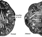
Fig. 5. Effect of laser micro-beam ablation of the cell body of the neuron ALD on the ultrastructure of its dendritic processes. The neuron pair ALD was ablated in the L1 and the worms were then incubated for 24 h at 36 °C. The resulting adult worms were recovered, fixed and sectioned for transmission electron microscopy. Normal adult worms (grown at 26 °C) were also examined. The section on the right, from an ALD-ablated adult worm, shows that the terminal lamellae of the complex dendrite of the ALD neuron are missing although, such visible damage to the ablated neuron is not always apparent by 24 h. The section on the left is from a normal adult worm; the lamellae of ALD are evident.
 You have
Access
- 29
- Cited by