INTRODUCTION
The order Leptostraca (Crustacea: Malacostraca: Phyllocarida) is characterized by the presence of a movable rostrum, six pairs of pleopods, striking uropods, and a bivalved carapace that covers eight pairs of thoracopods and partially covers the lateral sides of seven pleonites. Recently, the order Leptostraca includes 60 extant species (including subspecies) that have been classified into 11 genera and four families. Among them, a great part of species (36 species) are included in the genus Nebalia Leach, Reference Leach1814 (Mees, Reference Mees2015).
In this paper, we report a new species of Nebalia collected from Cambodia: Nebalia cambodiana sp. nov. Prior to this study, no leptostracan species had been recorded from South-east Asia, including Cambodia. Therefore, N. cambodiana is the first species of Leptostraca reported from South-east Asia, and the fifth from the Asian continent, along with N. dahli Kazmi & Tirmizi, Reference Kazmi and Tirmizi1989, reported from Pakistan; N. mortoni Lee & Bamber, Reference Lee and Bamber2011, from Hong Kong; N. koreana Song et al. Reference Song and Min2012, N. pseudotroncosoi Song et al. Reference Song, Moreira and Min2013 and N. dolsandoensis Song & Min, Reference Song, Moreira and Min2016, from South Korea. This species inhabits very clean oligotrophic sand and gravel sediments similar to N. daytoni Vetter, Reference Vetter1996.
Here, we provide a detailed description of the diagnostic characteristics of the new species including illustrations.
MATERIALS AND METHODS
Sample collection
A single specimen was collected from a diving deck (Koh Rong Samloem Island, Cambodia), using light traps in 2012 and preserved in 95% ethyl alcohol. The substrate at the sampling site was fine sand and gravel within an oligotrophic environment. The holotype of Nebalia cambodiana sp. nov. was deposited in the National Institute of Biological Resources (NIBR), Incheon, Korea.
Morphological analysis
The specimen was observed and dissected under a stereomicroscope (Model SZX-7; Olympus, Tokyo, Japan). Illustrations of dissected appendages were made with a drawing tube connected to a light microscope (Model DM 2500; Leica, X50–630, Wetzlar, Germany). Drawings of the entire body were made using a drawing tube attached to a stereomicroscope (Olympus SZX-12). Measurements of the appendages and whole body were taken using a stage micrometer (Leica, Germany) and an ocular micrometer.
Total length (TL) was measured from the articulation between the rostrum and carapace to the posterior end of the uropods, excluding setation. Dorsal carapace length (DCL) was measured as the distance between the articulation of the rostrum and the margin of the posterodorsal cleft. Lateral carapace length (LCL) was measured as the distance along the lateral surface of the carapace, between the anterior-most and posterior-most margins. Carapace height (CH) was measured between the dorsal and ventral margins. Rostrum length (RL) was measured along the midline.
RESULTS
SYSTEMATICS
ORDER LEPTOSTRACA Claus, Reference Claus1880
Family NEBALIIDAE Samouelle, Reference Samouelle1819
Genus Nebalia Leach, Reference Leach1814
Nebalia cambodiana sp. nov.
(Figures 1–4)
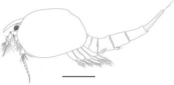
Nebalia cambodiana sp. nov., female holotype, lateral view. Scale bar: 1 mm.

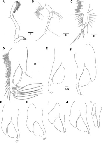
Nebalia cambodiana sp. nov., adult female holotype: (A), rostrum, dorsal view; (B), eye, lateral view; (C), antennule, lateral view; (D), antenna, lateral view; (E), antenna, third article, external side, lateral view. Scale bars: 0.1 mm.

Nebalia cambodiana sp. nov., adult female holotype: (A), mandibular palp; (B), first maxilla (palp somewhat broken); (C), second maxilla; (D), thoracopod 1, showing setation; (E), thoracopod 2; (F), thoracopod 3; (G), thoracopod 4; (H), thoracopod 5; (I), thoracopod 6; (J), thoracopod 7; (K), thoracopod 8. Scale bars: 0.1 mm.

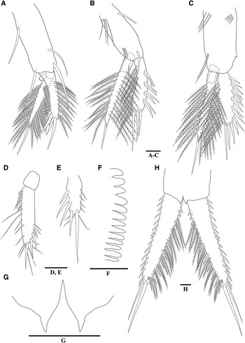
Nebalia cambodiana sp. nov., adult female holotype: (A), pleopod 1, lateral view; (B), pleopod 2, lateral view; (C) pleopod 4, lateral view; (D), pleopod 5, ventral view; (E), pleopod 6, ventral view; (F), pleonites 5–7, posterior dorsal margin, denticles; (G), anal plates; (H) uropods, ventral view. Scale bars: 0.1 mm.

TYPE LOCALITY
Koh Rong Samloem Island (10°37′23″N 103°17′42″E), Thai Bay, Cambodia, depth of ~3–5 m.
TYPE MATERIAL
Holotype (NIBRIV0000331463): Adult female (Figure 1), RL 0.8 mm, DCL 1.4 mm, LCL 2.2 mm, CH 1.5 mm, TL 5.3 mm. Collected on 4 November 2012, by T. Park & Y. Eun.
DIAGNOSIS OF FEMALE HOLOTYPE
Carapace small compared to whole body, covering lateral sides of pleonite 2 and part of pleonite 3. Rostrum very long, length nearly 3.5 times width. Eye with one distinct dorsal papilla and four small lobes on distal margin. Antennular flagellum shorter than peduncle. Antennular scale sub-elliptical, 2 times longer than wide. Only one distal robust seta arising from fourth antennular article. Denticles along pleonites 5–7 posterior dorsal margins distally rounded. Pleopod 4 protopod with even posterior margin, lacking serrations; posterolateral corner with small projection. Anal plates with distinct lateral ‘shoulder’. Uropods short, about 0.8 times as long as pleonite 7 and anal somite combined.
DESCRIPTION OF FEMALE HOLOTYPE (NIBRIV0000331463)
Carapace (Figure 1) oval, covering lateral sides of pleonite 2, and some of lateral sides of pleonite 3. LCL 2.2 mm; about 1.5 times longer than high.
Rostrum (Figures 1 & 2A) very long, clearly extending beyond distal margin of eyestalk; about 3.5 times as long as wide; with more or less parallel margins.
Compound eye (Figure 2B) oval, with one distinct dorsal papilla and four small lobes on distal margin; ommatidial part extending about three-fifths of total length of eyestalk, dark red pigmentation present on almost entire ommatidial part after fixation.
Antennule (Figure 2C) peduncle composed of four articles. First article stout, shorter than eyestalk. Second article with (1) one plumose seta on anterior margin of proximal fourth; (2) cluster of seven plumose setae on lateral surface; and (3) subterminal cluster of about 11 short and long simple setae and one thick seta. Third article shorter than the second, widest distally, with cluster of about six simple setae on anterior distal margin, three simple setae on posterior margin, one long thick seta on posterior distal margin. Fourth article shorter than third, with (1) seven simple setae on anterior margin; (2) one thick robust seta distally; (3) lateral row of four simple setae; and (4) one long simple seta on posterior distal margin, longer than antennular scale. Antennular scale elliptical, 2 times as long as wide, with (1) row of five simple setae along medial surface; and (2) numerous simple setae of different lengths. Flagellum well-developed, with nine articles, shorter than peduncle; each article with (1) 3–4 aesthetascs on anterodistal margin; (2) 1–2 simple setae arising from anterodistal margin, one much longer than the other, oriented backwards.
Antenna (Figure 2D) peduncle composed of three articles. First article without acute process. Second article slightly rectangular, with distal large acute process. Third article longer than second, with different rows of setae along medial anterior margin (Figure 2E): (1) proximal row of about 4–5 simple setae and one long plumose seta on inner surface, (2) seven spines along proximal half, the distalmost being the longest, (3) seven simple setae each associated with spines of (2), (4) 10 simple setae thinner than those of (3), (5) five plumose setae and three spines, and (6) terminal row of six spines, increasing distally in length, the distalmost next to four simple setae; one long plumose seta on posterior margin; cluster of plumose setae along distal inner margin. Flagellum well-developed, composed of 10 articles; each article with four terminal setae, one shorter and thinner than the others, oriented posteriorly.
Mandibular palp (Figure 3A) composed of three articles; second article about 1.5 times longer than first with two setae: (1) one at mid-length on lateral side; (2) other subterminally on anterior margin. Third article slightly shorter than second, with margins slightly parallel; proximal half of anterior margin with row of setae shorter than others of palp; posterior margin with three types of setae: (1) row of setae arising from ending of proximal third extending to proximal half, distally plumose; (2) row of longer setae than those of (1) extending along distal half, distally plumose; and (3) about six curved setae on distal margin.
First maxilla (Figure 3B) proximal endite with rounded margin bearing distally plumose setae. Distal endite larger than proximal endite; medial margin with one plumose seta and two types of setae: (1) eight setae of increasing size with three notable teeth along distal posterior margin also provided with several smaller teeth; (2) six spatulate setae. Palp well-developed, with proximal cluster of four setae and three widely spaced setae, numbering possibly about 7–8 along its whole length; apex strongly curved distally.
Second maxilla (Figure 3C) protopod composed of four endites with plumose setae, endites 1 and 3 being the largest nearly subequal in size; endites 2 and 4 smaller than both 1 and 3. Fourth endite the smallest, with four plumose setae. Endopod composed of two articles, longer than exopod, first article 1.4 times as long as second one; lateral and medial margins with numerous plumose setae; second article with three terminal setae distally. Exopod as long as first article of endopod; medial margin with numerous plumose setae, two distalmost setae slightly shorter than exopod.
Thoracopods (Figure 3D–K): endopod longer than exopod; with numerous plumose setae along anterior margin. Distal article of endopod distinctly expanded, slightly oriented backwards, with about 7–8 long plumose setae, with recurved apex. Exopod with 5–17 plumose setae along posterior margin. Thoracopod 8 epipod noticeably smaller than those of thoracopods 1–7.
Pleonites (Figure 4F) 5–7 with denticles along posterior dorsal margin; denticles with somewhat parallel sides, distally rounded.
Pleopod 1 (Figure 4A) composed of protopod, endopod and exopod. Protopod with posterior margin lacking serrations; three basal simple setae: (1) one long seta arising subdistally; (2) one long, thicker seta near the base of endopod; and (3) longer seta near the base of exopod, almost reaching the distal end of exopod spine-row; posterior margin with one long simple seta on proximal third. Endopod composed of two articles, proximal article shorter, with appendix interna with three short recurved hooks. Distal article longer than exopod; lateral and medial margins of second article each with about 24–26 plumose setae, distal margin with acute projection at apex and one long, stout terminal seta. Exopod about 0.7 times length of protopod, with row of about 11–12 serrate spines along lateral margin (‘spine-row’), each spine with tiny tridentate tip, central tooth bifid; four stout spines on distolateral margin, two long alternating with other two short spines; row of long plumose setae along medial margin.
Pleopod 2 (Figure 4B) protopod with (1) one simple thick setae along posterior margin on proximal third, (2) four simple setae on anterior proximal margin, (3) three long setae subdistally near the base of endopod, and (4) one slightly thick seta near the base of exopod; posterior margin even. Endopod slightly longer than exopod, composed of two articles; distal article, lateral and medial margins of distal article each with about 22–24 plumose setae, ending in one long robust seta. Exopod with a row of four pairs of long thick setae, each pair composed of one long and one relatively shorter seta; medial margin with row of long plumose setae, three distal stout spines, increasing distally in length.
Pleopod 3 similar to pleopod 2.
Pleopod 4 (Figure 4C) protopod with three simple setae along anterior margin on proximal third; two long setae subdistally near the base of endopod; posterior margin lacking serrations, posterolateral corner with small projection. Setation and pattern of spine pairs on margins of endopod and exopod nearly same as pleopods 2–3.
Pleopods 5–6 (Figure 4D, E): fifth pleopod uniramous, composed of two articles; second article long, about 3.9 times as long as wide, with four stout spines along distolateral and terminal margin, increasing distally in length; 22 simple setae along medial and distal margin, setae appearing more or less jointed at midlength. Sixth pleopod uniramous, composed of one article, with five stout spines along distolateral and terminal margin, increasing distally in length. Medial and terminal margins with ‘jointed’ setae similar to those of fifth pleopod.
Anal somite, anal plates and uropod (Figures 1 & 4G): anal somite short, slightly longer than pleonite 7. Anal plates with distinctly lateral ‘shoulder’. Uropods short, about 0.8 times as long as pleonite 7 and anal somite combined. Uropods (Figure 4H) with about 15–17 robust setae along lateral margin increasing distally in length, about 9–11 simple setae and 15–17 plumose setae on lateral inner margin, cluster of three short spine-like setae on distolateral inner border.
HABITAT
This species was collected from a depth of ~3–5 m, on sediment composed of fine sand and gravel within an oligotrophic environment.
DISTRIBUTION
Nebalia cambodiana sp. nov. is only known from the type locality.
ETYMOLOGY
The species name ‘cambodiana’ is derived from the name of the country where the specimen was collected.
REMARKS
Nebalia cambodiana sp. nov. differs from previously reported species of Nebalia in the following unique combination of characteristics: rostrum very long, length nearly 3.5 times width; a compound eye with one distinct dorsal papilla and four small lobes on distal margin; an antennular flagellum with 9 articles; the fourth article of the antennular peduncle has only one distal thick seta; the antennular scale is elliptical, with a length that is 2 times the width; exopod of the second maxilla is the same as first article of its endopod; pleonites 5 to 7 have rounded denticles along their posterior dorsal margins; the protopod of pleopod 4 lacks serration along the posterior margin; and anal plates bear a distinct lateral ‘shoulder’.
Among the aforementioned characteristics, the shape of the compound eye is the most defining characteristic in the genus Nebalia because eye morphology tends to be relatively conservative, with most species having an eye that is slightly dilated and not subdivided or lobed (Haney et al. Reference Haney, Hessler and Martin2001). On the contrary, five species of Nebalia including the new species described here, have unusual eye morphology: N. daytoni Vetter, Reference Vetter1996 has an eye with a flat anterior margin bordered by a superior and inferior protuberance; N. schizophthalma Haney et al. Reference Haney, Hessler and Martin2001 has an eye that is distinctly invaginated distally; N. troncosoi Moreira et al. Reference Moreira, Cacabelos and Domínguez2003 and N. pseudotroncosoi Song et al. Reference Song, Moreira and Min2013 possess two to three small distal lobes on the compound eye; and N. cambodiana sp. nov. has a compound eye with one distinct dorsal papilla and four small lobes on distal margin.
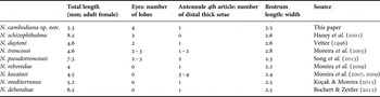
Nebalia cambodiana sp. nov. is most similar to N. daytoni Vetter, Reference Vetter1996, with respect to external morphology. The new species, however, differs from N. daytoni in (1) the relative length to width ratio of the rostrum and (2) the number of lobes on the compound eye. In N. daytoni, the rostrum is relatively short with a length nearly 2.6 times its width and the compound eye is distally bilobed. In the new species, the rostrum is distinctively very long (nearly 3.5 times as long as wide); and the compound eye has one distinct dorsal papilla and four small lobes on distal margin. Also, the new species is similar to N. kocatasi Moreira et al. Reference Moreira, Koçak and Katagan2007, N. mediterranea Koçak & Moreira, Reference Koçak and Moreira2015, N. reboredae Moreira & Urgorri, 2009 and N. deborahae Bochert & Zettler, Reference Bochert and Zettler2012. However, they are different from the new species in some diagnostic characteristics (Table 1).
Comparison based on selected diagnostic characteristics among N. cambodiana sp. nov. and related species (females).

ACKNOWLEDGEMENT
We thank Ye Eun (NIBR) for collection of the type specimen.
FINANCIAL SUPPORT
This work was supported by a grant from the National Institute of Biological Resources (NIBR) funded by the Ministry of Environment (MOE) of the Republic of Korea (NIBR201201002, NIBR201601111), and a grant from Inha University. It was also supported by the programme on Management of Marine Organisms causing Ecological Disturbance and Harmful Effects, funded by KIMST/MOF.