Introduction
Aquaporins are transmembrane proteins that facilitate water transport and play an essential role in most water-transporting tissues, including the eye, lung and kidney.Reference Beitz, Golldack, Rothert and von Bülow1 The protein and messenger RNA (mRNA) of numerous types of aquaporins have been reported to be expressed in the inner ear of experimental animals.Reference Sawada, Takeda, Kitano, Takeuchi, Kakigi and Azuma2–Reference Dong, Kim, Kim and Yeo6 Notably, both the protein and mRNA of aquaporin 2 and vasopressin type 2 receptor, expressed mainly in the cochlea and endolymphatic sac, are hypothesised to be involved in inner-ear endolymph regulation.Reference Dong, Kim, Kim and Yeo6–Reference Teggi, Carpini and Zagato8
As there are histological similarities between the cell types and their disposition in the epithelia of the renal collecting duct and the inner-ear endolymphatic sac,Reference Dahlmann and von Düring9 the function of the endolymphatic sac is often compared with that of the kidney. Previous studies demonstrated that the reabsorption of around 20 per cent of water by the renal collecting duct, regulated by arginine vasopressin type 2 receptor stimulation, results in aquaporin 2 translocation to the luminal membrane via cyclic adenosine monophosphate (cAMP) signalling, thus resulting in increased water reabsorption.Reference Bichet10,Reference Conner, Bill and Conner11 However, in the endolymphatic sac, arginine vasopressin induces aquaporin 2 translocation to the basolateral epithelial membrane in a contrasting manner to the kidney,Reference Maekawa, Kitahara, Kizawa, Okazaki, Kamakura and Horii12 suggesting that arginine vasopressin exhibits differing effects in the inner ear and kidney.
The water homeostasis of inner-ear fluid has been confirmed to be regulated using the arginine vasopressin–aquaporin 2 system in a rat model,Reference Takeda, Takeda, Kitano, Okada and Kakigi13,Reference Takeda, Sawada, Takeda, Kitano, Suzuki and Kakigi14 and the protein or mRNA of aquaporin 2 and vasopressin type 2 receptor has been reported to be expressed in human endolymphatic sac tissues.Reference Couloigner, Berrebl, Teixeira, Paris, Florentin and Grayeli15,Reference Taguchi, Takeda, Kakigi, Takumida, Nishioka and Kitano16 However, the specific physiological role of arginine vasopressin–aquaporin 2 in the human endolymphatic sac is yet to be fully elucidated. Given the difficulty in obtaining endolymphatic sac tissue samples containing well-preserved epithelial cells, functional studies on the human endolymphatic sac remain insufficient. Notably, the distribution and subcellular localisation of vasopressin type 2 receptor and aquaporin 2 expression in the epithelium of the endolymphatic sac are yet to be fully understood. Therefore, the present study aimed to determine the expression levels of aquaporin 2 and vasopressin type 2 receptor in epithelial cells of the endolymphatic sac, using well-preserved samples of human endolymphatic sac collected during acoustic neurinoma removal using the translabyrinthine approach.
Materials and methods
Ethics statement
The present study was approved by the medical ethics committee of the Second Xiangya Hospital (approval number: S525), and informed consent was obtained from all participants according to the national legislation and the Declaration of Helsinki. All patients provided informed consent for collection of their endolymphatic sac tissue samples.
Tissue preparation
Sac specimens were obtained from patients with acoustic neurinoma. Fresh tissue samples of the human endolymphatic sac were collected during acoustic neurinoma surgery using the translabyrinthine approach. Samples included the intraosseous and extraosseous parts of endolymphatic sac tissue, with the exception of the endolymphatic duct and part of the endolymphatic sac located on the sigmoid sinus. A total of 10 endolymphatic sac specimens from 10 patients were collected between January 2019 and June 2020 at our university hospital.
Immunohistochemistry
Tissues were fixed with 4 per cent paraformaldehyde solution for 12 hours at room temperature and transferred sequentially to 50, 70, 95 and 100 per cent ethanol baths (1 hour in each bath) for dehydration. Samples were infiltrated using molten paraffin wax in the oven for 1 hour before being embedded into paraffin wax blocks. Paraffin embedded endolymphatic sac tissues were cut into 5 μm sections for subsequent immunohistochemical analysis.
The slides were deparaffinised and rehydrated in 3 per cent hydrogen peroxide for 5 minutes at room temperature. Following antigen retrieval using 1 per cent sodium dodecyl sulphate in phosphate buffered saline for 10 minutes, the sections were blocked in 10 per cent normal horse serum (Abcam, Cambridge, UK) in phosphate buffered saline for 1 hour, and then incubated with primary antibodies (100 μl; 1:400) against aquaporin 2 (catalogue number 108 AB233742, mouse monoclonal antibody (clone 7H8B5); Abcam) and vasopressin type 2 receptor (catalogue number 110 AB273021, rabbit monoclonal antibody (EPR24555-59); Abcam) in 0.1 per cent Triton–phosphate buffered saline overnight at room temperature. Negative control samples were processed simultaneously in an identical manner, with Triton–phosphate buffered saline replacing the primary antibody.
The slides were incubated with horseradish peroxidase conjugated secondary antibody (at 1:200; Beyotime Institute of Biotechnology, Haimen, China) for 1 hour at room temperature, and then washed three times in phosphate buffered saline. The samples were subsequently incubated in diaminobenzidine solution (at 1:50; FujiFilm Wako Pure Chemical, Osaka, Japan) for visualisation.
After final wash steps using phosphate buffered saline, the samples were dehydrated and mounted with cover glasses. The positive immunoreactivity localised at the plasma membrane of the epithelial cells was evaluated.
Immunofluorescence double labelling
For immunofluorescence staining, the immunohistochemical procedure was followed as previously described; however, following incubation with the primary antibodies, the samples were instead incubated with the Invitrogen™ fluorescent secondary antibodies (at 1:400, catalogue number A-11034; Thermo Fisher Scientific, Waltham, Massachusetts, USA) for 2 hours at 37°C. After washing with 0.1 M phosphate buffered saline, the slides were mounted using antifading medium with DAPI (4′,6-diamidino-2-phenylindole).
Confocal images were acquired with a laser scanning confocal microscope (Leica TCS SP5; Leica Microsystems, Wetzlar, Germany) using 561 nm and 633 nm lasers for excitation, and a 63× magnification oil immersion objective lens (1.4 numerical aperture; Leica Microsystems). Confocal microscopy analysis was used to demonstrate subcellular localisation in the membrane, cytoplasm and nucleus for the expression of aquaporin 2 and vasopressin type 2 receptor. Some sections were stained with haematoxylin and eosin, and observed under a light microscope for morphological study.
Results
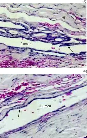
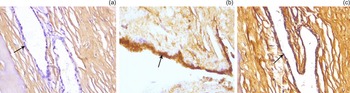
Using a light microscope, the results of the haematoxylin and eosin staining demonstrated that the morphology of all specimens was well preserved, with an intact folded epithelium lining in the lumen of the intraosseous part of the endolymphatic sac (Figure 1a) and a simple cuboidal epithelium lining in the lumen of the extraosseous part of the endolymphatic sac (Figure 1b). Immunoreactivity to antibodies against aquaporin 2 and vasopressin type 2 receptor was present in all 10 samples. Moreover, immunoreactivity was not observed in sections incubated with phosphate buffered saline instead of the primary antibodies (Figure 2a).
An intact folded epithelial lining in the lumen of the (a) intraosseous part of the endolymphatic sac (arrow) (H&E; scale bar 200 μm), and a simple cuboidal epithelium lining in the lumen of the (b) extraosseous part of the endolymphatic sac (arrow) (H&E; scale bar 100 μm).

(a) Immunoreactivity was not observed in the epithelium (arrow) in sections incubated with phosphate buffered saline instead of primary antibodies on immunohistochemical analysis (scale bar 200 μm). (b) Aquaporin 2 (scale bar 100 μm) and (c) vasopressin type 2 proteins were expressed in the epithelial layer of the endolymphatic sac (arrows), but were not observed in the connective tissue surrounding the endolymphatic sac (scale bar 200 μm).

Representative images of the immunohistochemical staining of aquaporin 2 and vasopressin type 2 receptor are presented in Figure 2. Aquaporin 2 and vasopressin type 2 receptor proteins were positively expressed in the epithelial layer of the endolymphatic sac, but were not observed in the connective tissue around the endolymphatic sac (Figures 2b and c).
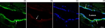
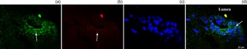
Confocal microscopy analysis demonstrated that vasopressin type 2 receptor labelling was present in both the apical and basolateral plasma membranes, and in the cytoplasm of the endolymphatic sac epithelium. Moreover, aquaporin 2 was strongly expressed at the basolateral site of the endolymphatic sac epithelium in both extraosseous (Figure 3) and intraosseous (Figure 4) parts of the endolymphatic sac.
A strong immunofluorescence signal was visualised for (a) aquaporin 2 (green) at the basolateral site of the endolymphatic sac epithelium (arrow) (scale bar 20 μm), and a moderate immunofluorescence signal was visualised for (b) vasopressin type 2 (red) in the apical and basolateral plasma membranes, and the cytoplasm of the epithelial cells (arrow) (scale bar 20 μm). (c) The nuclei were stained blue in the extraosseous part of the endolymphatic sac using DAPI (scale bar 20 μm). (d) Co-localisation of aquaporin 2 and vasopressin type 2 (blue) was mainly observed at the basolateral site of endolymphatic sac epithelium (arrows) (scale bar 20 μm).

A strong immunofluorescence signal was visualised for (a) aquaporin 2 (green) at the basolateral site of the endolymphatic sac epithelium (arrow) (scale bar 20 μm), and a moderate immunofluorescence signal was visualised for (b) vasopressin type 2 (red) in the apical and basolateral plasma membranes, and the cytoplasm of the epithelial cells (arrow) (scale bar 20 μm). (c) The nuclei were stained blue in the intraosseous part of the endolymphatic sac using DAPI (scale bar 20 μm). (d) Co-localisation of aquaporin 2 and vasopressin type 2 (blue) was mainly observed at the basolateral site of the endolymphatic sac epithelium (arrow) (scale bar 20 μm).

Discussion
Systemic osmotic pressure is maintained by vasopressin-mediated feedback regulation of body fluid. In collecting ducts of the kidney, vasopressin induces the expression of aquaporin 2, resulting in increased water reabsorption.Reference Bichet10,Reference Conner, Bill and Conner11 In the inner ear of rodents, water homeostasis of the inner-ear fluid was confirmed to be regulated using the vasopressin–aquaporin 2 system.Reference Sawada, Takeda, Kitano, Takeuchi, Kakigi and Azuma2,Reference Maekawa, Kitahara, Kizawa, Okazaki, Kamakura and Horii12–Reference Takeda, Sawada, Takeda, Kitano, Suzuki and Kakigi14 The present study showed expression of aquaporin 2 and vasopressin type 2 receptor proteins along the epithelial lining of the endolymphatic sac. Notably, this was found in both the intraosseous and extraosseous parts of the human endolymphatic sac, but was not observed in the connective tissue around the sac, which is consistent with the results of previous studies.Reference Couloigner, Berrebl, Teixeira, Paris, Florentin and Grayeli15,Reference Taguchi, Takeda, Kakigi, Takumida, Nishioka and Kitano16 The same distribution pattern has also been reported in the kidney from the cortex through the inner medulla,Reference Nielsen, Frøkiær, Marples, Kwon, Agre and Knepper17,Reference Cohen18 where a large amount of water is filtrated and reabsorbed. The water channel composed of aquaporin 2 is mediated by arginine vasopressin, and its driving force is the osmotic gradient. Although the potential role of aquaporin and vasopressin type 2 receptor in kidney function remains elusive, the expression levels of aquaporin 2 and vasopressin type 2 receptor in the endolymphatic sac epithelium highlight the active transport of water, similar to that in the kidney.Reference Taguchi, Takeda, Kakigi, Takumida, Nishioka and Kitano16
Administration of arginine vasopressin or cholera toxin, a protein that increases the intracellular concentration of cAMP, the second messenger of arginine vasopressin–vasopressin type 2 receptor, has been shown to induce endolymphatic hydrops in guinea pigs.Reference Takeda, Takeda, Kitano, Okada and Kakigi13,Reference Kumagami, Loewenheim, Beitz, Wild, Schwartz and Yamashita19 In the same species, vasopressin type 2 receptor antagonists were confirmed to decrease cochlear- and endolymphatic sac-associated aquaporin 2 mRNA, and to reduce the endolymphatic hydrops observed following endolymphatic sac obliteration.Reference Takeda, Sawada, Takeda, Kitano, Suzuki and Kakigi14 Based on the longitudinal theory of endolymph flow or absorption, it was previously hypothesised that aquaporin 2 mediated the reabsorption of endolymph via the transepithelial transport of water from the apical to the basolateral plasma membrane in the inner ear.Reference Kakigi, Nishimura, Takeda, Taguchi and Nishioka20 However, using the endolymphatic sac obtained from rats, Kumagami et al.Reference Kumagami, Loewenheim, Beitz, Wild, Schwartz and Yamashita19 demonstrated that arginine vasopressin mediated an inhibition of membrane turnover in the endolymphatic sac epithelium, compared with the stimulation found in the kidney. These authors also demonstrated a decrease in the exposure of aquaporin 2 water channels to the surface membrane, suggesting decreased water reabsorption in the endolymphatic sac caused by arginine vasopressin, which may in turn cause endolymphatic hydrops. Using homogenised endolymphatic sac tissue culture, Maekawa et al.Reference Maekawa, Kitahara, Kizawa, Okazaki, Kamakura and Horii12 demonstrated that aquaporin-2-like immunoreactivity was translocated from the luminal to the basolateral side with endosomal trapping in the endolymphatic sac during arginine vasopressin exposure, suggesting that the vasopressin type 2 receptor–aquaporin 2 activation and endosomal trapping of aquaporin 2 may alter the luminal membrane's permeability to water and reduce the endolymph efflux. This may result in a restriction of water movement from the sac lumen across the epithelium.
By contrast, the results of the present study demonstrated that, at a steady state, aquaporin 2 was strongly expressed at the basolateral site of the epithelium in both the intraosseous and extraosseous parts of the endolymphatic sac, highlighting that the suggested function of aquaporin 2 in the endolymphatic sac epithelium may differ from that in the kidney. The change in distribution and subcellular localisation of aquaporin 2 in the endolymphatic sac epithelium also supports a contrasting effect of arginine vasopressin between the endolymphatic sac and the kidney.Reference Maekawa, Kitahara, Kizawa, Okazaki, Kamakura and Horii12,Reference Kumagami, Loewenheim, Beitz, Wild, Schwartz and Yamashita19
In addition, results from a previous study demonstrated that aquaporin 2 was present in the ribosomal-rich cells performing secretory functions in the endolymphatic sac.Reference Kumagami, Loewenheim, Beitz, Wild, Schwartz and Yamashita19 The luminal (apical) epithelium is in contact with endolymph, and the basolateral membrane is in contact with connective tissue and blood vessels in the endolymphatic sac. Thus, we hypothesised that a high expression level of aquaporin 2 at the basolateral site of the epithelial cells in the endolymphatic sac may be involved in the secretion of endolymph. This may maintain the homeostasis of endolymph by generating a transepithelial water movement from the connective tissue and blood vessels to the endolymphatic space in a contrasting manner in the kidney. Therefore, the development of endolymphatic hydrops following the administration of arginine vasopressin may occur because of the activation of vasopressin type 2 receptor–aquaporin 2, resulting in an increasing influx of water to the endolymphatic space, while inhibition results in a reduced influx of water to the endolymphatic space.
Interestingly, a clinical study reported that the plasma levels of arginine vasopressin were significantly higher in patients suffering from Ménière's disease.Reference Aoki, Asai, Nishihori, Mizuta, Ito and Ando21,Reference Kitahara, Doi, Maekawa, Kizawa, Horii and Kubo22 As a result, the vasopressin type 2 receptor antagonist has been proposed to be a promising drug in the treatment of Ménière's disease.Reference Takeda and Taguchi23 However, specific interaction between vasopressin type 2 receptor–aquaporin 2 and other channels is crucial for maintaining normal homeostasis of the inner-ear fluid.Reference Asmar, Gaboury and Saliba24 Further studies are required to better understand the inner-ear fluid homeostasis and the pathogenesis of diseases caused by its disturbance.
However, limitations exist in the present study. Theoretically, the use of endolymphatic sac samples from healthy individuals with normal hearing as a control would have improved our study. Yet, harvesting endolymphatic sac samples from patients with normal inner-ear function is almost impossible for ethical reasons. Furthermore, additional studies on endolymphatic sac samples from patients with Ménière's disease are necessary to clarify the specific involvement of aquaporin 2 in the pathophysiology of Ménière's disease.
• This study aimed to determine the distribution and subcellular localisation of aquaporin 2 and vasopressin type 2 receptor in the human endolymphatic sac
• Immunohistological staining of endolymphatic sac samples from acoustic neurinoma patients was conducted using aquaporin 2 and vasopressin type 2 receptor monoclonal antibodies
• Aquaporin 2 and vasopressin type 2 receptor were only expressed in epithelium, not in connective tissue
• Vasopressin type 2 receptor was localised in the apical and basolateral plasma membranes, and cytoplasm of epithelium; aquaporin 2 was strongly expressed at the basolateral site of epithelium
• Strong basolateral expression of aquaporin 2 in endolymphatic sac epithelium differs to corresponding expression in the kidney
• This suggests that the function of aquaporin 2 may be different between the endolymphatic sac and kidney
Conclusion
The results of the present study demonstrated that both aquaporin 2 and vasopressin type 2 receptor were expressed in the epithelial cells of the human endolymphatic sac, suggesting that this water channel is involved in inner-ear fluid homeostasis. The results of the present study also highlighted that aquaporin 2 was strongly expressed at the basolateral site of the endolymphatic sac epithelium, in contrast to its expression localisation in the kidney. This indicated that the function of aquaporin 2 in the endolymphatic sac epithelium may differ from that in the kidney.
Competing interests
None declared.