In 1966, the eminent cell biologist Lord Florey described vascular endothelial cells as a ‘sheet of nucleated cellophane’ (Ref. Reference Florey1). Using light microscopy, the technology available at the time, vascular endothelium appeared as a uniform, passive barrier interposed between circulating blood and the underlying arterial wall (Ref. Reference Florey1). With the advent of modern vascular biology, and the ability to isolate and culture endothelial cells, an image of a metabolically active, highly synthetic cell that is a key participant in inflammatory, immune and endocrine processes has emerged (Refs Reference Gimbrone, Cotran and Folkman2, Reference Nachman and Jaffe3). Endothelial cells display heterogeneity in morphology, molecular components and functional output (Ref. Reference Aird4). Studies using gene expression profiling have demonstrated tissue-specific expression patterns in microvascular endothelial cells (Ref. Reference Chi5). The brain endothelium is a highly specialised endothelial cell type that has an active role in the physiology and pathology of the brain (Refs Reference Zlokovic6, Reference Guo and Lo7, Reference Pardridge8).
The concept of a blood–brain barrier (BBB) evolved from the early work of the German scientist Paul Ehrlich, who in 1885 showed that intravascular dyes that freely permeate peripheral tissue are unable to penetrate the brain (Ref. Reference Ribatti9). It is now recognised that the BBB is not an impermeable wall, but rather an active, dynamic interface that permits selective entry of solutes into the central nervous system (CNS) (Refs Reference Zlokovic6, Reference Pardridge8). In 1967, Reese and Karnovsky demonstrated that the cerebral endothelium is the anatomical site of the BBB (Ref. Reference Reese and Karnovsky10). Brain endothelia possess structural, biochemical and functional characteristics that are adapted for this purpose. The presence of highly organised tight and adherens junctions between adjacent endothelial cells, as well as the relative paucity of vesicular transport, restricts the passage of non-lipid-soluble substances from the blood to the brain. The presence of enzymes such as 3,4-dihydroxyphenylalanine (DOPA) decarboxylase and monoamine oxidase on the surface of brain endothelial cells prevents unregulated entry of intravascular amines into the CNS (Refs Reference Hardebo11, Reference Kalaria and Harik12). The cerebral endothelium actively controls the passage of polar solutes into the CNS through energy-dependent, carrier-mediated systems that transport hexose sugars, amino acids, monocarboxylic acids, nucleosides and vitamins (Refs Reference Zlokovic6, Reference Pardridge8, Reference Gerhart13, Reference Smith, Stoll and Pardridge14, Reference Tsuji15, Reference Ohtsuki and Terasaki16, Reference Spector and Johanson17).
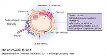
The confines of the cranial space, as well as the need to preserve the functional relationships between elements of the CNS, stress the importance of the BBB in the maintenance of the brain milieu. The haemodynamic communication between neurons and the cerebrovasculature is necessary to efficiently couple cerebral blood flow (CBF) to neuronal activation (Ref. Reference Attwell18). The cerebral endothelium is a key component of the neurovascular unit, an emerging concept that emphasises the interactions among glial, neuronal and vascular elements (Refs Reference Zlokovic6, Reference Guo and Lo7, Reference Park19, Reference Lok20) (Fig. 1). Pericytes are integral members of the neurovascular unit (Ref. Reference Lai and Kuo21). These cells are important players in vascular-mediated events in the CNS. However, a discussion of pericytes is beyond the scope of this review; the reader is referred to informative publications on this important and understudied cell type (Refs Reference Zlokovic6, Reference Lai and Kuo21, Reference Krueger and Bechmann22). Homeostatic signalling within the neurovascular unit is crucial for normal brain function. Dysfunctional cell–cell signalling in the neurovascular unit is increasingly implicated as a characteristic feature of CNS diseases (Ref. Reference Guo and Lo7). Several excellent papers have addressed the importance of the neurovascular unit for brain health (Refs Reference Iadecola23, Reference Benarroch24, Reference Zlokovic25, Reference Zacchigna, Lambrechts and Carmeliet26); the role of the brain endothelial cell in the development of diseases of the CNS is the focus of this review.
The neurovascular unit. The neurovascular unit (NVU) consists of astrocytes, pericytes, neurons and the endothelium. Endothelial cells are linked by tight junctions and encircled by the basal lamina. The figure illustrates the close relationships among the NVU, microglia and neurons. Note that in the figure only the capillary vessel is shown with its adjacent pericyte and nearby microglia and neurons. The factors released from endothelial cells in this vessel could impact all these cell types. In larger smooth muscle containing venules and arterioles, smooth muscle cells might also be affected (not shown). The following is a partial listing of the secretory products released by highly synthetic endothelial cells (Ref. Reference Grammas96). Anti- and procoagulation proteins: antithrombin III, factor V, heparin sulfate, plasminogen activator inhibitor-1, plasminogen activator inhibitor-2, platelet-activating factor, prostacyclin, thrombomodulin, thromboxane A2, thromboplastin, von Willebrand factor. Extracellular matrix proteins: collagen I–XVIII, fibronectin, laminin, proteoglycans. Growth factors: endothelium-derived growth factor, fibroblast growth factor, granulocyte colony-stimulating factor, granulocyte macrophage colony-stimulating factor, insulin-like growth factor, platelet-derived growth factor, transforming growth factor beta. Inflammatory mediators: cell adhesion molecules, interleukin-1, interleukin-6, interleukin-8, leukotriene B4, leukotriene C4, leukotriene D4, leukotriene E4, major histocompatibility complex class II, monocyte chemoattractant protein-1, monocyte chemoattractant protein-2. Proteases: matrix metalloproteinases, thrombin, tissue plasminogen activator. Vasoconstrictor/dilation factors: endothelin, endothelium-derived contracting factor, free radicals, leukotrienes, thromboxane, A2/prostaglandin F2a, endothelium-derived hyperpolarising factor, nitric oxide, prostacyclin/prostaglandin E2.

Markers of endothelial dysfunction and CNS disease
The activated endothelial cell in various clinical states has become a major target of human therapeutics (Refs Reference Schnitzer27, Reference Versari28, Reference Tang and Vanhoutte29). A dysfunctional endothelium contributes significantly to systemic vascular disorders such as atherosclerosis (Ref. Reference Gimbrone30). Increasingly, endothelial dysfunction has been implicated as a link between neurological dysfunction and vascular disease and as a crucial player in the development of CNS disorders, including Alzheimer disease (AD), Parkinson disease (PD), amyotrophic lateral sclerosis (ALS), multiple sclerosis (MS), human immunodeficiency virus (HIV)-1-associated neurocognitive disorder and traumatic brain injury (TBI) (Refs Reference Zlokovic6, Reference Barcia31, Reference Banks, Ercal and Price32, Reference Morel33, Reference Zhong34, Reference Altman and Rutledge35, Reference Alexander36). Vascular risk factors, including cholesterol, homocysteine and hypertension, have been shown to be important or relevant for the development of neurodegenerative diseases such as AD (Ref. Reference Altman and Rutledge35). Using post-occlusive reactive hyperaemia and spectral analysis of skin vasomotion as markers of endothelial function, a recent study documents peripheral endothelial dysfunction in unmedicated patients with schizophrenia (Ref. Reference Israel37). Markers of endothelial injury or dysfunction are demonstrable in several neurodegenerative disorders, as discussed below.
Endothelial microparticles
Endothelial microparticles (EMPs) are submicrometre-sized vesicles shed from plasma membranes in response to cell activation, injury or apoptosis. EMPs are an emerging marker of endothelial dysfunction and are also considered to have a major biological role in inflammation, vascular injury, angiogenesis and thrombosis (Ref. Reference Chironi38). EMPs are known to be increased in acute coronary syndromes, severe hypertension with end organ damage, and thrombotic thrombocytopenic purpura, which are all conditions associated with endothelial injury (Ref. Reference Boulanger39). Several publications implicate EMPs in the development of several CNS disorders (Ref. Reference Jung40). A study examining the cerebrospinal fluid (CSF) and plasma of patients with severe TBI shows increased generation of EMPs in the CSF of patients with TBI compared with controls (Ref. Reference Morel33). Sustained generation of these microparticles in TBI patients is associated with a poor clinical outcome, suggesting that EMPs drive deleterious processes that contribute to TBI-induced functional decline (Ref. Reference Morel33).
The release of EMPs has also been associated with endothelial dysfunction in patients with MS (Refs Reference Minagar41, Reference Jy42, Reference Jimenez43). A study examining the plasma from 50 patients with MS (30 in exacerbation and 20 in remission) and from 48 controls showed that endothelial dysfunction, demonstrated by shedding of EMPs that express platelet-endothelial cell adhesion molecule-1 (CD31), is a consistent feature during exacerbation of MS. Furthermore, EMPs from activated cerebral endothelial cells in MS promote the inflammatory reaction characteristic of MS and enhance transmigration of activated leukocytes into the brain (Ref. Reference Jimenez43).
Blood-based biomarkers of microvascular pathology
Several well-defined markers of endothelial injury are detected in CNS disease states. In schizophrenia, markers of endothelial dysfunction (von Willebrand factor and C-reactive protein) are correlated with the severity of clinical symptoms in attack-like schizophrenia (Ref. Reference Shcherbakova44). Elevated levels of soluble adhesion molecules, including intracellular adhesion molecule-1 (ICAM-1) and vascular cell adhesion molecule-1 (VCAM-1), have been reported as markers of BBB damage in MS and AD (Refs Reference Ukkonen45, Reference Ewers, Mielke and Hampel46). Also, endothelial-derived vasoactive proteins, including endothelin and atrial natriuretic peptide, are significantly altered in mild AD and in mild cognitive impairment (Ref. Reference Ewers, Mielke and Hampel46), which suggests that these proteins could be useful in disease staging and diagnosis.
Endothelial progenitor cells
An emerging marker of vascular function that has been little explored in neurodegenerative disease is the endothelial progenitor cell (EPC). Recent evidence has shown that vascular function not only depends on cells that reside within the vessel wall, but also appears to be significantly modulated by circulating cells derived from the bone marrow. A specific subset of these stem cells, termed EPCs, has been shown to promote vascular repair and improve endothelial function (Refs Reference Dzau47, Reference Van Craenenbroeck and Conraads48). Since their original discovery in 1997 (Ref. Reference Asahara49), circulating EPCs have emerged as an important biomarker for cardiovascular disease (Ref. Reference Möbius-Winkler50). Evidence is accumulating that EPCs contribute substantially to preservation of a structurally and functionally intact endothelium (Ref. Reference Van Craenenbroeck and Conraads48). A decrease in the number of EPCs is a predictor of morbidity and mortality in cardiovascular diseases (Refs Reference Möbius-Winkler50, Reference Umemura and Higashi51). A recent study documents a decrease in EPCs in patients with AD (Ref. Reference Lee52). Further research into EPCs in CNS disorders is warranted.
Data from EMPs, blood-based proteins and EPCs support the idea that endothelial cells are injured in CNS diseases. At the gene level, endothelial dysfunction is suggested by transcriptional profiling, which shows low expression of the homeobox gene MEOX2 (also known as GAX) in brain endothelial cells in AD (Ref. Reference Wu53). In mice, deletion of this gene results in vascular regression and altered amyloid beta (Aβ) transport (Ref. Reference Wu53). The consequences of brain endothelial dysfunction for BBB permeability, BBB transport and neurodegenerative disease pathogenesis are described below.
Implications of BBB dysfunction in CNS disease
Pathological changes in BBB permeability
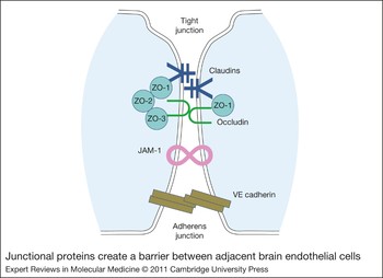
Brain capillaries exhibit a number of anatomical and biochemical characteristics seemingly unique to membranes known to regulate water and electrolyte permeability such as renal proximal tubules, including the presence of tight junctions and a high mitochondrial content (Ref. Reference Oldendorf, Cornford and Brown54). The major components of brain endothelial junctions are occludin, claudins, VE-cadherin, zona occludens proteins ZO-1 to ZO-3 (also known as TJP1–TJP3) and junctional-associated molecules (Refs Reference Zlokovic6, Reference Förster55) (Fig. 2). Although tight junctions serve as the main anatomic barrier, restricting paracellular traffic of hydrophilic molecules, lipophilic compounds move freely along their concentration gradients and enter the CNS. BBB breakdown as a result of disruption of tight junctions, which might initiate or contribute to a vicious circle of disease resulting in progressive synaptic and neuronal dysfunction and loss, has been demonstrated in MS, ALS and HIV-1-associated neurocognitive disorder (Refs Reference Zlokovic6, Reference Förster55, Reference Arhart56).
Junctional proteins create a barrier between adjacent brain endothelial cells. Main microvascular endothelial cells form the primary anatomical barrier between the blood and the brain. The endothelium is relatively impermeable to polar molecules as a result of tight and adherens junctions between cells. The junctional complexes are composed of several proteins. The primary proteins that form the tight junctions are the claudins (3, 5 and 12), occludin, zona occludens proteins ZO-1, ZO-2 and ZO-3, and junctional adhesion molecule-1 (JAM-1). Adherens junctions are primarily formed by VE cadherin, which is connected to actin within the cells.

Multiple sclerosis
Endothelial cell dysfunction might contribute to the pathogenesis of MS (Ref. Reference Alexander36). MS is a demyelinating disease characterised by breakdown of the BBB and accumulation of inflammatory cell infiltrates in the CNS. In MS, finger-like projections of demyelination extend into the white matter alongside blood vessels (Ref. Reference Weir57). In experimental autoimmune encephalomyelitis (EAE), an animal model of MS, there is a significant relocalisation of the tight junction protein ZO-1, which precedes overt clinical disease and correlates with sites of inflammatory cell accumulation (Ref. Reference Bennett58). Modifications in the integrity and organisation of junctional molecules and BBB permeability are thought to contribute to the pathogenesis of MS (Refs Reference Plumb59, Reference Kirk60). Dysregulation of junctional molecular complexes permits progressive immune cell entry into the CNS. Also, under normal physiological conditions, resting leukocytes do not cross the BBB in significant numbers because nonactivated leukocytes do not avidly bind to or penetrate the healthy BBB. In MS, increased expression of several chemokines and chemokine receptors on reactive cerebral endothelial cells facilitates transmigration of leukocytes across the BBB (Refs Reference Alexander36, Reference Minagar and Alexander61, Reference Holman, Klein and Ransohoff62). Dysregulation of the BBB and transendothelial migration of activated leukocytes are among the earliest cerebrovascular abnormalities seen in MS and parallel the release of inflammatory cytokines or chemokines in this disease (Ref. Reference Minagar and Alexander61).
Amyotrophic lateral sclerosis
ALS is a chronic neurodegenerative disorder of motor neurons in the brain, brainstem and spinal cord that results in progressive paralysis and death (Ref. Reference Garbuzova-Davis63). A breach of the BBB has been suggested in ALS because of the increased levels of albumin, IgG and complement proteins found in the CSF and spinal cord in ALS patients and in animal models of the disease (Refs Reference Arhart56, Reference Engelhardt and Appel64). Mouse models of ALS, which express mutant forms of the antioxidant enzyme superoxide dismutase-1 (SOD1), show vascular abnormalities before motor neuron degeneration (Refs Reference Zhong34, Reference Garbuzova-Davis63, Reference Nicaise65). Specifically, there is a reduction in the level of tight junction proteins ZO-1, occludin and claudin-5 (Refs Reference Arhart56, Reference Nicaise65). Also, Prussian Blue staining, an indicator of haemosiderin deposition and microhaemorrhages, in presymptomatic SOD1 mutants suggests that barrier breakdown is an early event in disease progression (Ref. Reference Zhong34). Endothelial damage accumulates before motor neuron degeneration and neurovascular inflammation in the SOD1 mutant, supporting a primary role for endothelial dysfunction in disease initiation (Refs Reference Arhart56, Reference Nicaise65).
HIV-1-associated neurocognitive disorder
HIV-1-associated neurocognitive disorder is the most common and clinically important CNS complication of late HIV-1 infection (Ref. Reference Ances and Clifford66). BBB-regulated cellular trafficking is crucial for the development of CNS pathology caused by HIV, as well as other neuroinflammatory conditions, including meningitis and encephalitis (Ref. Reference Zlokovic6). Exposure of the BBB to the HIV-1 virus or virus-infected monocytes affects several aspects of BBB function as well as brain endothelial cell protein expression (Ref. Reference Kanmogne67). HIV-1 virus or virus-infected monocytes activate brain endothelial cells, enabling leukocyte passage across the BBB through highly regulated processes such as diapedesis and adsorptive endocytosis (Ref. Reference Banks, Ercal and Price32). High levels of soluble CD40 ligand (CD40L) are found in CSF and plasma, whereas CD40, a receptor for CD40L, is highly expressed in the brain endothelial cells of patients with HIV-1 encephalitis (Ref. Reference Ramirez68). Exposure of human brain endothelial cells to CD40L upregulates expression of leukocyte adhesion molecules (ICAM-1, VCAM-1), which causes a fourfold increase in monocyte adhesion to, and migration across, brain endothelial cells in an in vitro BBB model (Ref. Reference Ramirez68). Brain endothelial cells also secrete neuroinflammatory molecules when stimulated by HIV-1 or HIV-derived proteins gp120 and Tat (Ref. Reference Yang69). A proteomic examination of human brain microvascular endothelial cells exposed to HIV-1-infected macrophages shows that over 200 proteins in brain endothelial cells are upregulated (Ref. Reference Ricardo-Dukelow70). Proteins in a wide range of functional categories are affected, including voltage-gated ion channel, heat shock, transport, cytoskeletal, regulatory and calcium-binding proteins. Also, HIV-1 virions induce transcriptional upregulation and expression of proinflammatory genes and the transcription factor STAT1 in brain endothelial cells (Ref. Reference Yang69). Overall, evidence that the BBB is impaired during progressive HIV-1 disease and the emergence of cognitive impairments during HIV-1-associated cognitive disorder support a major role for brain endothelial cells in establishing and maintaining the virus within the CNS.
Pathological changes in BBB transport
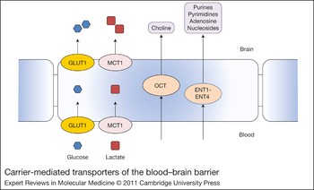
The high energy and nutrient demands of the brain, as well as the need to tightly regulate the electrolyte milieu for optimal neuronal function, highlight the importance of BBB transport systems. Specific carrier-mediated transport systems facilitate transport of nutrients such as hexose sugars (glucose, galactose), neutral, basic and acidic amino acids, monocarboxylic acids (lactate, pyruvate, ketone bodies), nucleosides (adenosine, guanosine, uridine), purines (adenine, guanine) and amines (choline) (Refs Reference Zlokovic6, Reference Ribatti9, Reference Gerhart13, Reference Smith, Stoll and Pardridge14, Reference Tsuji15, Reference Ohtsuki and Terasaki16, Reference Spector and Johanson17) (Fig. 3). The concentration gradients for nutrients are generally in the direction from the blood to the brain. These are regulated by brain metabolic needs and by the concentrations of substrates in plasma (Ref. Reference Zlokovic6). The complexity of the BBB is further indicated by the polarity of cerebral endothelium with regard to transport, with differential localisation of carriers in luminal and abluminal plasma membranes (Ref. Reference Shen and Zhang71). Accumulating evidence suggests that these transport systems are dysregulated in CNS disease and, indeed, might significantly contribute to disease initiation and progression.
Carrier-mediated transporters of the blood–brain barrier. The high energy and nutrient demands of the brain, as well as the need to tightly regulate the electrolyte milieu for optimal neuronal function, highlight the importance of blood–brain barrier transport systems. Specific carrier-mediated transport systems facilitate transport of nutrients such as hexose sugars, neutral, basic and acidic amino acids and monocarboxylic acids, purines and amines. Transporters and prototypical substrates are shown. Glucose 1 transporter (GLUT1) (glucose); equilibrative nucleoside transporters (ENTs) (purines); monocarboxylic acid transporter 1 (MCT1) (lactate); organic cation transporters (OCTs) (choline).

Alzheimer disease
Because of the importance of BBB transporters for the maintenance of the CNS milieu, abnormalities in brain endothelial cell function that result in altered transporter activities have consequences for brain function and disease development. In AD, a consistent finding evident early in the disease process is region-specific decrease in glucose utilisation (Ref. Reference Ferreira72). Analysis of positron emission tomography scans from patients with mild cognitive impairment shows diminished glucose uptake in brain areas susceptible to the development of AD (Refs Reference Chetelat73, Reference Gibson and Shi74). It is likely that the decreased expression of GLUT1, the primary glucose transporter at the BBB, documented in AD patients contributes to this energy deficit (Ref. Reference Kalaria and Harik75).
Also important in the pathogenesis of AD is the Aβ protein. The amyloid cascade hypothesis of AD postulates a central role for this protein in disease pathogenesis (Ref. Reference Pimplikar76). Levels of Aβ in the brain are regulated by active processes at the BBB. Individuals with late-onset AD do not have increased production of Aβ and are thought to have increased Aβ accumulation as a result of its faulty clearance (Ref. Reference Zlokovic6). The receptor for advanced glycation end products (RAGE) is the major influx transporter for Aβ across the BBB. Low-density lipoprotein receptor-related protein 1 (LRP1) is a major efflux transporter for Aβ across the BBB (Ref. Reference Deane, Bell, Sagare and Zlokovic77). LRP1 is a member of the LDL receptor family and acts as a multifunctional scavenger and signalling receptor (Ref. Reference Deane78). Binding of Aβ to LRP1 at the abluminal side of the BBB initiates Aβ clearance from brain to blood by transcytosis across the BBB (Ref Reference Deane, Bell, Sagare and Zlokovic77). The importance of LRP for Aβ clearance is highlighted by several studies in transgenic AD mice and AD patients. In animals deficient in the homeobox gene MEOX2, impaired Aβ efflux is caused by reduced LRP levels (Ref. Reference Wu53). In AD patients and AD mouse models, a non-Aβ clearing phenotype results from downregulation of LRP1 (Ref. Reference Bell79). Aβ deposition in AD individuals with cerebral amyloid angiopathy is controlled through the LRP1 receptor (Ref. Reference Bell79). Finally, the Dutch mutant form of Aβ is poorly cleared from the brain because of low-affinity binding of LRP to Aβ (Ref. Reference Deane80). In AD and transgenic models, RAGE expression has been shown to increase, whereas the number of LRP receptors decreases (Ref. Reference Yan81). In mouse models of AD, dense plaques develop initially on blood vessels. It is believed that plaques are generated on blood vessels because of deficient Aβ clearance across the BBB (Ref. Reference Kumar-Singh82). Reduced expression of LRP1 has been associated with positive staining of cerebral vessels for Aβ during normal ageing in rodent and nonhuman primates, and in individuals with AD (Ref. Reference Shibata83).
RAGE-dependent BBB transport of circulating Aβ results in expression of proinflammatory cytokines in the brain and production of endothelin-1, which causes decreased CBF. Systemic administration of a recombinant truncated form of RAGE reduced the accumulation of AB in the brain parenchyma of transgenic APP mice (Ref. Reference Deane80). Finally, inflammatory proteins have been shown to impair LRP activity and increase RAGE, thereby promoting increased Aβ in the AD brain (Ref. Reference Jaeger84).
Parkinson disease
PD is a neurodegenerative disorder that is characterised by resting tremor, rigidity and bradykinesia and involves progressive degeneration of dopaminergic neurons in the substantia nigra leading to dopamine deficiency (Ref. Reference Qian, Flood and Hong85). The BBB expresses transporters that affect both PD drug therapy and dopaminergic neuronal function. l-DOPA, but not dopamine, is transported across the BBB in humans by the L1 facilitative transporter. After transport across the BBB, l-DOPA is converted to dopamine at nerve terminals that contain DOPA decarboxylase (Ref. Reference Hawkins86). Impairment of BBB function has been implicated in the pathogenesis of PD (Refs Reference Carvey87, Reference Kortekaas88). In PD individuals, capillaries are mis-shapen and normal contacts between nigral neurons and capillaries are lost at an early stage of disease. Capillary basement membrane thickening and collagen accumulation have also been shown in PD (Ref. Reference Farkas89). Focal disruption of the BBB induces in vivo degeneration of dopaminergic neurons, which is a characteristic feature of PD (Ref. Reference Carvey87). In an animal model of PD, administration of 1-methyl-4-phenyl-1,2,3,6-tetrahydropyridine results in motor deficits and loss of dopaminergic neurons, and is temporally associated with a significant reduction in mRNA for the BBB l-type amino acid transporter for tyrosine, a precursor for dopamine, which enters the brain through the l-type amino acid transporter (Ref. Reference Ohtsuki90).
It has also been suggested that absorption or metabolism of putative PD toxins and/or their faulty elimination across the BBB has a role in the pathogenesis of PD (Ref. Reference Zlokovic6). The ATP-binding cassette superfamily of transporter genes regulates the bioavailability of xenobiotics. The multidrug transporter of P-glycoprotein (P-gp) is the best characterised member of this family (Ref. Reference Lee91). Low activity of the P-gp efflux transporter at the BBB in the midbrain of individuals at risk for PD supports a mechanistic role for this transporter in the retention of putative PD toxins. Imaging studies have demonstrated impairment in the ability of BBB efflux pumps to remove neurotoxins in patients with PD (Ref. Reference Bartels92). Furthermore, inflammation has also been shown to reduce the activity of P-gp (Ref. Reference Hartz93). The importance of P-gp in the pathogenesis of PD is derived from gene polymorphism studies. Some polymorphisms might be protective and reduce risk, whereas others might predispose carriers to the damaging effects of pesticides, leading to a PD phenotype (Refs Reference Lee91, Reference Funke94, Reference Westerlund95). This transporter system is a significant modulator of susceptibility to PD among male ethnic Chinese who are more than 60 years old (Ref. Reference Lee91).
Brain endothelial cells as regulators of neuroinflammation and oxidative stress in neurodegenerative disease
Neuroinflammation and oxidative stress are both implicated in the pathogenesis of neurodegenerative disease. The brain endothelium is a highly dynamic, reactive interface that serves as both a source of, and a target for, inflammatory proteins and reactive oxygen species (ROS) (Ref. Reference Grammas96). Using laser capture microdissection, a recent study documents that endothelial samples showed differences in gene expression between schizophrenics and controls. Functional profiling revealed that the genes affected are related to inflammatory processes (Ref. Reference Harris97). Vascular-derived products of a dysfunctional endothelium could result in neuronal injury in neurodegenerative disease states (Ref. Reference Grammas, Moore and Weigel98). Thus, beyond overt disruptions in BBB permeability or transport functions, abnormal endothelial function can affect the CNS milieu and neuronal viability or survival. For example, in the AD brain, an injured or altered brain endothelial cell releases a large number of bioactive molecules, some of which are directly toxic to neurons (Ref. Reference Grammas99). Because of the intimate communication among cells in the neurovascular unit, endothelial cell products have important paracrine effects on astrocytes and microglia (Refs Reference Zlokovic6, Reference Lok20). In this regard, the neurotoxic protease thrombin, which is synthesised and released by brain endothelial cells in AD (Ref. Reference Yin100), has significant proinflammatory effects on both microglia and astrocytes. In microglia, thrombin increases transcription of inflammation-associated genes tumour necrosis factor alpha (TNFα) and inducible nitric oxide synthase (Ref. Reference Huang101). In astrocytes, activation of the thrombin receptor (PAR-1) leads to increased expression of matrix metalloproteinase 9 (MMP-9) (Ref. Reference Choi102). Thus, vascular-derived thrombin might directly injure neurons or affect neuronal viability indirectly by activation of microglia and astrocytes in the neurovascular unit.
Neuroinflammation: a key role for matrix metalloproteinases in the inflammatory cycle
The cerebromicrocirculation is a rich source of cytokines and chemokines. In neurodegenerative diseases, a myriad of inflammatory-associated proteins have been documented (Refs Reference Zlokovic6, Reference Ramirez68, Reference Yang69, Reference Jaeger84, Reference Grammas96, Reference Rosenberg103, Reference Grammas and Ovase104, Reference Grammas, Samany and Thirumangalakudi105). Rather than list these factors, we focus on MMPs, central players in the inflammatory response, in order to explore how their expression and effects on BBB function might contribute to pathological events in the diseased brain.
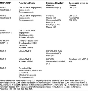
The major group of enzymes controlling basement membrane and extracellular matrix turnover are the gelatinases (MMP-2 and MMP-9), collagenases, stromelysins and membrane-type MMPs. MMPs are usually secreted in an inactive proenzyme form and can be activated extracellularly by other proteinases, including active MMPs and some tissue inhibitors of metalloproteinases (TIMPs) (Refs Reference Rosenberg103, Reference Bugno106) (Table 1). Several physiological mechanisms prevent uncontrolled MMP activation. One of these is binding to MMPs by TIMPs. By inhibition of MMP activity, TIMPs contribute to the regulation of ECM remodelling associated with various physiological and pathological conditions (Refs Reference Vilalta107, Reference Zhang108). TIMPs can also aid in the activation of MMPs. For example, TIMP-2 forms a trimolecular complex with the pro-form of MMP-2 and membrane type (MT)-MMP-1, thereby activating MMP-2 at the cell surface (Ref. Reference Rosenberg103). MMPs can cleave virtually all structural ECM molecules. In addition to providing structural scaffolding, ECM molecules act as binding reservoirs for various growth factors and cytokines (Ref. Reference Sternlicht and Werb109). MMPs increase the permeability of the BBB by attacking the ECM, basal lamina and tight junctional proteins, leading to neuroinflammation (Ref. Reference Rosenberg103). MMPs are expressed by all cell types of the neurovascular unit, including endothelial cells (Refs Reference Rosenberg103, Reference Bugno106, Reference Visse and Nagase110, Reference del Zoppo111); however, the expression of MMPs by brain endothelial cells is relatively unexplored.
Matrix metalloproteinases and tissue inhibitors of metalloproteinases involved in neurodegenerative disordersa

Abbreviations: AD, Alzheimer disease; ALS, amyotrophic lateral sclerosis; BBB, blood–brain barrier; CSF, cerebrospinal fluid; EBM, endothelial basement membrane; EC, endothelial cell; ECM, extracellular matrix; IL-1β, interleukin-1 beta; MMP, matrix metalloproteinase; MT-MMP-1, membrane-type metalloproteinase-1; PD, Parkinson disease; TIMP, tissue inhibitor of metalloproteinases; TNFα, tumour necrosis factor alpha.
Inflammatory cytokines can regulate expression of MMPs, and these enzymes can, in turn, affect the level and activity of inflammatory proteins. The major proinflammatory cytokines interleukin 1β (IL-1β), TNFα, interferon-γ (IFNγ) and IL-6 are important regulators of MMP and TIMP expression (Refs Reference Bugno106, Reference Ries and Petrides112). All these cytokines have been shown to be upregulated in neurodegenerative disease. In this regard, the inflammatory protease thrombin, which is upregulated in AD and other brain diseases (Refs Reference Yin100, Reference Sokolova and Reiser113), causes a time- and dose-dependent decrease in the attachment of endothelial cells to basement membrane components, with a concomitant increase in MMP-2 activation in endothelial cells (Ref. Reference Maragoudakis, Tsopanoglou and Andriopoulou114). Exposure of cultured brain endothelial cells to both IL-1β and TNFα causes a shift in the expression of TIMP-1 and TIMP-3; TIMP-1 expression is upregulated, whereas levels of TIMP-3 decrease (Ref. Reference Bugno106). This imbalance in TIMP expression has implications for the regulation of MMPs in brain microvasculature (Ref. Reference Bugno106). TNFα and IL-1β also induce the transcription of MMP-3 and MMP-9, which are both involved in acute and chronic neuroinflammation (Ref. Reference Rosenberg103). MT-MMP-1, a cell surface MMP, acts as a broad-spectrum ECM proteinase and can regulate activation of other MMPs. Because MT-MMP-1 is efficiently inhibited by TIMP-3 but not by TIMP-1, the ‘reprogramming’ of the TIMP-1–TIMP-3 balance during brain endothelial cell activation by inflammatory proteins could result in a local increase in MT-MMP-1 activity, with consequences for microvascular basement membrane proteolysis.
MMPs contribute to the propagation and regulation of neuroinflammatory responses to injury. The development of tissue damage that follows transient ischaemia suggests the crucial interplay between MMPs and mediators of neuroinflammation (Refs Reference Cunningham, Wetzel and Rosenberg115, Reference Amantea116). MMPs are involved in the processing of proinflammatory cytokines, such as IL-1β, into biologically active forms through a caspase-1-independent pathway (Ref. Reference Schönbeck, Mach and Libby117). MMPs and TIMPs can contribute to immunoregulatory processes by affecting cytokine and cytokine receptor turnover (Ref. Reference Sternlicht and Werb109). For example, MMPs can activate recombinant soluble proTNFα (Ref. Reference Woessner and Nagase118). In addition to causing the activation and release of cytokines and growth factors, MMPs can also cleave their cell surface receptors (Ref. Reference Sheu119).
The relevance of MMPs in the development of injury in several chronic neurodegenerative disorders, including AD, PD, ALS, MS and HIV-1-associated neurocognitive disorder (Refs Reference Rosenberg103, Reference Visse and Nagase110), is supported by studies documenting altered MMP and TIMP levels, relative to controls, in the CSF, brain-derived microvessels and plasma. It is important to note that there are some contradictory data in the literature regarding changes in the levels of MMPs and TIMPs in the CSF and plasma in neurodegenerative diseases (see Table 1 for a more detailed breakdown of MMP and TIMP changes in various neurodegenerative diseases).
MMPs regulate lymphocyte and monocyte transmigration across the BBB
MS is a disease of the CNS that is characterised by multifocal infiltration of T cells from the systemic immune system across the BBB (Ref. Reference Alexander36). MMPs disrupt the BBB and facilitate the remodelling of the ECM, they are therefore heavily implicated in MS because changes in the barrier function of brain microvessels allow T cells to invade the CNS more readily (Ref. Reference Rosenberg120). In EAE, inhibitors of MMPs slow down the progression of symptoms (Ref. Reference Rosenberg103). This indicates that MMP-facilitated endothelial cell dysfunction might be part of the mechanism by which leukocytes penetrate the brain tissue and damage the myelin sheath of white matter neurons in MS.
Exposure of human brain microvascular endothelial cells to HIV-infected monocytes results in decreased expression of tight junction proteins (Ref. Reference Persidsky121). Alterations of protein expression in tight junctions are associated with increased endothelial permeability and increased transendothelial migration of HIV-infected monocytes across an in vitro model of the BBB (Ref. Reference Huang122). The HIV-induced decrease in the expression of junctional adhesion molecule-1 (JAM-1), occludens and ZO-1 is restored by inhibition of MMP activity (Ref. Reference Huang122). This suggests that downregulation of MMP activity constitutes a novel mechanism for protection against HIV-induced BBB disruption (Ref. Reference Huang122). The increased permeability of human microvascular endothelial cell monolayers to monocytes caused by HIV-1 infection is inhibited by TIMP-1 and TIMP-2, suggesting that MMP-2 and MMP-9 are primary mediators of enhanced permeability (Ref. Reference Dhawan123).
MMPs in vascular inflammation and direct neurotoxic effects in AD and PD
In addition to altered levels of MMPs in CSF and plasma, MMPs have been localised to intraparenchymal lesions found in neurodegenerative diseases (Ref. Reference Rosenberg103). This implies that in addition to their known function degrading ECM, MMPs might serve as mediators of apoptotic or necrotic cell death.
The cerebral microvasculature could be central to a destructive cycle of events where inflammatory factors stimulate MMP expression, which in turn promotes the release of inflammatory mediators. Brain microvessels derived from patients with AD release IL-6, TNFα, IL-8, IL-1β, thrombin, and MMP-2 and MMP-9 (Refs Reference Yin100, Reference Grammas and Ovase104, Reference Grammas, Samany and Thirumangalakudi105, Reference Thirumangalakudi124). Through the effects of MMPs on cytokines and stimulation of MMPs by inflammatory proteins, MMPs could be key amplifiers of local inflammation. The close proximity of blood vessels to AD lesions suggests intimate communication between the neurovascular unit and neurons (Ref. Reference Miyakawa125). In PD, mutation or overexpression of α-synuclein protein has a pivotal role in the pathogenesis of PD (Ref. Reference Lee126). α-Synuclein-stimulated microglia release MMPs that subsequently activate the thrombin receptor, amplifying microglial inflammatory signals in an autocrine manner (Ref. Reference Lee126). Similar pathways might operate in brain endothelial cells because MMPs, thrombin and α-synuclein have all been identified in these cells (Refs Reference Yin100, Reference Thirumangalakudi124, Reference Tamo127).
Finally, MMPs might have a role in neurodegenerative disease by both direct and indirect neurotoxic mechanisms. MMP-3 has been implicated as a neurotoxic factor that contributes directly to the death of dopaminergic neurons in PD (Ref. Reference Kim128). In AD, MMP-9 toxicity in Aβ-mediated animal models is supported by experiments where inhibition of MMPs improves Aβ-mediated cognitive impairment and neurotoxicity in mice (Ref. Reference Mizoguchi129). Recent evidence indicates that protein aggregation, and in particular the formation of toxic protein oligomers, is a key mechanism in synucleinopathies such as PD (Ref. Reference Levin130). In vitro models of pathological α-synuclein aggregation indicate that MMP-1 and MMP-3 can influence the pathogenesis of PD by the generation of specific aggregation-enhancing α-synuclein fragments that result from limited proteolysis (Ref. Reference Levin130).
Mitochondria as mediators of oxidative stress
The human brain, although it constitutes only 2% of total body weight, accounts for 20% of oxygen consumption in the body, reflecting its high rate of metabolic activity and the high level of ATP consumption by neurons (Ref. Reference Pimenta de Castro, Martins and Tufi131). Mitochondria have a crucial role in the supply of energy to the brain. The mitochondrial hypothesis of neurodegeneration postulates that defects in mitochondrial metabolism lead to mitochondrial dysfunction and eventually to depletion of cellular ATP, increased free radical generation, calcium dysregulation and, potentially, defective mitochondrial fission or fusion (Ref. Reference Reeve, Krishnan and Turnbull132). Mitochondrial membrane lipids are highly susceptible to ROS, especially the long-chain polyunsaturated fatty acid components (Ref. Reference Müller133). Inner mitochondrial membrane proteins such as complexes I, II and III are themselves directly susceptible to effects of oxidative stress, leading to membrane depolarisation and subsequently, impaired mitochondrial function (Ref. Reference Müller133). Disorders of mitochondrial function, caused by mutations in mitochondrial DNA, proteins or the presence of mitochondrial inhibitors, appear to have a role in several neurodegenerative diseases (Ref. Reference Reeve, Krishnan and Turnbull132).
Cerebrovascular mitochondria, ageing and oxidative stress
Diseases of the CNS are for the most part age-associated disorders (Ref. Reference Sakar and Fisher134). It is well established that mitochondrial DNA accumulates mutations with ageing (Ref. Reference Müller133). A decrease in the mitochondrial antioxidant MnSOD (superoxide dismutase) has also been found in the cerebrovasculature with increasing age (Ref. Reference Tripathy135). The physiology and pathophysiology of endothelial cells are closely related to the functioning of mitochondria, and mitochondrial dysfunction could be an important mediator of vascular lesions (Refs Reference Victor and Rocha136, Reference Puddu137). Endothelial mitochondria might serve as sensors of the local environment (i.e. oxygen concentrations) (Ref. Reference Davidson and Duchen138). This is especially important for cerebrovascular endothelial cells because of the interactions between endothelia, astrocytes and neurons within the neurovascular unit. Also, because of the highly metabolic nature of the BBB and the numerous transport systems it supports, brain endothelial cells have an extensive mitochondrial network and, relative to other endothelial cell types, an increased mitochondrial mass (Ref. Reference Oldendorf, Cornford and Brown54). Many injuries or insults thought to initiate BBB dysfunction result in ROS, resulting in oxidative damage, tight junction disruption and activation of MMPs (Refs Reference Kolev139, Reference Pu, Lu and Moochhala140). Exposure of brain endothelial cells to oxidised lipids increases nitric oxide and ROS production and stimulates translocation of the proapoptotic protein Bax (Refs Reference Hamdheydari141, Reference Chang142). Activation of the protease pathway involving Bax, mitochondrial cytochrome c and caspase in cerebral endothelial cells is attenuated by the antioxidant resveratrol (Ref. Reference Chang142). The brain vasculature appears to be especially sensitive to oxidative stress. This sensitivity might in part be due to higher levels of NAD(P)H-oxidase in brain endothelial cells compared with endothelial cells in peripheral vessels (Ref. Reference Closhen143). In this regard, a recent study shows that the inflammatory protein cysteine and glycine-rich protein 1 (CRP) evokes NAD(P)H-oxidase-dependent functional derangements in endothelial cells derived from the brain but not from the aorta (Ref. Reference Closhen143). The higher concentration of mitochondria in cerebrovascular endothelial cells, relative to other endothelial cell types (Ref. Reference Oldendorf, Cornford and Brown54), might also account for the sensitivity of the BBB to oxidant stressors.
Mitochondrial dysfunction in sporadic neurodegenerative diseases
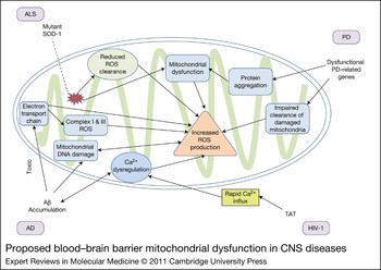
Cerebral vasculopathy is a central aspect of most, if not all, inherited mitochondrial diseases. Although these are rare diseases, this demonstrates that disorders whose cause is known to be mitochondrial in origin result quite specifically in cerebrovascular pathology. There is increasing evidence that acquired mitochondrial defects promote a similar vasculopathy in sporadically occurring disorders such as PD, AD and ALS (Refs Reference Gibson and Shi74, Reference Martin144) (Fig. 4). The literature suggests pathogenic links among mitochondrial malfunction, ROS and chronic inflammatory diseases (Ref. Reference Fernandez-Checa145). Genes associated with autosomal recessive PD, including Parkin (PARK2), PINK1 (PARK6) and DJ-1 (PARK7), all encode proteins with functional effects on mitochondria (Refs Reference Pimenta de Castro, Martins and Tufi131, Reference Lopez and Sidransky146). Both PINK1 and Parkin are crucial in the removal of damaged mitochondria by mitophagy, whereas DJ-1 is localised to mitochondria and has a role in oxidative stress protection (Ref. Reference Pimenta de Castro, Martins and Tufi131). Approximately 90% of cellular ROS production is attributable to mitochondria (Ref. Reference Müller133). Oxidative stress has been consistently linked to neurodegenerative diseases (Ref. Reference Casetta, Govoni and Granieri147). ROS production in mitochondria can be induced by blocking complex I, a key enzyme of the respiratory chain, resulting in superoxide production. Administration of rotenone, a complex I inhibitor, to experimental animals results in loss of mitochondrial membrane potential, robust ROS production, and PD-like symptoms and pathology (Ref. Reference Tanner148). Exposure of experimental animals to high levels of manganese results in neurodegenerative changes that are similar to those described in PD. Manganese induces direct injury to mitochondria in brain endothelial cells (Ref. Reference dos Santos149). Altered mitochondrial function probably underlies the ensuing impairment in energy metabolism, redox status and compromised BBB function. The central role of mitochondrial ROS-mediated signalling in endothelial cells implies that mitochondrial dysfunction might lead directly to vascular pathology.
Proposed blood–brain barrier mitochondrial dysfunction in CNS diseases. In CNS disease states, the mitochondria become dysfunctional and produce large amounts of ROS. AD is characterised by the formation of Aβ plaques. Aβ disrupts the electron transport chain and calcium homeostasis, leading to ROS production in mitochondria. ALS is thought to be caused by damage or mutation to SOD1. Mutant SOD1 results in reduced clearance of mitochondrial-produced ROS. In HIV-1-associated neurocognitive disorder, HIV-1-infected cells produce the TAT protein. This viral-encoded protein induces apoptosis in cells by causing proapoptotic proteins to translocate to the mitochondrial cell membrane, leading to mitochondrial dysfunction, the release of ROS and eventual cell death. Genes associated with autosomal recessive PD encode proteins with functional effects on mitochondria. These genes are crucial in the removal of damaged mitochondria by mitophagy, have roles in oxidative stress protection, and can form aggregates that cause mitochondrial dysfunction and eventual ROS production. Abbreviations: Aβ, amyloid beta protein; AD, Alzheimer disease; ALS, amyotrophic lateral sclerosis; CNS, central nervous system; HIV-1, human immunodeficiency virus-1; PD, Parkinson disease; ROS, reactive oxygen species; SOD1, superoxide dismutase-1.

Oxidative stress, vascular abnormalities and metabolic energy defects are early features thought to have a crucial role in AD pathogenesis (Ref. Reference Aliev150). Mitochondria, as both a major source of ROS and energy-generating organelles, are probable contributors to the disease process. Studies of the BBB in AD patients have shown decreased mitochondrial content (Ref. Reference Claudio151). Studies of Aβ-peptide-induced cerebral endothelial cell death have demonstrated mitochondrial dysfunction and caspase activation (Ref. Reference Xu152). Aβ disrupts the integrity of nuclear and mitochondrial DNA, activates caspase-8 and caspase-3, and causes apoptosis in brain endothelial cells (Ref. Reference Xu152). Furthermore, Aβ-induced cell death can be prevented by zVAD-fmk, a broad-spectrum caspase inhibitor, or by the antioxidant N-acetyl-cysteine (Ref. Reference Xu152).
In ALS, morphological and functional defects in mitochondria are found in both human ALS patients and ALS mice overexpressing mutant SOD1 (Refs Reference Wong153, Reference Kong and Xu154). In SOD1 mice, mitochondrial degeneration precedes disease symptoms, arguing for a causal role in disease progression. In these mouse models, BBB abnormalities, such as reduced numbers of tight junction proteins, also occur before motor neuron degeneration. SOD1 mice show dysfunctional mitochondria with reduced ATP production, oxidative phosphorylation and calcium buffering capacity (Ref. Reference Zhong34). Although SOD1 is a cytoplasmic protein, a segment of this protein partitions into the mitochondria. Mitochondrial accumulation of misfolded mutant SOD1 has been proposed as one possible trigger of cell death in this disease. A recent study shows that in ALS mice, mutant SOD1 damages mitochondria by promoting conformational changes in the apoptosis protein Bcl-2 (Ref. Reference Pedrini155).
Taken together, these studies highlight the role of vascular oxidative stress and BBB mitochondria and suggest that strategies aimed at improving mitochondrial function or ROS scavenging are of potential clinical relevance for a wide variety of CNS diseases.
Clinical implications
Diseases of the nervous system pose a significant health challenge, affecting one in three Americans at some point in life, with a cost estimated by the Alzheimer's Association to exceed $500 billion per year. Although these diseases are a diverse group, they share many features and common mechanisms. Increasingly, the brain vasculature appears to be an active player in the initiation and progression of these disorders. Indeed, with over 400 miles of capillary length in the brain and the intimate communication among cells of the neurovascular unit, a key role for the brain endothelium in mediating brain disease processes is not surprising (Ref. Reference Zlokovic6). The endothelium is a common target for all cardiovascular risk factors, and functional impairment of vascular endothelial cells occurs long before overt disease. Neuroinflammation and oxidative stress, invariant features of neurodegenerative diseases, are associated with endothelial dysfunction. If therapeutic strategies to combat cardiovascular disease prove useful in treating or preventing neurodegenerative disorders, novel and effective therapies for devastating CNS disorders could be possible in the near future.
Research in progress and outstanding research questions
Although clinical manifestations of neurodegenerative disorders have long been described, identification of the cellular, biochemical and molecular bases for CNS disease is more recent. Many gaps remain in our knowledge and understanding of both the interplay of genetics and environment and the chronology of disease evolution. The specialised nature of BBB endothelium has evolved to protect the brain from injury; thus, even minor disturbances of endothelial function have important consequences for neuronal function. Despite the importance of brain endothelial cell function for CNS health, the idea that the cerebral vasculature could be an active participant in the pathogenesis of CNS disease is an evolving concept. Because the vascular–neuronal axis is still relatively unexplored, there is great potential for new discoveries in the areas of new disease markers and animal models.
To understand the contribution of brain endothelial function to CNS disease pathogenesis, neurovascular-derived markers such as EMPs, bloodborne proteins or EPCs could be used to investigate neurovascular pathophysiology in these diseases and could highlight mechanisms that might be common among disparate brain diseases. The use of neurovascular-derived markers for the diagnosis and monitoring of neurodegenerative disorders could provide new insights into the disease processes in the brain. There is a concerted research effort to develop peripheral, accessible markers for neurodegenerative diseases. This is crucial for the development of noninvasive approaches that would allow in-office screening protocols as well as early disease diagnosis. The use of BBB endothelial biomarkers in the staging of CNS disorders, especially EPCs, is in its infancy and warrants further study.
The finding that vascular-based processes are contributory to CNS diseases opens up new possibilities for the development of unique CNS disease animal models. Currently, most animal models of CNS disease focus on the overexpression of specific proteins that are thought to have a role in disease pathogenesis. Although considerable useful information has been derived from these studies, there are limitations to this approach. Results are skewed to processes that involve those proteins and are based on a priori assumptions that these proteins or processes are the most relevant for disease progression. The development of new animal models where the inciting stimulus is ‘vascular injury’ could produce animals that show defects in proteins or signalling cascades not previously identified. This unbiased approach could identify new targets for therapy. It should be borne in mind that because endothelial injury is increasingly identified in CNS diseases, efforts will need to focus on whether these vascular changes drive the pathological events in the brain or are secondary bystander effects. Also, a role for vascular or endothelial abnormalities in the development of CNS diseases is an emerging field. It is therefore difficult to determine from the literature how ‘common’ these findings are, because until recently they had not been explored. Thus, the significance of these findings is open to discussion.
Finally, when considering the cerebromicrovasculature and diseases of the CNS, the BBB has traditionally been considered a hindrance for disease treatment. Indeed, an intact BBB is a major obstacle for the delivery of drugs into the CNS parenchyma. Approximately 98% of small-molecule drugs and all large-molecule neurotherapeutics are normally excluded from the brain (Refs Reference Pardridge156, Reference Agarwal157). However, the location of the cerebrovascular endothelium as an interface that is readily accessible to intravascular therapeutics, suggests that the BBB itself could be a target for both novel and existing drugs that might otherwise have been overlooked as neurotherapeutics because of their inability to cross the BBB. Understanding the functional BBB could lead to new therapeutic approaches for diseases of the CNS that target the dynamic properties of brain endothelial cells.
Acknowledgements and funding
This work was supported in part by grants from the National Institutes of Health (AG15964, AG020569 and AG028367). Dr. Grammas is the recipient of the Shirley and Mildred Garrison Chair in Aging. Dr. Miller is the recipient of the CH Foundation Chair. The authors gratefully acknowledge the assistance of Terri Stahl and Lauren Wyshywaniuk and appreciate the helpful suggestions of the peer reviewers.