Mutations in filamin A (FLNA) gene cause an X-linked form of myxomatous valvular dystrophy along with a constellation of other phenotypic disorders involving cellular cytoskeleton formation and function. The association between these gene mutations and mitral valve prolapse was first described in 2007;Reference Kyndt and Gueffet 1 however, there have been few reports since then.Reference Lardeux and Kyndt 2 , Reference Bernstein, Bernstein, Hehr and Hudgins 3
Here, we report a case of a young Japanese man undergoing successful mitral valve replacement and tricuspid annuloplasty for prolapse of both his atrioventricular valves. In the follow-up, the patient was diagnosed as having hypermobility type Ehlers–Danlos syndrome (EDS type III) because of his clinical manifestations, but was revealed to have a mutation in the FLNA gene after the operation.
Case
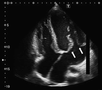
A 21-year-old man underwent mitral valve replacement and tricuspid annuloplasty for prolapse of both his atrioventricular valves. On account of having associated pectus excavatum, hypermobility of the wrist joint and thumb proximal interphalangeal, and elasticity of the skin, he was diagnosed with EDS type III. The patient’s mitral valve prolapse was observed at 3 years of age, and it gradually progressed over time. Tricuspid valve prolapse subsequently developed. Despite administration of an angiotensin-converting enzyme inhibitor, diuretics, and digoxin since 9 years of age, the patient has recently become aware of dyspnoea on effort. Echocardiography showed that the left ventricular diastolic diameter was 63.5 mm and the ejection fraction was 71%. Both mitral and tricuspid valve leaflets were thickened and billowed (Fig 1). Mitral regurgitation was severe, and tricuspid regurgitation was mild to moderate. In addition, the aortic annulus was 27 mm in diameter and aortic regurgitation was slight. As the other remarkable pre-operative examination findings, the brain magnetic resonance imaging revealed malformations of periventricular nodular heterotopias and hypoplastic corpus callosum.
Pre-operative echocardiography showed thickened and elongated mitral (white arrow) and tricuspid leaflets with a mxyomatous change. The thickness of the mitral leaflet is more than 4 mm.

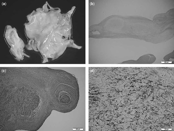
Mitral valve repair was not indicated because of the unusual deformity of the mitral valve leaflets, and thus valve replacement with a 29-mm mechanical valve was performed at 21 years of age while preserving the posterior mitral leaflet. Tricuspid annuloplasty was concomitantly performed with a 28-mm rigid complete ring. Pathological examination showed that the mitral valve leaflet and chordae tendineae were thickened with a myxomatous change and contained a high amount of collagen fibre (Fig 2).
Pathological findings of the resected anterior mitral leaflet. The macroscopic picture shows the thickened and elongated leaflet with a myxomatous change ( a ). Haematoxylin eosin stains show the myxomatous deposit in the leaflet ( b ) and also chordae tendinae ( c ). The scale bar=200 μm. The Elastica van Gieson (EVG) stain shows the proliferation of collagen fibres ( d ). The scale bar=100 μm.

Since post-operative day 33, intermittent pulseless heart beating was observed on the monitor, and fluoroscopy of the prosthetic valve detected intermittent posterior mitral leaflet entrapment. Therefore, the previously preserved posterior mitral leaflet and chordae were completely removed and an implanted mechanical valve was replaced with a mechanical valve of the same size and type on post-operative day 63.
Echocardiography revealed no mitral and tricuspid regurgitation 10 months after the second operation. Exonic sequencing analysis detected the mutation of the FLNA gene; hence, the patient is now being regularly monitored at an outpatient clinic. Following a genetic test, the patient’s mother, who had been treated for periventricular nodular heterotopias, idiopathic dilated cardiomyopathy, and functional mitral regurgitation, was revealed to have the same genetic disorder.
Comment
The presented case was the first Japanese patient to demonstrate atrioventricular dystrophy in both valves associated with a mutation within the filamin A gene. To our knowledge, only a large French family,Reference Kyndt and Gueffet 1 British family,Reference Newbury-Ecob, Zuccollo, Rutter and Young 4 Black African infant, two Chinese brothers in Hong Kong,Reference Lardeux and Kyndt 2 and two brothers of European descentReference Bernstein, Bernstein, Hehr and Hudgins 3 have been identified as having FLNA-related mitral valve dystrophy.
Owing to the fact that male patients are homozygous, mitral leaflets were clearly dystrophic in all male patients, as demonstrated by an increase in the anterior mitral leaflet and posterior mitral leaflet thickness and leaflet length.Reference Bernstein, Bernstein, Hehr and Hudgins 3 Hence, both patients who required surgical intervention for mitral regurgitation were male. Of those, a 20-year-old male patient underwent mitral valve plasty for severe mitral regurgitation without any follow-up data. Whereas valve repair should be attempted in a young patient with mitral regurgitation, the indication of valve repair for patients with severely redundant and elongated myxomatous mitral valve leaflets is still a matter of concern.
On the other hand, long-term outcomes after tricuspid valve replacement were not sufficient and there were no patients associated with FLNA mutation who required surgical intervention for tricuspid regurgitation. Thus, the application of tricuspid annuloplasty for the presented case was justified. Tricuspid regurgitation was not observed by echocardiography at 10 months after the operation; however, careful follow-up sessions to detect the recurrence of tricuspid regurgitation and also other valves were recommended.
To this day, a standard genetic test for EDS type III has not been established. periventricular nodular heterotopias, which has been recently revealed to be one of the phenotypes of FLNA mutations, has been rarely but previously reported to be associated with an EDS variant.Reference Sheen, Jansen and Chen 5 Both diseases represent the same type of cardiac valve dystrophy, and thus the diagnosis of each should overlap.Reference Bernstein, Bernstein, Hehr and Hudgins 3 We cannot suggest that all patients with heart valve prolapse diagnosed with EDS type III should undergo a genetic test to identify FLNA mutation, but this test may be beneficial if periventricular nodular heterotopias is confirmed by a brain magnetic resonance imaging.
In summary, a successful surgical repair of prolapse of both atrioventricular valves secondary to an FLNA mutation has been reported. Surgical repair for valvular dystrophy associated with an FLNA mutation has been rarely reported, and the long-term survival and freedom from reoperation are still a matter of concern. Although mitral valve prolapse is a well-observed phenotype of this genetic disorder, degenerative prolapse may occur in all cardiac valves. Careful observation should be mandatory to detect the progression of the regurgitation of other valves.
Acknowledgement
The authors thank Dr Hiroko Morisaki for her contribution to gene analysis.
Financial Support
This research received no specific grant from any funding agency, commercial nor non-profit sectors.
Conflicts of Interest
None.