Introduction
Evactinopora, a bryozoan genus with a star-shaped morphology of young colonies, is a distinctive component of Mississippian (Osagean; late Tournaisian) marine biotas in the midcontinent and Appalachian regions of North America. It is also reported to occur in Europe and Australia, however those specimens have been reassigned to other genera or lack diagnostic characters of Evactinopora. The main occurrence of the genus is in the middle Mississippi Valley region of North America (Fig. 1). Although Evactinopora has a distinctive morphology and produced major change in zooarial form during growth, there is little description of its structure and astogeny (colony development) and no meaningful determination of its environmental occurrence. This paper presents a detailed study of evactinoporid growth and evaluates species included in the family.
Location of fossil sites in the middle Mississippi Valley region of North America and Osagean stratigraphic units containing evactinoporids. (1) Location of sites with fossils examined in this study or recorded to contain Evactinopora fossils. Site marked with X is the Fern Glen Formation near St. Louis, Missouri, the source of most specimens studied. (2) Osagean formations on the southwestern and eastern portions of the Burlington platform and western edge of the Appalachian Basin. Stratigraphic column adapted from Greb and Potter (Reference Greb and Potter2013), Kincade (Reference Kincade2016), and Bridges and Mulvany (Reference Bridges and Mulvany2018).

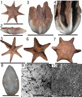
Four novel aspects of colony growth deserve study and documentation: (1) the radial growth pattern; (2) the action of detaching the colony from a hard surface after larval attachment; (3) the formation of a distinctive star-shaped colony with slender radiating rays during early astogeny; and (4) major change during growth from radiating rays to later development of high, vertical vanes (Fig. 2). The term rays/vanes refers to the radial outgrowths from the colony axis that change from slender rays of young colonies to vertical radial sheets on mature colonies.
(1–10) Astogeny of the Evactinopora radiata colony. The reddish color results from inclusion of red clay of the matrix. (1, 2) Top surface and side views of young colony ISGS-3522 with well-developed outrigger rays; (3) side view of mature colony ISGS-3506 with partial vanes.; note wide central gap at base of vanes; (4) side view of mature colony ISGS-3529 with nearly complete vanes; (5–7) top views showing range in ratio of ray diameter to center disk diameter, ISGS-3515, ISGS-3512, and ISGS-3505; (8) accretionary growth lines of outer tip of ray on ISGS-3503, in area without autozooecial chambers; (9) growth layers of prismatic calcite on basal surface of ISGS-3518, converging on mesotheca (median wall of ray) extending across the center of image; (10) fine scale recrystallization fabric of ray skeleton of ISGS-3507. (1, 2) Scale bars = 5mm; (3–7) scale bars = 1 cm; (8) scale bar = 1 mm; (9) scale bar = 200 μm; (10) scale bar = 50 μm.

Following detachment, the colony produced distinctive characters associated with stabilization and feeding activity on a soft-sediment substrate. There are no known modern bryozoan equivalents with stellate growth, and the stellate or cruciform growth form appears to be limited to the Carboniferous and Permian in the genera Evactinopora Meek and Worthen, Reference Meek and Worthen1865 and Evactinostella Crockford, Reference Crockford1957. Documentation of the colony growth of evactinoporids is based on study of the oldest known species, Evactinopora radiata Meek and Worthen, Reference Meek and Worthen1865, but this growth pattern is consistent with known specimens in all other species in the family.
Evactinopora had a free-living life habit that contrasts with three other well-known types of free-living among bryozoan colonies: (1) passive free-living by massive overgrowth, (2) passive free-living without adaptive modifications, and (3) free-living small disks that have the ability to move. Free-living trepostome and cystoporate bryozoans with sheet-like or domal forms are common in Ordovician (Männil, Reference Männil1961) to Mississippian strata (Ulrich, Reference Ulrich1890; Ross, Reference Ross1988), and the form is also developed in Eocene cheilostomes (McKinney and Taylor, Reference McKinney and Taylor2003). This growth form is produced by overgrowth of the attachment surface and spreading onto surrounding areas of the substrate (McKinney and Jackson, Reference McKinney and Jackson1989). They tend to have semi-radial to irregular growth forms and large mass to stabilize themselves on shifting sediment substrate. Secondly, the fenestrate lyre-shaped Mississippian bryozoans, Lyropora Hall, Reference Hall1857 and Lyroporella Simpson, Reference Simpson1895, probably became detached and settled onto sediment, producing a thickened lyre-shaped skeleton lacking stabilization structures (McKinney, Reference McKinney1977). Their growth propagated sideways along the sediment surface (McKinney, Reference McKinney1977; McKinney et al., Reference McKinney, Taylor and Zullo1993) rather than having vertical growth like Evactinopora. Free-living bryozoan colonies having the ability of self-movement are limited to the lunulitid and cupuladriid cheilostomes that occur from the Late Cretaceous to the Recent (Cook and Chimonides, Reference Cook and Chimonides1983). The larvae of lunulitids and cupuladriids settle on fragments of shell and other hard surfaces before overgrowing settlement particles as the colony expands to a thin, rounded disk of usually <1 cm in diameter with a low mass. They are able to propel themselves over the substrate by use of long mandibles of avicularia (McKinney and Jackson, Reference McKinney and Jackson1989, p. 197), like echinoids that utilize spines as mobile stilts for movement. Although most Evactinopora colonies also have diameters averaging 1 cm, they differ from lunulitids and cupuladriids in secreting a dense skeletal base that keeps the colony in a set position on the substrate and in having a vertical growth direction. Evactinopora also develops a complex colony form to optimize feeding. Detachment of the colony from the settlement surface, in contrast to overgrowing, is rare among bryozoans and producing lateral skeletal expansions that stabilize colonies and maintain their vertical growth is unique to evactinoporids.
Evactinopora is the most distinctive fossil in the Fern Glen Formation where it crops out in the vicinity of St. Louis, Missouri, USA (Weller, Reference Weller1909) (Fig. 1), and the genus is best known from that occurrence. The Fern Glen Formation has yielded most of the specimens available for this study.
Several morphotypes of Evactinopora were described in the late 1800s as distinct species, distinguished by the number of rays present on young colonies, but the genus has had no comprehensive study since that time. Published literature contains few details of the generic and specific characteristics of Evactinopora other than gross morphology, with little information provided on zooecial parameters, colony growth, or environmental occurrence (Meek and Worthen, Reference Meek and Worthen1865; Meek and Worthen, Reference Meek and Worthen1868; Ulrich, Reference Ulrich1890). The fragility of the vanes characteristic of fully developed Evactinopora species and post-burial loss of vanes on mature colonies has inhibited documentation of the genus. The current study is made possible by a sample set of >230 specimens that provides a record of changing colony morphology, life habits, and environmental occurrence of Evactinopora radiata. A collection of over 225 specimens obtained from a single locality provides data on morphologic variation present within the species.
Distribution of evactinoporids
The genus Evactinopora in North America is distributed over an area including the middle Mississippi Valley region from the states of Iowa to Arkansas, eastward to Illinois and Kentucky, and southeastward to the southern Appalachian Mountains (Fig. 1). It is abundant in some deposits of the Fern Glen Formation of eastern Missouri (Weller, Reference Weller1909; Unklesbay, Reference Unklesbay1955) and it occurs in the Reeds Spring Formation of southwestern Missouri (Kaiser, Reference Kaiser1950) and the upper Boone Formation of northern Arkansas (Adams et al., Reference Adams, Purdue, Burchard and Ulrich1904; Walter Manger, personal communication, 2018), and the Burlington Formation of Iowa, Missouri, and Illinois (Meek and Worthen, Reference Meek and Worthen1868; Ulrich, Reference Ulrich1890; Crook, Reference Crook1911). The lower part of the Boone Formation of northern Arkansas is coeval with the calcareous shales of the Fern Glen Formation in eastern Missouri. In Kentucky, the genus is recorded from Marion County (Knott, Reference Knott1885) and the New Providence Formation in Lincoln County (Ulrich, Reference Ulrich1890; Butts, Reference Butts1922). Shales of the New Providence Formation are coeval with the Fern Glen Formation in Missouri. In the southern Appalachian Mountains, the genus occurs in the Fort Payne Formation of Alabama (F.K. McKinney, personal communication, 2010).
Reports of Evactinopora outside of North America are limited to Europe and Australia. Ernst (Reference Ernst, Moyano, Cancino and Wyse Jackson2005, p. 51) reported an unnamed Evactinopora species in Germany, but this record was later revised and the material placed in the genus Volgia (Ernst et al., Reference Ernst, Wyse Jackson and Aretz2015). Evactinopora incerta Morozova, Reference Morozova1981, from European Russia, is known only from small cross sections that do not show colony form and, as a result, cannot be verified to be Evactinopora. Evactinopora castletoniensis described from the Mississippian of the Isle of Man in the United Kingdom by Barnes (Reference Barnes1903) was shown by Bancroft (Reference Bancroft1988) to be unrelated to Evactinopora. It consists of radial spinose outgrowths from the reverse surfaces of fenestellid bryozoan colonies. Similarly, Bancroft (Reference Bancroft1988) showed that the genera Palaeocoryne Duncan and Jenkins, Reference Duncan and Jenkins1869 and Claviradix Ferguson, Reference Ferguson1961, named from the Mississippian of England and originally compared to Evactinopora, are also skeletal outgrowths of fenestellid bryozoan colonies, and as such the generic names applied are invalid. A similar misidentification was made when Warren (Reference Warren1927) named two new species of radial fossils having long slender rays with a circular cross-section, collected in the Banff area of Canada. These were later transferred by Nelson and Bolton (Reference Nelson and Bolton1980) to a new genus, Macgowanella, but like the English material, these are radial outgrowths of fenestellid bryozoan colonies and Macgowanella is not considered to have any legitimate taxonomic basis.
In Australia, species of Evactinopora were described by Hudleston (Reference Hudleston1883), Crockford (Reference Crockford1947), and Campbell and Engel (Reference Campbell and Engel1963). Evactinopora crucialis Hudleston, Reference Hudleston1883 is a four-vaned specimen similar to North American E. grandis, but was selected as the type species of the new genus Evactinostella Crockford (Reference Crockford1957) and is distinct from Evactinopora. Evactinostella occurs in Permian strata of Western Australia (Hudleston, Reference Hudleston1883; Crockford, Reference Crockford1957; Håkansson et al., Reference Håkansson, Key and Ernst2016) and the Northern Territory of Australia (Wass, Reference Wass1967). Despite the considerable difference in age and geographic location, there is similarity in colony morphology and environmental setting for Evactinopora grandis and Evactinostella crucialis. A second Evactinopora species described by Hudleston (Reference Hudleston1883), Evactinopora dendroidea, was assigned to Hexagonella by Waagen and Wentzel (Reference Waagen and Wentzel1886), but considered by Hinde (in Nicholson and Hinde, Reference Nicholson and Hinde1890, p. 203) to be an unusual colony growth form of Evactinostella crucialis. Other species from Australia assigned to Evactinopora by Crockford (Reference Crockford1947) and Campbell and Engel (Reference Campbell and Engel1963) include E. irregularis Crockford, Reference Crockford1947, which possesses maculae, and E. vesicularis Campbell and Engel, Reference Campbell and Engel1963. These differ in morphological character and are therefore excluded from the genus.
Preservation
Evactinopora colonies are composed of a skeleton with a center mound of sturdy, strong calcite and distal rays/vanes that are weak and fragile. The center part of the colony remained intact during sediment compaction and seafloor disturbances long after death, and many specimens consist solely of this remnant part of the colony. The thin, weak rays of juveniles and vanes of mature colonies were easily broken from the colony base. Consequently, there are few adult specimens that preserve the distinctive vanes and most specimens look like stubby stars. Determining growth stage is difficult when vanes are missing, producing a bias towards identifying specimens as young growth stage colonies.
Degradation after death occurred by biogenic boring and dissolution as well as by breakage. Evactinopora colonies are commonly marked by Trypanites borings and/or microborings, similar to borings produced by macrobenthos such as annelid polychaete worms and by shell-boring cyanobacteria. Distinct borings are readily recognized, whereas generalized boring and dissolution is associated with irregular or roughened surfaces. No evidence of in-vivo boring could be determined; this would be evidenced by skeletal regrowth around borings and their entrances (cf., Wyse Jackson and Key, Reference Wyse Jackson and Key2007, fig. 3D).
Evactinopora colonies obtained from the Fern Glen Formation at Imperial, Missouri all have skeleton recrystallized to tiny crystals in the 10–50 μm size range. Skeletal microstructure of these specimens is lost as a result of fine-scale recrystallization (Fig. 2.9, 2.10), although larger features, such as accretionary layers of prismatic calcite crystals aligned perpendicular to growth line surfaces, are preserved (Fig. 2.8, 2.9). Despite the recrystallization, detailed structures of autozooecial chamber walls, vesicular structure, growth layers, and the mesotheca remain visible as color variation in the colony skeleton or as a result of sediment infilling of autozooecial chambers.
Evactinopora colonies obtained from the Burlington Formation and equivalent upper Boone Formation in Arkansas commonly have skeleton replaced by diagenetic silica or occur as molds in cherty matrix. Molds of Evactinopora from northern Arkansas preserve fine details of skeleton surface morphology and internal molds of autozooecial chambers, showing the position of mesotheca and the bifoliate arrangement of the autozooecial chambers in vanes.
Materials and methods
Two hundred and thirty Evactinopora radiata colonies were collected from shale of the Fern Glen Formation (Mississippian) in east-central Missouri to determine their morphological characters and variability. Two collections of 180 specimens and of 45 specimens were collected from strata exposed at Imperial, Missouri, a town a short distance south of the city of St. Louis. The larger collection was obtained by taking bulk samples of recently exposed Fern Glen Formation shale and washing the material on 1 mm sieves to remove the fines. The washed shale was picked to collect all recognizable Evactinopora specimens, regardless of preservation quality. Because the basal part of the colony is composed of solid calcite secreted onto the surface of the colony by the bryozoan, all specimens recovered from the rock strata preserve the colony base. Many of them are small, young colonies preserving early growth stages. Five specimens come from small exposures of the Fern Glen Formation from sites near Barnhart, Missouri.
Because of the stellate form of the colonies, specimens collected from the Fern Glen Formation have shale sediment adhering to them. This was removed by mechanical means of scraping the specimens with a blade while being viewed at high magnification under a microscope. Frequent immersion of specimens in water loosened calcareous clays. The slender rays of juvenile colonies and the thin vanes of adult colonies are fragile and commonly fractured by sediment compaction, making preparation difficult. Many colonies prepared in this manner show areas of weakness and breakage caused by sediment compaction and boring by endoskeletozoan organisms (sensu Taylor and Wilson, Reference Taylor and Wilson2002). All specimens were studied and photographed. The damaged specimens often reveal microstructure and subtle features not easily seen on more complete specimens. The collections from Imperial, Missouri provide a good record of survivorship of colonies and growth stages.
Photographs of colonies were obtained by taking multiple images at different focal levels and combining them with the stacking function of Adobe Photoshop. Photographs of the colony top, side, and base and close-up views of diverse morphological characters and skeletal ultrastructure provide an archive on which documentation of colony growth is based.
Repository and institutional abbreviation
Types, figured, and other specimens examined in this study are deposited in the paleontology collections of the Illinois Natural History Survey (ISGS).
Systematic paleontology
Phylum Bryozoa Ehrenberg, Reference Ehrenberg1831
Class Stenolaemata Borg, Reference Borg1926
Superorder Palaeostomata Ma, Buttler, and Taylor, Reference Ma, Buttler, Taylor, Rosso, Wyse Jackson and Porter2014
Order Cystoporata Astrova, Reference Astrova1964
Family Evactinoporidae new family
Genera
Evactinopora Meek and Worthen, Reference Meek and Worthen1865 (type genus herein designated); Evactinostella Crockford, Reference Crockford1957.
Diagnosis
Cystoporate bryozoa with a free-living colony form composed of thickened proximal skeletal tissue and slender rays and/or bifoliate vanes of varying number radiating from a central axis.
Description
Radial pattern of growth after ancestrula attachment to a hard surface, followed by early detachment of basal disk from substrate and dropping to sediment surface; slender outrigger rays containing bifoliate tissue are produced during early growth; vertical radial vanes are produced from top of rays during later growth; vanes joined at a central axis; thick solid carbonate secreted onto base and outer margin of rays and vanes.
Remarks
Family Evactinoporidae n. fam. has characters and morphology determined by a radial growth pattern from a central axis at all phases of life and having a growth phase that allowed detachment of the colony from a settlement surface to become free-living. The growth of slender rays as outriggers on the young colony provided stability and allowed upward growth of vanes containing autozooecia. Mature growth produced a vertical array of radial bifoliate vanes. Colony stability was enhanced by producing heavy thick accretionary stereom on base of the colony. Secretion of solid stereom on lower and outer margins of vanes provided strength for them. Radial growth is the basic design, an adaptation for maintaining an upright orientation, with very limited variation from a simple radial pattern.
Evactinopora was first assigned to the family Cystodictyonidae by Ulrich (Reference Ulrich1884, p. 34), members of which generally possess strap-like bifoliate or trifoliate branches (Utgaard, Reference Utgaard and Robison1983, p. G422), but was later reassigned to the family Hexagonellidae Crockford, Reference Crockford1947. The family Evactinoporidae n. fam. shares characters of bifoliate growth from a mesotheca, the presence of some solid stereom, lunaria, and well-developed vesicular tissue with the family Hexagonellidae Crockford, Reference Crockford1947. The genus Evactinostella Crockford, Reference Crockford1957, from the Permian of Australia, possesses many gross morphological characteristics in common with Evactinopora, and appears to have had a free-living habit following a sessile attached phase.
Genus Evactinopora Meek and Worthen, Reference Meek and Worthen1865
Type species
Evactinopora radiata Meek and Worthen, Reference Meek and Worthen1865 by original designation from the Mississippian of Missouri, USA.
Emended diagnosis
Evactinopora is characterized by a radial growth pattern; colony detachment of the early growth stage from a hard surface, followed by growth of long, slender outrigger rays from the center mound and later development of thin radial vanes grown upwards from the tops of the rays; accretionary growth of solid prismatic calcite on the base and lower and outer surface of the rays/vanes; on large specimens the lower part of the vanes may be curved into low-amplitude folds; autozooecial chambers are recumbent near the mesotheca and curve outward to an orientation nearly normal to the surface; small hemisepta (shelf-like projections) are present in the autozooecial chambers.
Occurrence
USA, Mississippian, Osagean Stage; late Tournaisian Stage. All known specimens in North America occur in Osagean Stage strata.
Remarks
An expanded generic diagnosis is provided for Evactinopora on the basis of need to document that the colony has a detachment scar and to illustrate the distal characteristics of vanes. Vanes rise upward above their attachment area and may became completely separated from each other in the upper portions of the colony, leaving an open space above the center mound. Evactinopora is a distinctive bifoliate cystoporate that is related to Evactinostella (=Evactinoporella). Evactinostella Crockford, Reference Crockford1957 differs from Evactinopora in having large maculae mounds on vane surfaces, which has implications relating to water flow interactions with the colony surface. Evactinopora has hemisepta in autozooecial chambers whereas complete diaphragms are present in Evactinostella.
The genus name Evactinoporella is an invalid name introduced by Mory and Haig (Reference Mory and Haig2011, p. 37) as an incorrect spelling of Evactinostella when they referred to E. crucialis from the Callytharra Formation (Permian) of the Dead Man's Gully locality in the Southern Carnarvon Basin of Western Australia. Evactinoporella constitutes an incorrect subsequent spelling, and as such is considered to be an unavailable name according to Article 33.3 of the International Code of Zoological Nomenclature (ICZN, 1999).
Growth and astogeny of the genus are best documented on E. radiata, the type species for the genus. Small species such as E. radiata were adapted to life on a soft mud substrate, whereas large species (e.g., E. grandis) were adapted to life on a coarser substrate.
Evactinopora radiata Meek and Worthen, Reference Meek and Worthen1865
Figures 2–5, 8–9
Reference Meek and Worthen1865 Evactinopora radiata Meek and Worthen, p. 165.
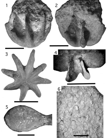
(1–6) Evactinopora radiata Meek and Worthen, Reference Meek and Worthen1865, holotype ISGS-10784. (1) Side view showing vanes with broken upper margins and bowl-shaped base of colony; (2) oblique side view showing thickened outer edge of vanes; (3) base view showing basal thickening, low thin ridge along centerline of vane base, and irregular, off-center detachment scar; (4) top view of part of colony showing thickened outer margins of vanes and thinning of vane toward top: thicker vane on right is broken at lower level than thinner vane on left; brown patch on inner margin of vane is old glue; (5) detail of thickened outer edge of a vane; (6) detail of autozooecial apertures on vanes; orientation the same as in (1). (1–4) Scale bars = 1 cm; (5, 6) scale bars = 1 mm.

(1–12) Comparison of individuals in population of Evactinopora radiata. Top and side views of specimens from the Imperial, Missouri collection site, arranged by number of ray/vane bases and showing thickness of the colony base. Thickness of base correlates with age of colony; the fragile vanes of mature colonies are lost due to preservation breakage. (1) ISGS-3508; (2) ISGS-3509; (3) ISGS-3510; (4) ISGS-3511; (5) ISGS-3504; (6) ISGS-3513; (7) ISGS-3516; (8) ISGS-3517; (9) ISGS-3519; (10) ISGS-3520; (11) ISGS-3521; (12) ISGS-3524. All specimens are scaled to appear as having the same diameter; measured diameters are (1) 5 mm; (2) 5 mm; (3) 5 mm; (4) 6 mm; (5) 6 mm; (6) 4 mm; (7) 4 mm; (8) 7 mm; (9) 5 mm; (10) 7 mm; (11) 8 mm; (12) 7 mm..

(1–6) Evactinopora radiata specimens from Imperial, Missouri collection site showing top (1 = ISGS-3533; 2 = ISGS-3532), side (3 = ISGS-3531; 4 = ISGS-3514), and base (5 = ISGS-3527; 6 = ISGS-3528) views illustrating vanes and top surfaces of the center mound. Note the large exhalent groove on the center mound of (1) (right portion) and bifurcating pattern of mesotheca edges. The appearance of uneven width of ray/vanes of (1) and (2) is an artifact of cropping photos of specimens embedded in matrix. (1, 2) Scale bars = 1 cm; (3–6) scale bars = 5 mm.

Reference Meek and Worthen1868 Evactinopora radiata; Meek and Worthen, p. 502, pl. 17, figs. 2a, 2b.
Reference Meek and Worthen1868 Evactinopora sexradiata Meek and Worthen, p. 502, pl. 17, fig. 3.
Reference Ulrich1884 Evactinopora radiata; Ulrich, p. 42, pl. 2, figs. 1, 1a–e.
Reference Ulrich1890 Evactinopora radiata; Ulrich, p. 509, pl. 73, figs. 3, 3a.
Reference Ulrich1890 Evactinopora sexradiata; Ulrich, p. 510, pl. 73, figs. 2a, 2b.
Reference Ulrich1890 Evactinopora quinqueradiata Ulrich, p. 510, pl. 73, fig. 1.
Reference Keyes1894 Evactinopora radiata; Keyes, p. 19.
Reference Utgaard and Robison1983 Evactinopora radiata; Utgaard, p. G417, fig. 199.2a, 199.2b.
Holotype
ISGS-10784.
Emended diagnosis
Small radial colony (1–3 cm) with slender rays attached to a stubby center mound of young colonies and mature colonies having thin (1–3 mm) vanes of varying number that grow upwards from tops of rays; base with a central detachment scar recording separation of colony from settlement surface; early growth an expanding cone shape with folds and grooves; folds extend into outrigger rays and autozooecial-bearing vanes grow upward from upper surfaces of rays to produce mature colony; vanes extend above the top of center mound; upper surface of center mound has grooves positioned below vane interspaces, with one groove larger and deeper than others; autozooecia surrounded by vesicular tissue; early growth portions of autozooecial chambers are recumbent near the mesotheca and turn toward the vane surface at a high angle; hemisepta present in chambers; base and outer margins of rays/vanes thickened with accretionary solid calcite stereom, with growth layers composed of prisms of calcite.
Occurrence
USA, Missouri, Arkansas; Reeds Spring, Fern Glen, and Burlington formations; Osagean (Tournaisian).
Study collection
Quarry site on north side Seckman Road E, between Interstate 35 and US Rt. 67, north of Rock Creek; Imperial, Jefferson County, Missouri: 38.3744°N, 90.3751°W; also, five samples from Barnhart quarry, beside Mississippi River, Barnhart, Jefferson County, Missouri: 38.l334°N, 90.3779°W.
Remarks
An expanded diagnosis is provided for E. radiata, the type species of the genus Evactinopora named by Meek and Worthen (Reference Meek and Worthen1865) that emphasizes the varied number of vanes developed in colonies and its zooecial and zooarial characteristics. Earlier diagnoses of E. radiata and species considered synonymous herein were short and concentrated on gross colony form. The holotype of E. radiata is a silicified fossil recovered from a chert nodule, collected at an unknown location in Missouri. The presence of crinoid columnals in the sample and occurrence in nodular chert suggest the sample is from the lower part of the Burlington Formation. The holotype of Evactinopora radiata is an eight-vaned colony with thickened outer margins on the vanes. Subsequently, Meek and Worthen (Reference Meek and Worthen1868) described E. sexradiata for a six-rayed colony and E. grandis for a much larger four-vaned colony. Instead of illustrating a six-rayed specimen for E. sexradiata, Meek and Worthen (Reference Meek and Worthen1868) illustrated a five-rayed specimen, an apparent discrepancy pointed out by Ulrich (Reference Ulrich1884). In 1890, Ulrich described Evactinopora quinqueradiata for colonies with five rays, perpetuating the practice of naming species based on the exact number of rays/vanes present in the colony. Weller (Reference Weller1909) stated that E. radiata has a variable number of rays, although he accepted E. quinqueradiata Ulrich, Reference Ulrich1884 as distinct from E. radiata on the basis of having more slender rays and a compact disk-like center mound.
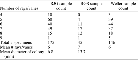
The normal practice for species determination in Bryozoa is based on characters of the autozooecial chambers, their arrangement and budding patterns, the presence or absence of heteromorphic zooids, and on wall microstructure, not on branch number in colonies. Weller (Reference Weller1909) presented data for a collection of colonies showing variation from four to nine rays (Table 1), but did not propose new species names for the more multi-rayed specimens, nor did he make recommendations on validity of named forms. Shimer and Shrock (Reference Shimer and Shrock1944) list E. radiata and E. grandis as species without reference to other species names. Utgaard (Reference Utgaard and Robison1983) presented a diagnosis of the genus similar to that of Meek and Worthen (Reference Meek and Worthen1868) as having 4–8 rays/vanes, but used the name E. sexradiata for specimens from the stratigraphic interval below the Burlington Formation in Missouri. Large-diameter specimens of Evactinopora observed in field outcrops typically have four or five rays/vanes, whereas smaller specimens have greater variation in number of rays/vanes. We conclude that the practice of recognizing species of Evactinopora based on an exact number of rays/vanes in the colony is of little value.
Number of rays/vanes in specimens of Evactinopora radiata in the two collections examined for this report (RJG = Richard J. Gottfried; BGS = Barry G. Sutton) compared with data published by Weller (Reference Weller1909).

This study of 230 Fern Glen Formation colonies of E. radiata supports the conclusion that species in the genus have a variable number of rays in the colony and that the size of the center disk changes during growth (Fig. 4). Mature colonies have characters consistent with growth patterns established on young specimens. The varied morphologies used by Meek and Worthen (Reference Meek and Worthen1865, Reference Meek and Worthen1868) and Ulrich (Reference Ulrich1890) for species definitions can be found within a large population of E. radiata. The size and shape of the center mound also varies within a population, and post-deposition dissolution produces even more variation to preserved colony shape. Without the identification of other characters that are consistent within a collection of many individuals, there is no basis for using ray/vane count as a character for species determination. Consequently, E. sexradiata and E. quniqueradiata are synonomized with E. radiata.
Evactinopora grandis Meek and Worthen, Reference Meek and Worthen1868
Figure 6
Reference Meek and Worthen1868 Evactinopora grandis Meek and Worthen, p. 503, pl. 15, figs. 2a, 2b.
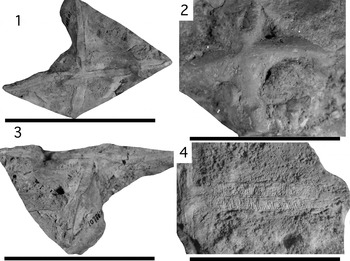
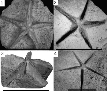
(1–4) Evactinopora grandis Meek and Worthen, Reference Meek and Worthen1868, syntype ISGS-10786-A. (1) Top view of eroded upper surface looking down along center axis; vane extending to lower edge of photo is eroded to lower level than other vanes and appears shorter and thicker than others, but has similar form; vane extending to upper edge of photo shows large curve on base of vane; (2) base view showing thickened base and lines along vane bases; vanes show minor curvature on lower portions; dimples close to center and vane bases produced by contact with bioclast crinoid columnals; (3) oblique top view showing large curve on vane in foreground that is largest on base and fades out toward top; (4) closeup of axis and lower part of vane showing growth lamellae, mesotheca, autozooecial chamber growth, and outward growth of vane with non-zooid bearing skeleton on outer margin. (1, 3) Scale bars = 10 cm; (2, 4) scale bars = 4 cm.

Reference Ulrich1890 Evactinopora grandis; Ulrich, p. 511, pl. 73, fig. 4.
Reference Keyes1894 Evactinopora grandis; Keyes, p. 19.
Cotypes
ISGS-10786-A and ISGS-10786-B.
Emended diagnosis
Large radial colony (to 12 cm diameter) with four vanes; vanes attached to thick center axis of the colony and taper outward; vane thickness greatest (to 0.8 cm) at or near center axis; center formed by merged inner margin of vanes and lacking center disk; lower portion of vanes may be curved into low amplitude folds that fade upward into a planar sheet; vanes covered on both sides with closely packed autozooecial chambers, except on outermost and basal margin of vanes; on inner part of vanes the autozooecial chambers extend out perpendicular to the outer surface of the vane.
Occurrence
USA, Iowa, Illinois; Burlington Formation; Osagean (late Tournaisian).
Remarks
Evactinopora grandis grew to a size of up to 12 cm in diameter and produced very thick vanes, but is known only from the lower part of the mature colony. It has the basic characteristics of evactinoporid radial growth and production of large vanes bearing many autozooids formed with a bifoliate growth pattern from mesotheca. Outer edges of vanes are formed of solid calcite lacking autozooecial chambers or vesicles, the base is formed of thick growth lamellae of solid calcite and is shaped like a shallow bowl. Vanes have a minor ridge on the base corresponding to the edge of the mesotheca. Specimens confidently identified herein as this species have four thick vanes, but five-vaned specimens also occur.
Evactinopora mangeri new species
Figure 7
Holotype
ISGS-3501, Osagean (late Tournaisian), upper Boone Formation, Goshen, Arkansas.
Paratype
ISGS-3502, Osagean (late Tournaisian), upper Boone Formation, Boone County, Arkansas.
Diagnosis
Large diameter (to 10 cm) Evactinopora with few (4–5), wide-spaced, low-arched vanes that extend to center axis of colony above base; vane width tapers outward at a low rate of change.
Occurrence
USA, Arkansas; upper Boone Formation; Osagean (late Tournaisian); holotype: Richland Creek, Goshen, Arkansas: 36.1004°N, 94.0010°W; paratype: Boone County, Arkansas: 36°N, 93°W.
Description
Large radial colony (to 10 cm diameter) with 4–5 thin vanes; vanes attached to small center axis and taper outward at low rate of change in thickness; vane thickness greatest (to 0.6 cm) at or near center axis, slightly narrower at attachment to center axis; center formed by merged inner margin of vanes, with minimal center mound and lacking center disk; vanes extend up in a low arch above center axis; vanes covered on both sides with closely packed autozooecial chambers except on outermost and basal margin of vanes; on inner part of vanes the autozooecial chambers extend out angled at a 60–70° to the outer surface of the vane, on distal part of vanes autozooecial chambers are oriented at a lower angle; autozooecial apertures circular with well-developed lunaria; base of colony has shape of a shallow bowl; slight ridge on base of vanes over the mesotheca margin; skeleton composed of thin growth lamellae.
Etymology
The species is named to honor Walter Manger of the University of Arkansas who provided well-preserved moldic fossils of Evactinopora from Arkansas and provided locality and stratigraphic information on these fossils.
Remarks
Evactinopora mangeri n. sp. is similar to E. grandis, but differs in having four or five vanes, thinner (max. 0.6 versus 0.8 cm) and shorter (max 5 versus 6 cm from axis) vanes, less-massive base, thinner growth lamellae, and lacking folds on the base of vanes. Autozooecial chambers grow at an angle to the outer vane surface instead of being perpendicular. Evactinopora mangeri n. sp. differs from E. radiata in having broader low-arched vanes that contrast with the high, narrow blade-like vanes of E. radiata and in having minimal development of a center mound.
The specimens examined are consistently smaller and less massive than E. grandis, with no intermediate examples known in collected specimens or field photographs of specimens. The distinct difference in vane thickness on nearly similar-diameter colonies suggests that E. mangeri n. sp. would not grow to the dimensions of E. grandis. There is also a geographic separation in distribution, with E. grandis occurrences known in states of Iowa and Illinois, USA, whereas E. mangeri n. sp. occurrences are known in the state of Arkansas, USA, of central North America (Fig. 1). This corresponds with locations closer to the Burlington Shelf margin for E. mangeri n. sp. and more inboard for E. grandis.
Evactinopora growth and functional morphology
A distinguishing feature of the genus is its radial growth morphology and distinctive rays and vanes developed during early and later stages of colony development (Figs. 2, 8). After larval settlement, a basal disk of zooid tubes formed by asexual budding from the ancestrula (cf.. McKinney and Wyse Jackson, Reference McKinney and Wyse Jackson2015, fig. 22). Growth from the basal disk was upward and, if settlement occurred on a non-horizontal surface, the new growth curved into an upright orientation, sometimes by producing a short stalk (Fig. 8). At this point in development, growth produced a cone shape with folds on the sides that expanded outward. The folds subsequently became the bases of radial rays of the young colony. Detachment of the basal disk from its substrate occurred at some time during this interval of cone growth.
Detachment produced a rounded scar on the base of the colony that remained visible during later growth (Fig. 8). Rare examples of the detachment surface show the bases of autozooecial chamber walls (Fig. 8.3) produced by earliest growth. Young colonies have clean but moderately irregular separation surfaces, suggesting weak attachment that allowed separation. The number of folds and rays that developed in the young colony is probably determined by the number of zooids budded from the ancestrula in the basal attachment disk (Pachut and Fisherkeller, Reference Pachut and Fisherkeller2011).
There is variation in the amount of accretionary skeleton deposited onto the detachment area during later growth. The separation scar tends to remain as a flattened or irregular area on the base of the colony, contrasting with the smooth curved layers on other parts of the thickened colony base. On some colonies the detachment scar was not completely covered and remains as a pit on the base. On many colonies the calcite that secreted over the detachment scar incorporates clay sediment of the substrate, giving it a color difference from surrounding uncolored white skeleton. Outside the boundary of the detachment scar, the center line of a ray/vane on the colony base is marked with a low ridge along the mesotheca margin.
Young Evactinopora colonies produced a raised center mound of skeletal tissue concurrent with growth of slender rays extending outward (laterally) into a distinct star-like colony (Figs. 4, 5). The rays are radial outrigger projections used for stabilization and maintaining an upright orientation to keep the upper feeding areas of the colony in the water column. The ray bases grew with a shallow upward curvature, giving the colony a rounded base. The base thickened as accretionary solid calcite was secreted onto it. The center of gravity of the colony is in the thickened base of the skeleton, allowing it to serve as a heavy anchor weight for stabilizing as well as strengthening the colony. During later growth, the curved bowl-shaped base of E. radiata changed from a shallow bowl shape to a deeper bowl shape. Accretionary layers added onto the base of the colony rays/vanes (Fig. 2.8, 2.9) continue up the outer edge of vanes, producing a strengthened margin for the rays/vanes (Fig. 3). This indicates the colony base remained covered by a layer of living tissue capable of secreting skeletal calcite. The accreted calcite layers contain no autozooecial chambers, although Utgaard (Reference Utgaard and Robison1983, p. G409, G411, fig. 2f) noted the presence of acanthostyles (skeletal spines cross-cutting stereom, herein termed microstylets) in thin-section samples. Microstylets have not been seen on the smooth outer surfaces of specimens in this study, but these fine skeletal cross-cutting features have probably been obscured by recrystallization.
Evactinopora grew vertically aligned radiating vanes from the tops of the slender rays of the young colony (Figs. 4, 5, 8). Colony growth filled in the proximal space between rays to produce a perimeter with scalloped margins (Figs. 2, 4, 5). The size of the center mound increased with growth, but varies, so the ratio of center-mound diameter to ray diameter ranges from ~0.1 to 0.8. Post-depositional breakage of rays/vanes of mature specimens also produces a continuum in the center-mound/ray ratio, making this measure unreliable for species determination unless based on well-preserved specimens.
The number of rays/vanes of colonies varies from four to nine (Table 1), similar to the count presented by Weller (Reference Weller1909), but specimens with nine are rare. No specimens were found with three rays. The number of rays/vanes rarely changed during colony growth, although rays/vanes are not all of the same size within a colony. Only one specimen among 230 colonies examined has a ray that branches at a point away from the center axis. This is assumed to be produced by a point mutation originating during ray growth after the initial ray branching from the colony center. Specimens with four rays are all quite small, suggesting that possessing a small number of rays inhibited chances of survival in the mud bottom environment favored by E. radiata.
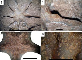
The colony center thickened at maturity into a mound, but does not extend to the highest part of the colony. It contains few autozooecial chambers. The upper surface of the center mound is sculptured with ridges corresponding with inner portions of rays/vanes and grooves radiating outward between the rays/vanes (Fig. 9). The crest of each ridge is marked with a thin dark line that is the upper edge of the mesotheca. On mature colonies the inner portions of ridges are deflected around grooves, changing the simple radial pattern of mesotheca growth from the center axis into a surface pattern of bifurcating branching mesotheca walls (Fig. 9.4). One groove on this upper surface of the center mound is broader and deeper than other grooves and is assumed to have functioned in providing a larger channel for directing the flow of exhalent water away from the colony, as an aid in feeding and the removal of waste.
The transformation from ray growth to vane growth occurred as low ridges developed on the upper surface of the rays and continued to grow upward into thin, bifoliate planar vanes that fit within the limits of a cylinder space (Fig. 8.5). In E. radiata, the vanes grew upward, but not outward, producing a colony that is taller than wide. The upper portion of vanes do not extend to the center axis. This leaves an open central space in the upper parts of the colony. In E. grandis, the vanes meet at the center of the mature colony and form a solid center axis. The outer and lower margins of vanes are thickened with solid calcite, while inner portions (toward the axis) are thinner and contain many autozooecial chambers arranged in a regular array on both sides of the vane. Vanes tend to have varying thickness from proximal to distal margins, and on E. radiata, the thinnest portion is located in mid-reaches of the vane (Fig. 9.2). On E. grandis, the vanes tend to taper from the center axis to the outer margin.
Autozooecial chambers are densely spread on the upper parts of the rays and vanes (Figs. 2–8) and originate from branching sheets of mesotheca. The arrangement of autozooecia is irregular on lower parts of the colony because of variation in shape and thickness of colony skeleton, but are arranged in rows of quincunx formation on vanes. Vesicular tissue, or ‘vesicular interstitial tissue’ of some earlier usage (Ulrich, Reference Ulrich1882, p. 127), separates the autozooecia and forms half or more of the skeleton volume in the lower part of the colony where autozooecia are irregularly arranged.
(1–3) Evactinopora mangeri n. sp. (1) Cast of upper part of holotype specimen ISGS-3501 showing vanes (tops are incomplete in cast) and three large exhalent grooves between vanes on center mound; (2) mold of upper part of holotype specimen ISGS-3501 showing dense array of sediment-filled autozooecial chambers and colony center; (3) side view of cast of upper part of holotype specimen ISGS-3501 showing low arch of incomplete vanes; upper Boone Formation, Goshen, Arkansas; (4) mold of lower part of paratype specimen of E. mangeri n. sp. ISGS-3502 showing base; upper Boone Formation, Boone County, Arkansas. Scale bars = 5 cm.

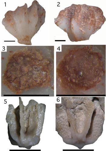
(1–6) Growth stages of Evactinopora radiata. (1, 2) Detachment stage of growth showing developing folds and detached base, with some breakage of vane margins; white is solid calcite; brownish areas contain some mud infill; specimens ISGS-3525 (left) and ISGS-3526 (right). (3, 4) Detachment scars of E. radiata colonies with subrounded to slightly irregular outline; scar on left specimen shows bases of chamber walls; ISGS-3525 (left) and ISGS-3526 (right). (5, 6) Small mature colonies with vanes showing autozooids; specimens ISGS-3530 (left) and ISGS-3531 (right). (1–4) Scale bars = 1 mm; (5) scale bar = 1 cm; (6) scale bar = 5 mm.

Autozooecial apertures are circular in shape, with a marginally elevated semi-circular lunaria situated on the proximal margins of apertures. Autozooecia chambers are recumbent for a short distance near the mesotheca, with a small hemiseptum on the outer zooecial wall at the point where the chambers bend towards the outer surface of the vane. The outer portions are oriented perpendicular to the surface on vanes. Autozooecia are spaced two or more diameters apart and separated by blister-like vesicles.
The presence of lunaria on the proximal side of the autozooecial aperture indicates the open, everted lophophore was inclined towards the top of the colony. Generation of a water current by the beating of cilia on lophophore tentacles would produce an inhalant current drawing water from the uppermost part of the colony before splitting into sub-flows between vanes, one in each intravane area, with water moving downward toward the center disk, becoming an exhalent waste-laden current. Grooves on the center mound directed depleted water away from the colony, with the deepest groove (Fig. 9) channeling most of the flow.
Vane separation is much greater on Evactinostella, so the pattern of water flow would be significantly different, drawing an inhalent current inward at a perpendicular angle toward the vane rather than laterally across the vane as in Evactinopora. Maculae on the vanes act as exhalent chimneys for depleted water that is directed away normal from the vane surface (Håkansson et al., Reference Håkansson, Key and Ernst2016). The need for macular development in Evactinostella is a result of the broader width and wide spacing of vanes that make it difficult to generate efficient colony-wide exhalent currents.
(1–4) Features of the center mound of Evactinopora radiata. (1, 2) Irregular surface of center mound with dominant and deeper groove in lower right and detail; variable thickness and height of ray/vane with thinnest part in mid area; ISGS-3533; (3) center mound of young colony; ISGS-3511; (4) detail of the branching pattern of mesotheca on crests of rays/vanes of mature colony; ISGS-3523. Mud-filled circles of autozooecial apertures and breached vesicles. (1, 3) Scale bar = 1 cm; (2, 4) scale bar = 1 mm.

Discussion
The production of thick, centrally located basal skeleton produced a low center of gravity for the evactinoporid colony and the thin, radiating rays spread the colony mass over a wider area of sediment, providing a mechanical means to prevent tipping and maintain an upright orientation, using the same principles for orientation as ballast and outriggers provide for boats. This acted to stabilize the young colony after detachment and helped maintain an upright orientation for the mature colony, a critical step in survival of evactinoporids under seafloor conditions. With stability, the colony was able to extend upwards and produce sheet-like vanes of bilaminate skeleton supporting autozooecia to feed in the bottom waters. The combination of early detachment, ray formation in young colonies, and heavy calcite secretion at the colony base are all interrelated features that produced a successful life style for these free-living bryozoans. There is little departure from this pattern of colony development among evactinoporid species. The production of outrigger rays to achieve stability for the young colony is a defining feature of evactinoporids.
Another defining feature is the detachment and separation of the colony from a solid substrate. Other free-living bryozoans, such as lunulitids and large trepostomes, attach and then engulf the settlement object, whereas evactinoporids consistently detached and dropped to the seafloor. It is possible that the separation was aided by secretions to weaken the attached portion of the skeleton, but that cannot be tested with the available material. A similar type of basal disk detachment was inferred for the flat-lying fenestellid Lyropora, but detachment is not shown to be a consistent part of the life cycle of the genus (McKinney, Reference McKinney1977). Detachment was the definitive action that led to origination of the distinctive radial characters of evactinoporids.
Species of Evactinopora occur in sediments ranging from fine-grained mudstone to muddy bioclastic sediment, similar to the range of substrate recorded for modern free-living lunulitid bryozoans (McKinney and Jackson, Reference McKinney and Jackson1989, p. 192). Evactinopora radiata Meek and Worthen, Reference Meek and Worthen1865, occurs in fine-grained mudrocks associated with normal low-energy water conditions and formed colonies that grew to ~1 cm in diameter, whereas the more robust species E. grandis Meek and Worthen, Reference Meek and Worthen1868 formed colonies up to 12 cm in diameter and occurs in coarser, bioclast-rich sediment associated with sporadic high-energy water conditions. Although colony dimensions and details of rays/vanes design are different for these species, they have the same overall colony morphology and growth habit.
The conditions associated with the origin of evactinoporids can be related to the long-term environmental stability of the Burlington platform in the Mississippi Valley region of North America (Lane and De Keyser, Reference Lane, De Keyser, Fouch and Magathan1980; Shelby, Reference Shelby1986; Kincade, Reference Kincade2016). The Burlington platform was a carbonate shelf that supported large populations of suspension-feeding organisms, especially crinoids. These organisms produced large volumes of bioclastic sediment and covered the platform surface with bioclastic carbonate deposits, while muddy sediment accumulated on the southern margin ramp of the platform (Lane and De Keyser, Reference Lane, De Keyser, Fouch and Magathan1980; Kincade, Reference Kincade2016). Areas of mud sediment cover on central and marginal portions of the platform would have been excellent places for opportunistic colonization of evactinoporids.
Conclusions
The radial growth pattern that characterizes Evactinopora is best explained as a result of adaptation to free-living on a soft substrate, produced by selection for colonies able to maintain an upright orientation on the sea floor after detachment of the bud mass from a hard substrate. Colonies that secreted thick, solid basal stereom for ballast and produced radial rays for outrigger stability would be favored to survive to reproductive maturity, allowing natural selection on successive generations to favor the novel characters of Evactinopora. The greatest change is the process of detachment from hard substrate, which is rare among Bryozoa. The need for stabilization on the soft surface was met by generating outrigger support with long slender rays radiating from the detached colony—another novelty. Growth in Evactinopora produced thin vanes of bifoliate skeleton grown upward from the tops of rays, providing a large surface area for feeding. Concurrent thickening of the center area of the colony and secretion of massive overgrowth on the base produced a low center of gravity for the colony—a mechanism to maintain stability and keep the feeding area orientated upward. These novel characters are related by function and retained as a character group in response to adaptation to life on a sediment surface with few hard surfaces for a bryozoan to use for attachment. Free-living became a way to overcome that limitation.
The star shape is one phase of colony growth of Evactinopora. The greater change involved growth of vertical sheets of skeleton densely packed with autozooecia that maximized food gathering and enabled colony growth. It is assumed that vane production occurred rapidly and abundant food probably led to rapid maturation and release of gametes for reproduction and formation of larvae. The small colony size and fragile nature of feeding vanes of E. radiata suggests a short-duration life cycle and opportunistic life style on the mud substrate seafloor. The larger thick-vaned species E. grandis adapted to life on coarse-grained sediment among crinoid meadows. Life on a coarser-grain substrate is associated with production of thicker vanes and a heavier skeleton, a relationship also noted in Evactinostella from the Permian of Western Australia, which lived on carbonate sand (Håkansson et al., Reference Håkansson, Key and Ernst2016).
There is no basis for recognizing species of Evactinopora defined as having an exact number or rays/vanes in the colony. The species E. quinqueradiata and E. sexradiata are synonomized with E. radiata. The larger-diameter E. grandis and E. mangeri n. sp. are distinct from E. radiata based on wider vane spacing, larger size and colony mass, low arching vanes, and minimal development of a center mound. The species names E. castletoniensis, E. incerta, E. irregularis, E. trifoliata, and E. vesicularis are excluded from the genus Evactinopora at this time. Evactinopora mangeri n. sp. is erected as a new species for larger-diameter Evactinopora with slender, slightly tapering vanes.
A new family, Evactinoporidae, is established for the evactinoporids, a unique group of free-living bryozoans that started life with detachment of the basal disk from the larval settlement surface. The surface of attachment is shown on some specimens, although there is skeletal overgrowth of the base on most specimens. This points to the spread of skeleton-secreting tissue covering the detachment surface. Upward radial growth produced changes in form starting with a cone shape changing to a radial star shape with slender rays and maturing to a tall cylindrical form with radiating vertical vanes. These characters are associated with stabilization and enhanced feeding ability, allowing evactinoporids to colonize and prosper on fine-grained mud substrate. Detachment probably occurred very soon after larval settlement and the initial growth of a basal disk, but may have occurred later at a young ray growth stage.
Acknowledgments
We are very grateful for the enthusiastic support of people who provided specimens for study and knowledge of the formations containing the fossils. C. Cook prepared and provided some specimens and stimulated much interest in fossils with his excellent work as a preparator. C.D. Cozart provided specimens collected in eastern Missouri and photos of specimens from northern Missouri and adjacent Illinois. J. and D. Stade donated specimens from the Mark Twain Reservoir area in Missouri. W. Manger, University of Arkansas, donated important specimens from the upper Boone Formation of Arkansas and provided information on the stratigraphic position and environment of deposition of early Mississippian strata in Arkansas. P. Mulvaney of the Missouri Department of Natural Resources and J. Devera of the Illinois Geological Survey provided information on geologic formations in those areas and about known occurrences of the genus in those units. J. Thomas and S. Heads of the Illinois Natural History Survey arranged for loan of type specimens that are illustrated and described in this report. K. Hollis, U.S. National Museum, provided data on samples with USNM collection numbers. In addition, B.G. Sutton would like to thank the many people who have been engaged in discussions and compared observations about these interesting fossils that are sparsely represented in formal collections. Details of his long-term collecting and studies of this bryozoan are documented in the webpage: Evactinopora Research Project—Mississippian bryozoan fossil; https://www.lakeneosho.org/Evactinopora/. Images of skeletal microstructure were provided by J. Reese of the Department of Geology and Geophysics, Texas A&M University, and the Microscopy Imaging Center of Texas A&M University.