Stress is a complex physiological and psychological response triggered by internal and external stimuli and is a significant global health challenge with a reported prevalence of 29·6 % in the general population and 44·9 % in healthcare workers(Reference Salari, Hosseinian-Far and Jalali1). Stressors can range from catastrophic natural disasters such as earthquakes, floods, storms and droughts to life stressors such as relocation, hazardous work environments, dehydration, sleep deprivation, extreme temperatures, financial difficulties and environmental factors such as light, humidity and vapours(Reference Goel, Basner and Rao2–Reference Sobolewski, Młynarczyk and Konarska4). Chronic exposure to stressors severely impacts physical, metabolic and mental health, manifesting as immunosuppression, hyperglycaemia, gut dysbiosis, villous atrophy, reduced enzyme activity (e.g. trypsin, chymotrypsin and amylase), oxidative stress, anxiety and sleep disturbance, all of which contribute to a decline in overall quality of life(Reference Ringseis and Eder5–Reference Yaribeygi, Panahi and Sahraei8).
The liver and intestine are particularly vulnerable to stress-induced damage because of their interrelated roles in the gut-liver axis, which is critical for maintaining metabolic and immune homeostasis. Chronic stress disrupts the integrity of the intestinal barrier, leading to adverse health effects. Stress can lead to uncoordinated epithelial cell desquamation, impaired tight junction function and decreased expression of mucin proteins (MUC-1 and MUC-2), resulting in compromised intestinal barrier integrity(Reference Yi, Xiong and Wu9,Reference Zhang, Zhao and Yang10) . These perturbations further impair nutrient absorption, villus morphology and transporter activity, leading to systemic metabolic and immune dysregulation(Reference Orhan, Tuzcu and Deeh11,Reference Yan, Tong and Tang12) . Specifically, stress alters the expression of key nutrient transporters responsible for cellular nutrient uptake. Fatty acid binding proteins (FABP) and fatty acid transport proteins (FATP) facilitate lipid absorption, GLUT and sodium-glucose co-transporters (SGLT) mediate carbohydrate absorption, and peptide transporters 1 (PepT1) and 2 (PepT2) regulate protein absorption(Reference Chen, Yong and Ju13). In addition, stress exacerbates liver dysfunction through mechanisms involving lipid accumulation, inflammation and apoptosis mediated by endoplasmic reticulum stress(Reference Chen, Yong and Ju13–Reference Ma, Xing and Li15). These adverse effects require effective stress management strategies. Conventional pharmaceutical treatments, including anxiolytics, sedatives, antidepressants and beta-blockers, are limited by their adverse side effects(Reference Sartori and Singewald16). This has led to increased interest in natural bioactive compounds with minimal side effects, such as magnesium and L-theanine, which have shown promise in alleviating stress-related symptoms. Magnesium, an essential mineral involved in various cellular processes, plays a key role in the regulation of synaptic plasticity, hippocampal neurogenesis and sleep through N-methyl-D-aspartate receptor antagonism(Reference Slutsky, Abumaria and Wu17–Reference Abbasi, Kimiagar and Sadeghniiat19). Similarly, L-theanine, a bioactive amino acid derived from green tea, exerts anxiolytic and neuroprotective effects by enhancing gamma-aminobutyric acid receptor activity, upregulating antioxidant enzymes and reducing oxidative stress-induced damage, thereby improving sleep quality and attenuating stress responses(Reference Egashira, Hayakawa and Osajima20–Reference Deb, Dutta and Phukan22).
Magnesium-L-theanine (Mg-T) is a novel compound formed by the chelation of magnesium ions with L-theanine, designed to leverage the individual and complementary benefits of its constituents in managing chronic stress. While both magnesium and L-theanine have established roles in stress regulation through modulation of the hypothalamic-pituitary-adrenal axis, neurotransmitter balance and redox homeostasis, recent studies suggest that their combination in a complex form yields superior outcomes. For instance, Boyle et al. (Reference Boyle, Billington and Lawton23) demonstrated that co-administration of magnesium and theanine was more effective in reducing stress-induced behavioural symptoms compared to either agent alone. Similarly, Dasdelen et al. (Reference Dasdelen, Er and Kaplan24) showed that Mg-T supplementation not only enhanced sleep quality but also increased brain levels of serotonin, dopamine and melatonin more significantly than individual treatments. These enhanced effects are thought to arise from the improved pharmacokinetic profile of the Mg-T complex, which facilitates better bioavailability, sustained neuromodulatory activity and greater central nervous system penetration, including elevated cerebrospinal fluid magnesium levels, as demonstrated in preclinical models(Reference Noah, Morel and Bertin25). Moreover, Mg-T’s potential has been recognised in recent patents(Reference Nelson and Komorowski26), further supporting its distinct mechanistic advantages over uncombined forms. Despite its emerging use for sleep and anxiety, the impact of Mg-T on liver metabolism, intestinal barrier integrity and stress-responsive protein expression remains under-investigated.
This study aims to investigate the effects of Mg-T supplementation in rats subjected to chronic variable stress (CVS). We assessed serum corticosterone, adrenocorticotropic hormone (ACTH) and insulin levels, as well as hepatic metabolic markers, including NAD+, nicotinamide (NAM) and nicotinic acid (NA). In addition, we analysed the expression of key metabolic regulators such as SIRT1, PPARγ and lipogenesis-associated proteins (SREBF1 and FASN) as well as insulin signalling proteins (GLUT2 and IRS1). To elucidate the impact of Mg-T on intestinal integrity, we examined intestinal morphology, mucosal barrier proteins (Claudin-1, Claudin-4, Occludin and ZO-1) and nutrient transporters (PepT1, LAT2, EAAT3, FABP2 and FATP4). Our results provide novel insights into the potential of Mg-T to mitigate CVS-induced metabolic dysfunction and intestinal barrier disruption.
Materials and methods
Animals
A total of 56 male Sprague-Dawley rats (8 weeks old, 180 ± 20 g) were obtained from the Firat University Experimental Research Center. Rats were housed in a controlled environment with a 12:12 h light/dark cycle at 22°C and a relative humidity of 55 ± 5 %. Animals had ad libitum access to standard rat chow and drinking water. All procedures were conducted in compliance with the ARRIVE guidelines. They were conducted in strict compliance with the relevant laws, the Animal Welfare Act, the Public Health Services Policy and guidelines established by the Institutional Animal Care and Use Committee of the Institute and the European Union guidelines (No. 2010/63/EU) for the protection of the laboratory animals used for scientific purposes. All the procedures involving animals were approved by the Local Ethics Committee for Animal Experiments of Firat University (Protocol number: 27.02.2024-22557).
Experimental design
Mg-T is a novel compound consisting of magnesium ions chelated with L-theanine ligands, forming a stable coordination complex that enhances solubility and biological activity compared to its individual components. Mg-T was formulated into controlled-release matrices, including hydrogels, rate-controlling membrane systems or soluble oral dosage forms (e.g. lozenges, tablets and sublingual liquids), all of which support gradual and targeted delivery across gastrointestinal environments (US20210308081A1). The Mg-T composition contains 6 % magnesium and 94 % L-theanine and was provided by JDS Therapeutics, LLC (NY, USA)(Reference Dasdelen, Er and Kaplan24). Since Mg-T has not previously been tested in a stress model in rats, the dose range used in this study was selected based on the effective doses of its components, magnesium and L-theanine, reported in various disease or oxidative stress models in rodents(Reference Zeng, Lin and Peng27–Reference Sumathi, Shobana and Thangarajeswari30). Accordingly, Mg-T was administered at 100, 200 and 400 mg/kg body weight in order to cover a range of biologically relevant doses and to determine the most effective dose for preventing stress-induced physiological and metabolic disturbances. The doses were selected to allow a linear escalation, enabling the assessment of potential dose-response relationships and to identify the optimal effective dose with minimal side effects.
The study was designed to evaluate the effects of different doses of Mg-T under normal and CVS conditions in a 2 × 4 factorial design with two factors: environmental condition (normal v. stress) and four doses of Mg-T (0, 100, 200 and 400 mg/kg body weight). The rats were randomly assigned to eight groups of seven animals each (n 7 per group): (1) control group, maintained under normal conditions and given 1 ml of water daily; (2) Mg-T 100 group, maintained under normal conditions and given 100 mg/kg of Mg-T; (3) Mg-T 200 group, maintained under normal conditions and given 200 mg/kg of Mg-T; (4) Mg-T 400 group, maintained under normal conditions and given 400 mg/kg of Mg-T; (5) stress group, exposed to CVS and given 1 ml of water daily; (6) stress + Mg-T 100 group, exposed to CVS and given 100 mg/kg of Mg-T; (7) stress + Mg-T 200 group, exposed to CVS and given 200 mg/kg of Mg-T and (8) stress + Mg-T 400 group, exposed to CVS and given 400 mg/kg of Mg-T. Mg-T was prepared freshly every day by dissolving in drinking water and administered to rats via oral gavage (1 ml/rat) once daily at the same time for 40 d throughout the CVS protocol. The same volume of drinking water was administered to the control groups (Mg-T 0) by oral gavage.
Stress protocol
Rodents treated with CVS were exposed to a series of stressors administered in an unpredictable manner over an extended period of time. The CVS protocol involved the daily application of variable stressors for 40 d, including cold exposure, water restriction, flashing lights, restricted movement, wet bedding and cage tipping (online Supplementary Table S1). The stressors were applied in a randomised order to minimise habituation, as recommended by Tagliari et al. (Reference Tagliari, Noschang and Ferreira31).
Laboratory analyses
At the end of the experiment, serum, liver and intestinal tissues were collected from the sacrificed rats by decapitation. Tissue samples for histological examination were stored in 10 % formaldehyde solution, while those for molecular analyses were stored at −80°C. Serum glucose, triglyceride, urea and creatinine concentrations and alanine aminotransferase (ALT) and aspartate aminotransferase (AST) activities were measured using a biochemical autoanalyser (LABGEOPT10V-Samsung, Labgeo, South Korea) with rat-specific kits.
Serum corticosterone, ACTH and insulin levels and liver NAD+, NAM, NA and NADPH were measured using an ELISA device (Elx-800; Bio-Tek, Vermont, USA) according to the procedures of commercial kits (SunRed, Shanghai, China). The ELISA kits used in this study demonstrated high sensitivity and reliability, with detection limits and assay ranges as follows: ACTH (5·226 ng/l; 7–1000 ng/l), corticosterone (5·005 ng/ml; 6–1000 ng/ml), insulin (0·102 mIU/l; 0·2–40 mIU/l), triglycerides (0·05 mmol/l; 0·05–15 mmol/l), NAD+ (2·256 nmol/ml; 5–900 nmol/ml), NAM (3·897 nmol/l; 5–1000 nmol/l), NA (2·362 µmol/ml; 2·5–700 µmol/ml) and NADPH (2·356 nmol/l; 3–600 nmol/l), all with intra-assay CVs < 10 % and inter-assay CVs < 12 %.
For the determination of magnesium in feed and serum, samples were processed using a closed-system microwave digestion method (Berrghorf, Eningen, Germany). For digestion, 250 mg of each sample was placed in digestion tubes together with 8 mL of nitric acid (65 %, v/v; Merck). The tubes were sealed and subjected to three microwave digestion steps: 145°C for 10 min at 80 % power, 160°C for 10 min at 80 % power and 190°C for 20 min at 80 % power. The digested samples were then diluted with a solution containing 0·2 % (v/v) nitric acid and 0·2 % (v/v) Triton X-100. Magnesium concentrations in the samples were measured by atomic absorption spectroscopy (Perkin-Elmer, Analyst 800, Norwalk, CT) in the same laboratory. Calibration curves were constructed using magnesium standards at concentrations of 0·5, 1, 2 and 5 ppm. Atomic absorption spectroscopy was set at a wavelength of 285·2 nm with a slit width of 0·7 nm, an oxygen flow of 17 L/min and an acetylene flow of 2·0 L/min. The calibration equation was calculated using a linear regression model(Reference Sahin, Orhan and Balci32).
Protein analysis
The protein expression levels of hepatic and intestinal markers were analysed in triplicate by sodium dodecyl sulfate-polyacrylamide gel electrophoresis followed by Western blotting to ensure reproducibility and accuracy of the results. The following primary antibodies were purchased from Santa Cruz Biotechnology, Inc. (Dallas, Texas, USA): PPARγ (catalogue no. sc-271392), GLUT2 (sc-518022), IRS-1 (sc-8038), SIRT1 (sc-74465), SREBF1 (sc-365513), FASN (sc-48357), Claudin-1 (sc-166338), Claudin-4 (sc-376643), Occludin (sc-133256), ZO-1 (sc-33725), MUC2 (sc-53381), PepT1 (sc-373742), LAT2 (sc-293242), FABP2 (sc-374482), FATP4 (sc-393309) and β-actin (sc-517582). The antibody against SGLT1 (catalogue no. DF7202–100) was obtained from Affinity Biosciences (Cincinnati, OH, USA), and the EAAT3 antibody (catalogue no. orb1161904) was purchased from Biorbyt Ltd. (Cambridge, UK). The secondary antibody, Horseradish Peroxidase (HRP)-conjugated anti-rabbit IgG (catalogue no. ab97023), was sourced from Abcam plc, Cambridge, UK. Analyses were performed using a Mini-Protean Tetra Cell electrophoresis system (BioRad, Hercules, California, USA) and a Pierce Power Blotter Turbo Blot system (Thermo Fisher Scientific, IL, USA)(Reference Orhan, Tuzcu and Deeh11,Reference Sahin, Orhan and Erten33,Reference Yabas, Orhan and Er34) . Tissues from the sacrificed rats were homogenised in lysis buffer to extract proteins. Homogenates were centrifuged at 15 000 rpm for 60 min at 4°C. Supernatants were mixed with sample buffer, heated at 95°C for 5 min to denature proteins and protein concentrations were quantified using a NanoDrop spectrophotometer (Maestro-Nano Maestrogen Inc., Las Vegas, NV). Equal amounts of protein from each group were loaded onto 12 % acrylamide gels for sulfate-polyacrylamide gel electrophoresis. Proteins were then transferred to nitrocellulose membranes using the Power Blotter System. Membranes were washed with PBS buffer and blocked with bovine serum albumin to prevent non-specific binding. Specific primary antibodies and their corresponding secondary antibodies were used to detect the target proteins. Membranes were incubated with primary antibodies for 16 h, followed by secondary antibodies for 90 min. Diaminobenzidine was used to visualise protein bands and their relative intensities were quantified using the ImageJ analysis software (ImageJ, National Institute of Health, Bethesda, USA). Western blot quantification was carried out using densitometric analysis by an investigator who was blinded to the treatment groups throughout image acquisition and data processing stages.
Histopathological analysis
To assess the effects of Mg-T on liver and intestinal histopathology, intestinal tissues were fixed in 10 % formalin, processed through a series of alcohol, xylene and paraffin embedding procedures and sectioned at 5 µm using a microtome. Sections were stained with haematoxylin and eosin and examined by light microscopy. Intestinal villus height was measured from the tip of the villus to the lamina propria and crypt depth was determined(Reference Onderci, Sahin and Sahin35,Reference Yalniz, Demirel and Orhan36) . All histological evaluations, including haematoxylin and eosin scoring and morphometric measurements, were performed by two independent observers blinded to the group allocations.
Statistical analysis
The sample size (n 56) was calculated using G * Power software (version 3.1.9.2) with an effect size of 0·70, type 1 error (α = 0·05) and 85 % power. Each group contained seven rats(Reference Cohen37,Reference Faul, Erdfelder and Lang38) . Data were analysed using the SPSS statistical software package (IBM SPSS version 22.0). Two-way ANOVA (General Linear Model (GLM) procedure) was used to evaluate the main effects of condition (normal v. CVS), Mg-T dose and their interaction. Tukey’s post hoc test was used for multiple comparisons of groups. Polynomial contrast analysis was used to assess linear, quadratic and cubic trends in response to increasing Mg-T levels. The statistical model used was:
yij = μ + Si + Mg-TDj + (S × Mg-TD)ij + eijy_{ij} = μ + S_i + Mg-TD_j + (S × Mg-TD)_{ij} + e_{ij}yij = μ + Si + Mg-TDj + (S × Mg-TD)ij + eij where yyy is the response variable, μ\muμ is the population mean, SSS is the main effect of environmental condition (stress), Mg-TDMg-TDMg-TD is the main effect of Mg-T dose, S × Mg-TDS\times Mg-TDS × Mg-TD is the interaction effect and eee is the residual error. Significance was defined when P < 0·05.
Results
Effects of Mg-T supplementation on biochemical and hepatic parameters in CVS-treated rats
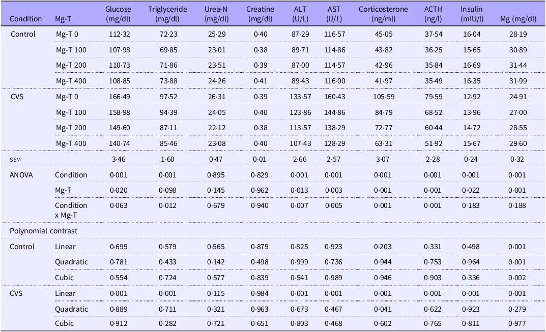
We first examined the biochemical parameters in the serum of rats and found that glucose (P < 0·001), corticosterone (P < 0·001) and ACTH (P < 0·001) concentrations were significantly increased in rats exposed to CVS compared to control rats housed under normal conditions (Table 1). Notably, Mg-T supplementation significantly reduced these elevations in the CVS group in a dose-dependent manner (linear; P = 0·001 for glucose, corticosterone and ACTH), whereas no significant change was observed in the control group. Similarly, CVS exposure significantly increased serum triglyceride levels (P < 0·001), ALT (P < 0·001) and AST activities (P < 0·001). Mg-T supplementation significantly attenuated these elevations in the CVS group (triglyceride: P = 0·001, ALT: P = 0·001, AST: P = 0·001; linear trends all P = 0·001), with a significant interaction effect between condition and Mg-T treatment (P = 0·012, 0·007 and 0·005, respectively), indicating that the treatment effect was specific to the stressed condition. In contrast, Mg-T had no significant impact on these parameters under normal conditions (P > 0·05 for all). Further analysis revealed no significant differences in urea (P = 0·895) and creatinine (P = 0·829) levels across groups or treatments, suggesting that renal function was not notably affected by either CVS or Mg-T supplementation. Importantly, serum insulin and magnesium levels were significantly decreased under CVS conditions (P < 0·001 for both), but Mg-T supplementation restored these levels in the CVS group (insulin: linear P = 0·001; Mg: linear P = 0·001). Although changes were less prominent in the control group, a significant increase in serum magnesium was observed in a dose-dependent manner even in non-stressed animals (P < 0·001).
Effect of Mg-T supplementation on biochemical parameters in rats subjected to CVS

Control: Housing under normal conditions; CVS: Exposure to chronic variable stressors; Mg-T: magnesium-L–theanine complex; Urea-N: Blood urea nitrogen; ALT: Alanine transaminase; AST: Aspartate transaminase; ACTH: Adrenocorticotropic hormone; Mg: Serum magnesium; sem: Standard error of the mean.
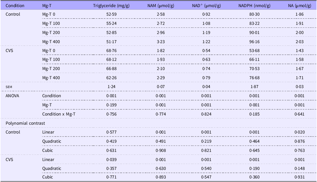
We also evaluated hepatic metabolic parameters and found that CVS significantly increased hepatic triglyceride levels compared to the control group (P < 0·001, Table 2). Interestingly, Mg-T supplementation did not alter hepatic triglyceride levels in the control group (linear P = 0·785). In contrast, a moderate increase was observed in the CVS group (linear P = 0·032), suggesting a distinct metabolic response to the compound under stress conditions. Furthermore, CVS exposure markedly reduced hepatic levels of NAM, NAD+, NADPH and NA (Table 2), indicating a disruption in redox balance and NAD+-related metabolic pathways. These reductions were significant for all parameters (P < 0·001 for condition effect). Notably, Mg-T supplementation significantly restored these metabolites in the CVS groups in a dose-dependent manner (linear P = 0·001 for all parameters), whereas no significant effect was observed in the control group (P > 0·05 for all). This suggests that Mg-T exerts its beneficial effects more prominently under stress-induced NAD+ depletion (P < 0·001), highlighting a direct impact of Mg-T on systemic magnesium availability. These findings provide mechanistic insight into the protective role of Mg-T against chronic stress-induced hepatic dysfunction, particularly through restoration of NAD+ metabolism and redox homeostasis, which are central to cellular stress responses.
Effect of Mg-T supplementation on liver parameters in rats subjected to CVS

Control: Housing under normal conditions; CVS: Exposure to chronic variable stressors; Mg-T: magnesium-L–theanine complex; NAM: Nicotinamide; NA: Nicotinic Acid, sem: Standard error of the mean.
Effects of Mg-T supplementation on the body weight of rats treated with CVS
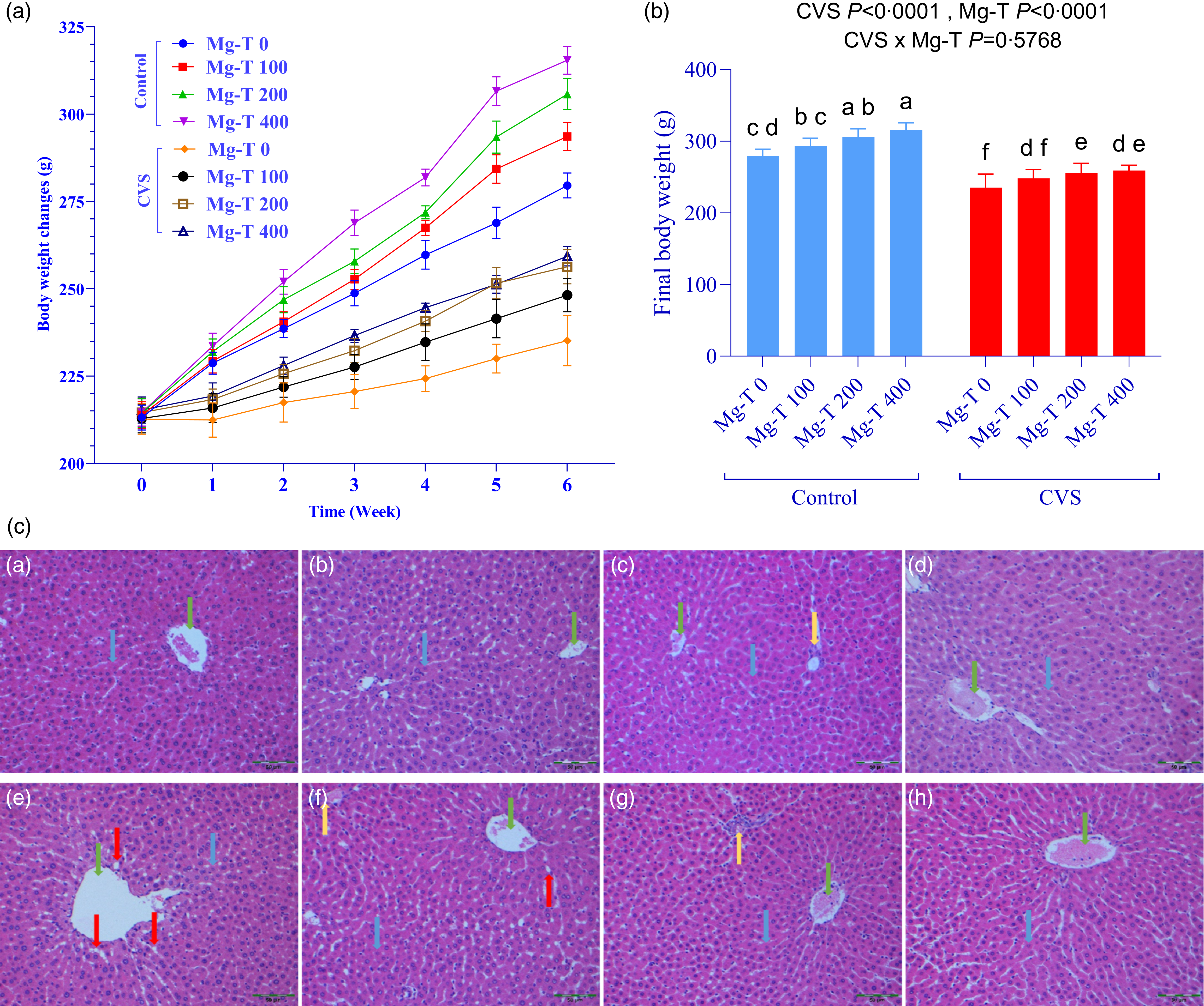
We tested changes in body weight in rats exposed to normal conditions and CVS and treated with different doses of Mg-T (0, 100, 200 and 400 mg/kg). While CVS-treated rats appeared to lose body weight, Mg-T supplementation significantly increased the body weight of rats housed in both normal and CVS conditions (Figure 1(a)), indicating its beneficial effect in alleviating stress-induced weight loss. Final body weight values of rats housed under normal and CVS conditions with or without Mg-T supplementation (0, 100, 200 and 400 mg/kg) are shown in Figure 1(b). As indicated by the ANOVA results, both the main effects of condition (CVS, P < 0·0001) and Mg-T treatment (P < 0·0001) were statistically significant, whereas the interaction effect (CVS × Mg-T) was not significant (P = 0·5768). This suggests that while both stress and treatment independently influenced final body weight, the effect of Mg-T was consistent across conditions without significant interaction between the two factors. Rats treated with 400 mg/kg Mg-T (Control-Mg-T 400) had the highest final body weight, significantly different from all other control groups (P < 0·05). Control-Mg-T 0 animals had the lowest body weight among controls, and each dose of Mg-T showed a stepwise improvement (Mg-T 0 < Mg-T 100 < Mg-T 200 < Mg-T 400). In the CVS group, although final body weight was significantly lower overall compared to controls, Mg-T supplementation still conferred a statistically significant benefit. The untreated CVS group (CVS-Mg-T 0) had the lowest final body weight, whereas Mg-T administration at 100, 200 and 400 mg/kg led to progressive increases in body weight, indicating a partial reversal of stress-induced weight loss. However, even the highest Mg-T dose in the CVS group did not fully restore body weight to control levels, as the letter groupings indicate separation between control and CVS animals.
Effects of CVS and different doses of Mg-T supplementation on body weight changes (a), final body weight (b) and the histopathological appearance of liver stained with haematoxylin-eosin (H&E) (c). Rats were randomly divided into eight groups: (a) control group (Mg-T 0), maintained under normal conditions and given 1 ml water daily; (b) Mg-T 100 group, maintained under normal conditions and given 100 mg/kg Mg-T; (c) Mg-T 200 group, maintained under normal conditions and given 200 mg/kg Mg-T; (d) Mg-T 400 group, maintained under normal conditions and given 400 mg/kg Mg-T; (e) CVS group (Mg-T 0), exposed to CVS and given 1 ml water daily; (f) CVS + Mg-T 100 group, exposed to CVS and given 100 mg/kg Mg-T; (g) CVS + Mg-T 200 group, exposed to CVS and given 200 mg/kg Mg-T and (h) CVS + Mg-T 400 group, exposed to CVS and given 400 mg/kg Mg-T. Each bar graph displays the data as the mean and standard deviation (n 7). The main effects and interactions are shown at the top of each chart. a–f Differences between groups with different letters on bars are statistically significant (analysis two-way ANOVA and post hoc Tukey test; P < 0·05). In addition, statistical analysis using two-way ANOVA and Polynomial Contrast is shown in online Supplementary Table S2. Blue arrows: hepatocyte; green arrows: central vein; yellow arrows: portal area and red arrows: fat vesicles (×200).

Effects of Mg-T supplementation on histological changes in the liver and jejunum of CVS-treated rats
On histopathological evaluation of liver tissue stained with H&E, the normal groups (Mg-T 0 to Mg-T 400) showed typical liver architecture with clear central veins (green arrows) and hepatocytes (blue arrows). In contrast, the CVS-Mg-T 0 group displayed histological abnormalities, including significant accumulation of fat vesicles (red arrows) and distortion of the portal areas (yellow arrows), indicating chronic stress-induced liver damage. However, Mg-T supplementation in the CVS-treated groups (CVS-Mg-T 0 – CVS-Mg-T 400) resulted in a dose-dependent reduction in fat vesicles and restoration of liver structure (Figure 1(c)).
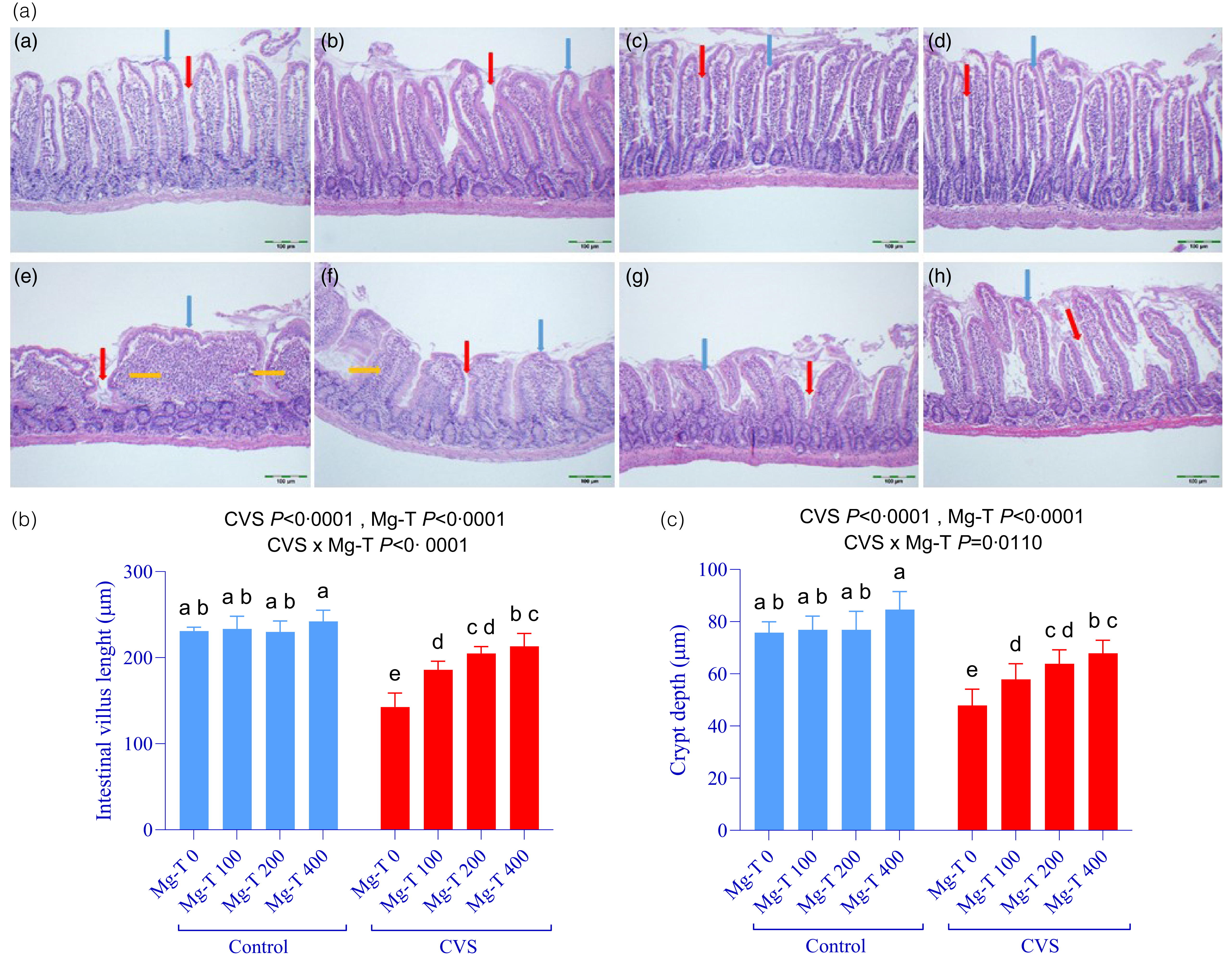
Histopathological examination of jejunal tissue revealed that exposure to CVS caused marked morphological damage, including shortened villi and shallower crypts, along with submucosal oedema and prominent inflammatory cell infiltration as clearly observed in the CVS-Mg-T 0 group (Figure 2(a); red and yellow arrows). In contrast, the control groups (Mg-T 0 to Mg-T 400) exhibited normal villus structure and crypt architecture, with well-aligned epithelial cells and no evidence of inflammation or structural disorganisation (blue and red arrows). Notably, Mg-T supplementation in CVS-exposed animals induced a dose-dependent improvement in intestinal morphology, as evidenced by longer villi, deeper crypts and reduced signs of inflammation (Figure 2(a)). At the highest dose (CVS-Mg-T 400), the intestinal architecture approached that of the control groups. Quantitative analysis supported these histological findings. Villus length was significantly affected by both CVS (P < 0·0001) and Mg-T treatment (P < 0·0001), with a highly significant interaction effect (Figure 2(b); P < 0·0001). Post hoc comparisons showed that villus length was significantly lower in the CVS-Mg-T 0 group compared to all other groups. Mg-T supplementation led to a progressive and significant increase in villus height in the CVS groups, confirming a linear dose-response effect under stress conditions. Similarly, crypt depth was significantly reduced by CVS (P < 0·0001), but increased with Mg-T treatment (P < 0·0001), again with a significant interaction (Figure 2(c); P = 0·0110). The shortest crypts were seen in the CVS-Mg-T 0 group, while Mg-T supplementation restored crypt depth in a dose-dependent manner, approaching control values.
Effects of CVS and different doses of Mg-T supplementation on the histopathological appearance of jejunum stained with haematoxylin-eosin (H&E) (a), jejunum villus length (b) and crypt depth (c). Rats were randomly divided into eight groups: (a) control group (Mg-T 0), maintained under normal conditions and given 1 ml water daily; (b) Mg-T 100 group, maintained under normal conditions and given 100 mg/kg Mg-T; (c) Mg-T 200 group, maintained under normal conditions and given 200 mg/kg Mg-T; (d) Mg-T 400 group, maintained under normal conditions and given 400 mg/kg Mg-T; (e) CVS group (Mg-T 0), exposed to CVS and given 1 ml water daily; (f) CVS + Mg-T 100 group, exposed to CVS and given 100 mg/kg Mg-T; (g) CVS + Mg-T 200 group, exposed to CVS and given 200 mg/kg Mg-T and (h) CVS + Mg-T 400 group, exposed to CVS and given 400 mg/kg Mg-T. Blue arrows: villus; red arrows: crypt; yellow arrows: inflammation ((h) & (e) ×100). Each bar graph displays the data as the mean and standard deviation (n 7). The main effects and interactions are shown at the top of each chart. a–e Differences between groups with different letters on bars are statistically significant (analysis two-way ANOVA and post hoc Tukey test; P < 0·05). In addition, statistical analysis using two-way ANOVA and Polynomial Contrast is shown in online Supplementary Table S3.

Possible molecular mechanisms underlying the effects of Mg-T supplementation in the liver and jejunum of CVS-treated rats
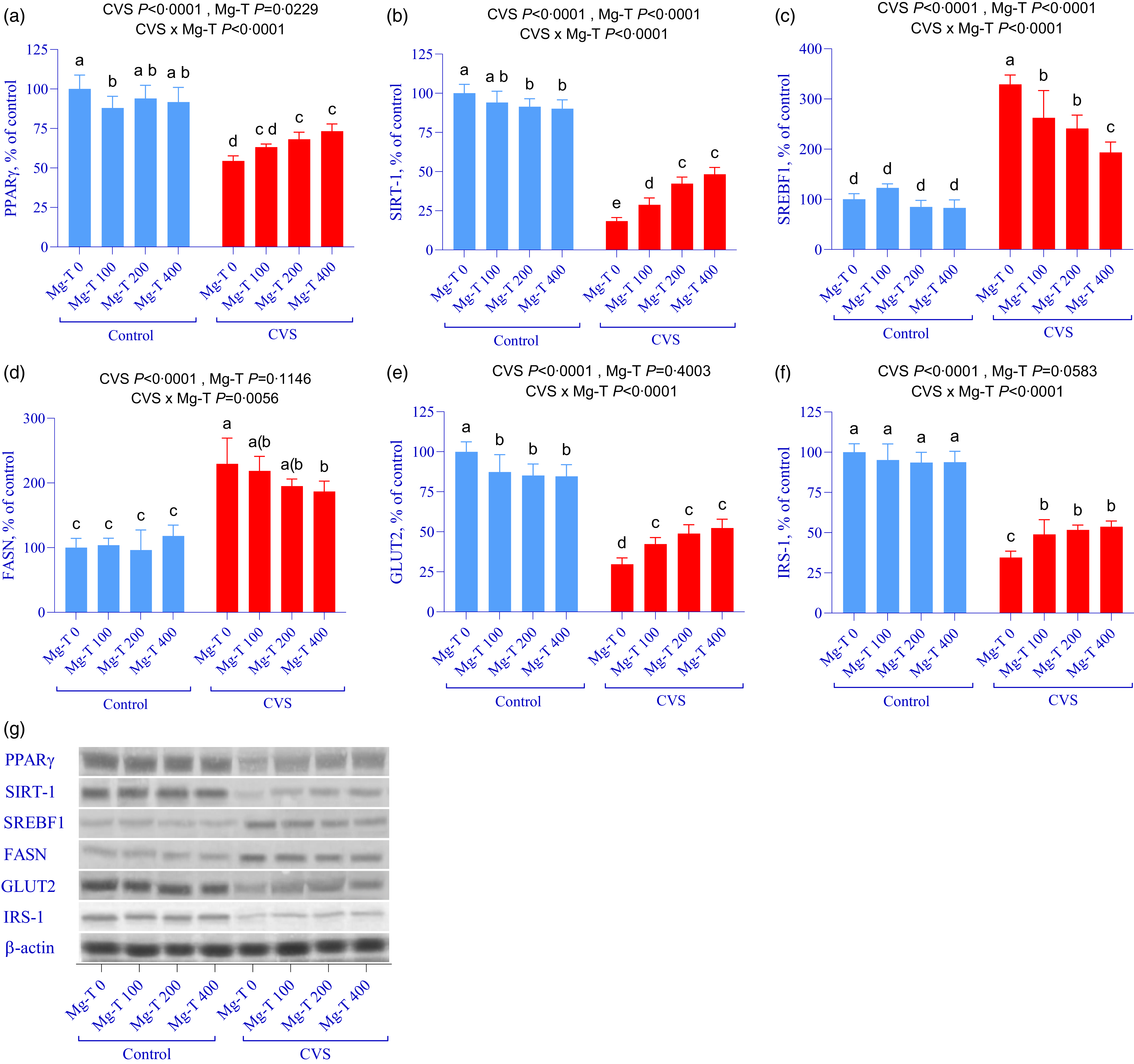
We next evaluated the effects of different doses of Mg-T on the hepatic expression of key metabolic and lipogenic proteins, including PPARγ, SIRT-1, SREBF1, FASN, GLUT2 and IRS-1, in rats exposed to CVS. As expected, CVS significantly reduced the levels of PPARγ, SIRT-1, GLUT2 and IRS-1, while markedly increasing the levels of the lipogenic regulators SREBF1 and FASN (Figure 3(a)–(f); P < 0·0001 for all). Mg-T supplementation effectively reversed these stress-induced alterations in a dose-dependent manner. Specifically, PPARγ and SIRT-1 levels increased linearly with Mg-T treatment (P < 0·0001), and GLUT2 and IRS-1 levels also improved significantly, though not fully reaching control levels. Conversely, SREBF1 and FASN levels showed a progressive and significant reduction with increasing Mg-T doses. Statistical analysis revealed significant interaction effects between CVS and Mg-T treatment for most markers (P < 0·0001), indicating that the regulatory effects of Mg-T were more prominent under stress conditions. Collectively, these results demonstrate that Mg-T supplementation mitigates stress-induced hepatic metabolic dysregulation by enhancing metabolic regulators and suppressing lipogenic pathways, supporting its potential as a therapeutic strategy for stress-related metabolic disorders.
Effects of CVS and different doses of Mg-T supplementation on protein expression of PPARγ (a), SIRT-1 (b), SREBF1 (c), FASN (d), GLUT2 (e) and IRS-1 (f) in the liver of rats. Protein levels were analysed by Western blot using β-actin as a loading control and representative bands are shown in (g). Rats were randomly divided into eight groups: (1) control group (Mg-T 0), maintained under normal conditions and given 1 ml water daily; (2) Mg-T 100 group, maintained under normal conditions and given 100 mg/kg Mg-T; (3) Mg-T 200 group, maintained under normal conditions and given 200 mg/kg Mg-T; (4) Mg-T 400 group, maintained under normal conditions and given 400 mg/kg Mg-T; (5) CVS group (Mg-T 0), exposed to CVS and given 1 ml water daily; (6) CVS + Mg-T 100 group, exposed to CVS and given 100 mg/kg Mg-T; (7) CVS + Mg-T 200 group, exposed to CVS and given 200 mg/kg Mg-T and (8) CVS + Mg-T 400 group, exposed to CVS and given 400 mg/kg Mg-T. Each bar graph displays the data as the mean and standard deviation (n 7). The main effects and interactions are shown at the top of each chart. Differences between groups with different letters on bars are statistically significant (analysis two-way ANOVA and post hoc Tukey test; P < 0·05). In addition, statistical analysis using two-way ANOVA and polynomial contrast is shown in online Supplementary Table S4.

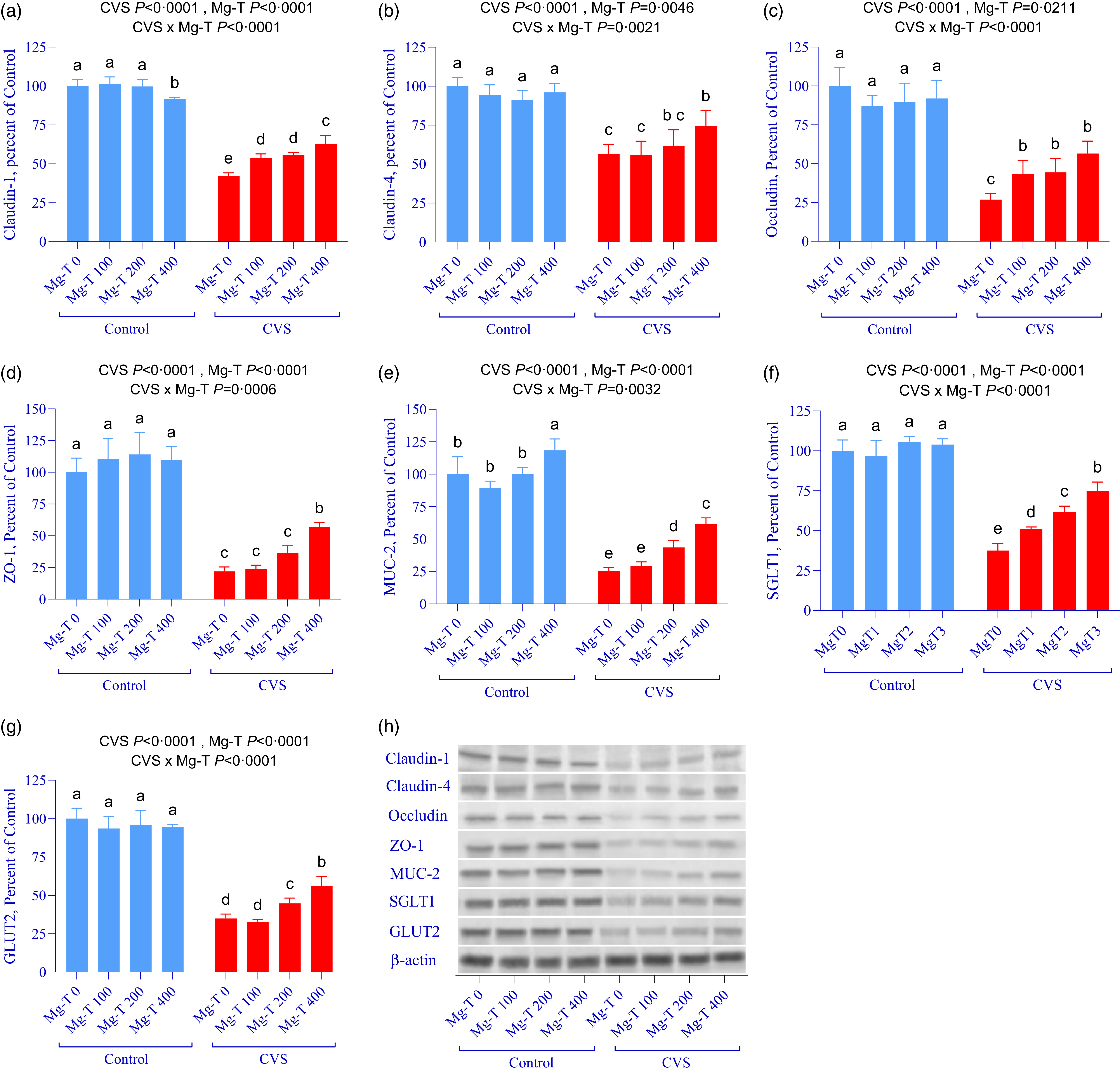
We also evaluated the effects of different doses of Mg-T on the expression of intestinal tight junction and barrier integrity proteins in the jejunum of rats exposed to CVS. As shown in Figure 4(a)–(g), CVS markedly decreased the expression of Claudin-1, Claudin-4, Occludin and ZO-1, as well as mucosal and transporter proteins including MUC-2, SGLT1 and GLUT2 (P < 0·0001 for all). Among these, Claudin-1, a critical component of epithelial tight junctions, was significantly suppressed in the CVS-Mg-T 0 group, but its expression progressively increased with Mg-T supplementation, demonstrating a significant dose-dependent effect (P < 0·0001 for Mg-T; P < 0·0001 for interaction). Similar patterns were observed for Claudin-4 (P = 0·0046 for Mg-T effect), Occludin (P = 0·0211) and ZO-1 (P < 0·0001), all of which showed significant interaction effects (CVS × Mg-T, P < 0·01), indicating that Mg-T supplementation specifically counteracted the stress-induced suppression of these genes. In each case, higher Mg-T doses (especially 200 and 400 mg/kg) significantly improved expression, approaching or partially restoring control values. In addition to structural markers, MUC-2, a key secretory mucin involved in barrier function, was drastically reduced under stress (group e), but significantly increased by Mg-T in a dose-responsive manner (P < 0·0001 for treatment and interaction). Similarly, SGLT1 and GLUT2, which are essential for nutrient transport and epithelial metabolism, were significantly downregulated in the CVS-Mg-T 0 group, but improved with increasing Mg-T doses, particularly at 400 mg/kg, with all comparisons showing highly significant differences (P < 0·0001).
Effects of CVS and different doses of Mg-T supplementation on protein expression of Claudin-1 (a), Claudin-4 (b), Occludin (c), ZO-1 (d), MUC-2 (e), SGLT1 (f) and GLUT2 (g) in the jejunum of rats. Protein levels were analysed by Western blot using β-actin as a loading control and representative bands are shown in (H). Rats were randomly divided into eight groups: (1) control group (Mg-T 0), maintained under normal conditions and given 1 ml water daily; (2) Mg-T 100 group, maintained under normal conditions and given 100 mg/kg Mg-T; (3) Mg-T 200 group, maintained under normal conditions and given 200 mg/kg Mg-T; (4) Mg-T 400 group, maintained under normal conditions and given 400 mg/kg Mg-T; (5) CVS group (Mg-T 0), exposed to CVS and given 1 ml water daily; (6) CVS + Mg-T 100 group, exposed to CVS and given 100 mg/kg Mg-T; (7) CVS + Mg-T 200 group, exposed to CVS and given 200 mg/kg Mg-T and (8) CVS + Mg-T 400 group, exposed to CVS and given 400 mg/kg Mg-T. Each bar graph displays the data as the mean and standard deviation (n 7). The main effects and interactions are shown at the top of each chart. Differences between groups with different letters on bars are statistically significant (analysis two-way ANOVA and post hoc Tukey test; P < 0·05). In addition, statistical analysis using two-way ANOVA and polynomial contrast is shown in online Supplementary Table S5.

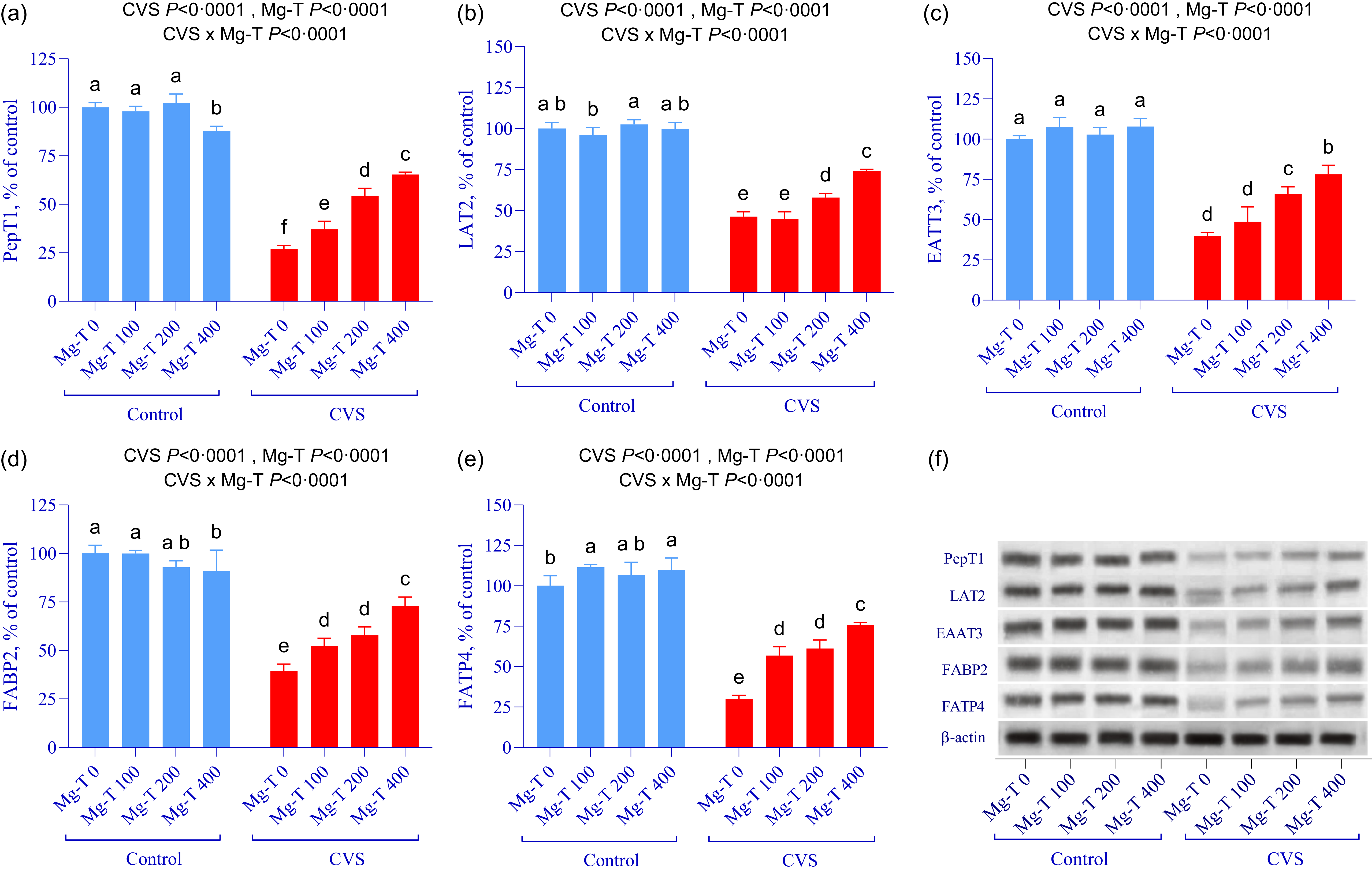
We further investigated the effects of Mg-T supplementation on intestinal amino acid and peptide transporters, focusing on PepT1, LAT1, EAAT1, EAAT3 and FATP4 expression levels in the jejunum of rats exposed to CVS. As shown in Figure 5(a)–(e), CVS significantly suppressed the expression of all five transporters compared to control groups (P < 0·0001 for condition effect across all markers). Notably, PepT1 and LAT1, which play key roles in the absorption of di-/tripeptides and large neutral amino acids, respectively, were drastically reduced in CVS-Mg-T 0 animals, while Mg-T supplementation restored their expression in a dose-dependent manner (P < 0·0001 for Mg-T and CVS × Mg-T interaction). The highest dose (Mg-T 400) brought expression levels significantly closer to controls, indicating robust compensatory effects. Similarly, EAAT1, BEAAT3 and FATP4, which are critical for excitatory amino acid and neutral amino acid transport, also showed significant reductions under stress but were progressively restored with increasing doses of Mg-T (P < 0·0001 for all treatment and interaction effects). Post hoc comparisons revealed distinct statistical groupings, with Mg-T 400 consistently showing higher expression than Mg-T 0 and Mg-T 100 within the CVS condition, reflecting a strong dose-response trend.
Effects of CVS and different doses of Mg-T supplementation on protein expression of PepT1 (a), LAT2 (b), EAAT3 (c), FABP2 (d) and FATP4 (e) in the jejunum of rats. Protein levels were analysed by Western blot using β-actin as a loading control and representative bands are shown in (f). Rats were randomly divided into eight groups: (1) control group (Mg-T 0), maintained under normal conditions and given 1 ml water daily; (2) Mg-T 100 group, maintained under normal conditions and given 100 mg/kg Mg-T; (3) Mg-T 200 group, maintained under normal conditions and given 200 mg/kg Mg-T; (4) Mg-T 400 group, maintained under normal conditions and given 400 mg/kg Mg-T; (5) CVS group (Mg-T 0), exposed to CVS and given 1 ml water daily; (6) CVS + Mg-T 100 group, exposed to CVS and given 100 mg/kg Mg-T; (7) CVS + Mg-T 200 group, exposed to CVS and given 200 mg/kg Mg-T and (8) CVS + Mg-T 400 group, exposed to CVS and given 400 mg/kg Mg-T. Each bar graph displays the data as the mean and standard deviation (n 7). The main effects and interactions are shown at the top of each chart. Differences between groups with different letters on bars are statistically significant (analysis two-way ANOVA and post hoc Tukey test; P < 0·05). In addition, statistical analysis using two-way ANOVA and polynomial contrast is shown in online Supplementary Table S6.

Correlation analysis
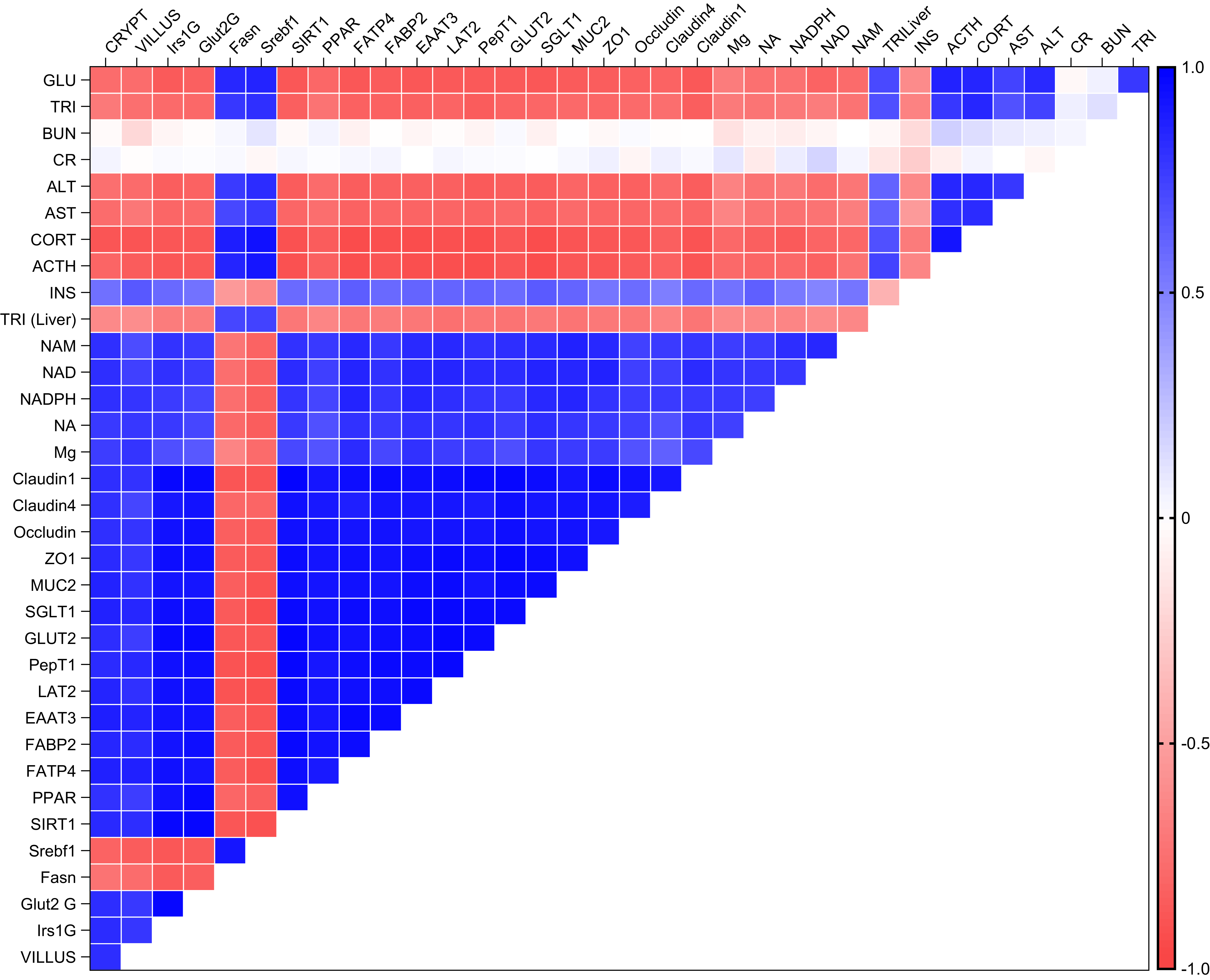
To better understand the interrelationships between physiological, biochemical, molecular and histological parameters, a correlation heatmap was generated (Figure 6). The analysis revealed a strong negative correlation between stress-related hormones (corticosterone, ACTH) and intestinal integrity markers such as Claudin-1, ZO-1, Occludin and MUC-2, as well as nutrient transporters including GLUT2, SGLT1, PepT1 and LAT1. This suggests that elevated stress hormone levels are closely linked to compromised intestinal barrier function and impaired nutrient absorption. Conversely, NAD+, NADPH, NAM and serum magnesium levels exhibited strong positive correlations with metabolic regulators, such as PPARγ and SIRT-1, as well as with intestinal structural markers and transporters, suggesting that restoration of redox and magnesium homeostasis supports gut-liver axis integrity. Additionally, villus length and crypt depth showed positive correlations with most tight junction proteins and nutrient transporters, reinforcing the link between mucosal morphology and functional protein expression. On the other hand, hepatic and serum triglyceride levels were negatively correlated with NAD+-related metabolites and intestinal transporters but positively associated with lipogenic markers such as FASN and SREBF1, further emphasising the contribution of lipid metabolic imbalance to intestinal dysfunction under chronic stress.
Correlation heatmap of physiological, biochemical, molecular and histological parameters. The heatmap illustrates the interrelationships between stress-related hormones, intestinal integrity markers, nutrient transporters and metabolic regulators.

Discussion
Chronic stress, particularly in forms such as CVS, disrupts the body’s balance by inducing oxidative stress, systemic inflammation and hormonal disruption. These changes adversely affect immune function, glucose metabolism, gut integrity and liver health(Reference Shafiq, Anjum and Hano6,Reference Yaribeygi, Panahi and Sahraei8) . The liver and intestines are particularly vulnerable due to their central role in metabolism and immune defence. Stress-induced disruption of tight junction proteins and mucins in the intestinal barrier allows luminal antigens to penetrate, exacerbating inflammation and dysbiosis(Reference Mayorga, Ross and Keating39). In this study, we investigated the therapeutic potential of Mg-T supplementation in counteracting stress-induced damage. The results showed that Mg-T significantly improved intestinal structure, restored nutrient transporter expression and alleviated liver dysfunction caused by CVS.
Biochemical analyses showed that CVS increased serum glucose, corticosterone, ACTH, ALT and AST levels while decreasing insulin and magnesium levels, reflecting a hypermetabolic state and liver dysfunction. Mg-T supplementation reversed these effects, lowering corticosterone and glucose levels while improving insulin sensitivity. It also improved levels of critical redox markers such as NAD+, NAM, NADPH and NA, highlighting the potential of Mg-T to mitigate stress-induced metabolic disturbances, consistent with previous findings(Reference Boyle, Billington and Lawton23). These beneficial effects may be partially explained by the capacity of Mg to act as a cofactor in NAD+ biosynthesis, which in turn activates SIRT1 signalling, a key regulator of lipid metabolism and oxidative stress(Reference Pandit, Kim and Oh40,Reference Li, Zhou and Pang41) . Mechanistically, NAD+-mediated activation of SIRT1, a key deacetylase involved in lipid metabolism, mitochondrial biogenesis and oxidative stress resistance, appears to be a central pathway through which Mg-T exerts its hepatoprotective effects. Upregulation of hepatic SIRT1 and PPARγ expression observed in this study further supports the engagement of this signalling axis. Moreover, emerging evidence highlights the importance of upstream regulators, particularly AMPK and mTOR, in modulating SIRT1/PPARγ activity. AMPK enhances NAD+ availability and directly promotes SIRT1 activation, facilitating fatty acid oxidation and insulin sensitivity(Reference Steinberg and Carling42). Concurrently, suppression of mTOR signalling by Mg-T may contribute to reduced lipogenesis and improved cellular energy homeostasis, as mTOR overactivation under stress is known to favour anabolic and inflammatory pathways(Reference Laplante and Sabatini43). In line with these mechanisms, L-theanine, a key component of the Mg-T complex, has been shown to activate AMPK and downregulate SREBP-1c and FASN expression in hepatic tissue, thereby attenuating lipid accumulation and oxidative stress(Reference Liang, Gu and Liu44). The combined action of magnesium and L-theanine may thus converge on the AMPK-SIRT1-PPARγ and mTOR-SREBP1c axes, representing a dual regulatory mechanism that underlies the observed improvements in hepatic metabolism, redox status and inflammatory control.
Consistent with our findings, studies have shown that L-theanine, a component of Mg-T, effectively reduces oxidative damage, lowers corticosterone levels and increases antioxidant activities (superoxide dismutase, CAT, GSH-Px) in rats and mice under chronic stress(Reference Tian, Sun and Gou45,Reference Liu, Gong and Lin46) . It also reduces serum markers of inflammation, including ALT, AST, TNF-α, IL-6 and IFN-γ (Reference Uyanga, Oke and Amevor47). Moreover, L-theanine is often used to improve sleep quality through its effects on inhibitory neurotransmitters, particularly in combination with magnesium, a natural N-methyl-D-aspartate antagonist and gamma-aminobutyric acid agonist. Studies suggest that Mg-L-theanine complexes enhance the expression of gamma-aminobutyric acidergic, serotonergic and glutamatergic receptors, reduce levels of inducible nitric oxide synthase and improve neurotransmitters such as dopamine, serotonin and melatonin, contributing to stress reduction and improved sleep quality(Reference Dasdelen, Er and Kaplan24). In addition, Mg-L-theanine compounds with higher magnesium concentrations have been shown to significantly increase delta wave activity during sleep, which is associated with deeper, more restorative sleep stages and memory consolidation.
Oxidative stress has been identified as a key factor negatively affecting intestinal mucosal morphology(Reference Song, Xiao and Ke48). Consistent with our findings, several studies have demonstrated that dietary supplementation with L-theanine can mitigate such damage. For instance, broilers subjected to oxidative stress and fed L-theanine-enriched diets exhibited improved jejunal villus height, suggesting enhanced mucosal morphology(Reference Liu, Li and Zheng49,Reference Wang, Tang and Long50) . Similarly, Wang et al. (Reference Wang, Tang and Long50) reported that dietary L-theanine significantly increased serum total antioxidant capacity, elevated superoxide dismutase and glutathione peroxidase activity, and reduced malondialdehyde levels. These systemic antioxidant effects are in agreement with our results observed under chronic stress exposure. Supporting this, Huang et al. (Reference Huang, Lu and Wu51) showed that L-theanine administration markedly increased hippocampal superoxide dismutase and GSH levels while decreasing malondialdehyde concentrations in sleep-deprived mice, thereby restoring redox homeostasis. Moreover, their study demonstrated that L-theanine upregulated Nrf2 protein expression, highlighting its potential to activate intrinsic antioxidant defence mechanisms. These findings align with our observations, suggesting that L-theanine mitigates stress-induced oxidative damage by enhancing endogenous antioxidant pathways, possibly through Nrf2 signalling.
In addition, L-theanine positively influenced nutrient absorption by increasing villus height and decreasing crypt depth, particularly in the jejunum, which is consistent with our findings. Further supporting our findings, Liang et al. (Reference Liang, Gu and Liu44) demonstrated that L-theanine significantly reduced hepatic steatosis and lowered serum ALT, AST, TG and LDL-C levels in a high-fat diet-induced non-alcoholic steatosis model. These hepatoprotective effects are likely to be mediated through the modulation of AMPK-mTOR signalling and SREBP-1c pathways, which regulate lipid metabolism. Although limited in number, comparative studies have suggested that Mg-L-theanine exhibits superior efficacy to either magnesium or L-theanine alone in stress-related models. For example, Mg-theanine significantly improved neurotransmitter profiles and antioxidant status compared to either compound alone in murine models of sleep deprivation and caffeine-induced stress(Reference Nelson and Komorowski26). Furthermore, a randomised preclinical study reported that the combination of Mg2 + and L-theanine synergistically suppressed corticosterone levels, elevated serotonin and melatonin and improved sleep parameters more effectively than individual treatments(Reference Dasdelen, Er and Kaplan24). Thus, the current findings provide critical mechanistic and translational insight into the added value of combining Mg²+ and L-theanine into a single compound. In addition, long-term Mg supplementation may lead to changes in pituitary-gonadal and pituitary-adrenocortical hormonal status(Reference Dmitrašinović, Pešić and Stanić52). Consistent with our findings, Zogovic et al. (Reference Zogović, Pešić and Dmitrasinović53) reported that Mg supplementation reduced cortisol and IL-6 levels. Furthermore, psychosocial stress exacerbates these health problems by increasing Mg excretion, leading to deficiencies(Reference Grases, Pérez-Castelló and Sanchis54). Boyle et al. (Reference Boyle, Billington and Lawton23) highlighted the synergistic effect of Mg, vitamin B6 and L-theanine in significantly reducing anxiety under social stress, further supporting their combined therapeutic benefits.
Chronic stress induced marked histopathological changes in the liver, including lipid accumulation, fat vesicle formation and structural disorganisation, indicative of hepatic steatosis. Similar observations have been reported in other stress models, where liver damage is associated with oxidative stress and inflammatory responses(Reference Zhang, Chen and Zhao55). Mg-T supplementation reversed these changes, improved liver histology, reduced fat accumulation and restored liver structure in a dose-dependent manner. These effects may be due to the modulation of metabolic pathways such as mTOR/SREBP1c-ACC1/FASN, which control fatty acid synthesis(Reference Lin, Han and Gong56). On the hepatic side, CVS increased lipogenic protein expression (SREBF1, FASN) and suppressed insulin signalling (GLUT2, IRS-1) and NAD+ metabolism (NAM, NA, NADPH). Mg-T reversed these changes and enhanced SIRT1 expression. These outcomes suggest a restoration of redox balance and insulin sensitivity, potentially via NAD+-dependent SIRT1 activation, a pathway known to play a critical role in hepatic lipid metabolism, oxidative stress adaptation and intestinal mucosal protection(Reference Pandit, Kim and Oh40,Reference Liu, Li and Zheng49) . Since magnesium is a key cofactor for NAD+ synthesis, and L-theanine has been shown to regulate hepatic oxidative stress markers and lipid metabolism,(Reference Liang, Gu and Liu44,Reference Tian, Sun and Gou45) their combination may synergistically support NAD+/SIRT1 axis function under stress.
CVS has a significant impact on gut health, causing morphological damage such as shortened villi, reduced crypt depth, oedema and inflammation. It also affects the expression of essential tight junction proteins and mucins, which are critical for maintaining the integrity of the intestinal barrier. These changes were associated with the downregulation of tight junction proteins (Claudin-1, Claudin-4, Occludin, ZO-1) and MUC-2, which are essential for maintaining barrier integrity. Stress-induced dysfunction of the intestinal barrier facilitates the translocation of antigens and pathogens, promoting systemic inflammation and dysbiosis(Reference Lian, Braber and Garssen57). The present data show that Mg-T supplementation significantly restored the expression of these barrier-associated proteins in the jejunum, suggesting enhanced epithelial stability. This aligns with recent evidence that L-theanine modulates intestinal epithelial function and suppresses inflammation through the regulation of linoleic and arachidonic acid metabolism, including suppression of proinflammatory markers such as COX-1 and ALOX-5(Reference Gu, Tan and Yang58). Mg-T supplementation effectively counteracted these effects by increasing villus height and crypt depth and restoring the expression of tight junction protein expression, consistent with previous findings demonstrating the protective effects of Mg and L-theanine on gut health(Reference Yan, Tong and Tang12,Reference Uyanga, Oke and Amevor47) . Both heat and chronic stress are known to be detrimental to intestinal health, with histological analyses revealing colonic mucosal erosion, epithelial cell loss and lymphocyte infiltration(Reference Liu, Li and Zheng49,Reference Van Der Pijl, Chen and Mulder59,Reference Xu, Lin and Liu60) . Liu et al. (Reference Liu, Li and Zheng49) also reported that L-theanine supplementation attenuated stress-induced weight loss and improved intestinal microbiota balance by promoting beneficial bacteria. Indicators such as villus height and crypt depth are widely used to assess gut function, with greater villus height and reduced crypt depth indicating improved nutrient absorption(Reference Mohammadsadeghi, Afsharmanesh and Ebrahimnejad61,Reference Ming, Wang and Huang62) . Stress disrupts these parameters by causing irregular epithelial desquamation and impairing the expression of tight junction proteins, leading to increased intestinal permeability(Reference Song, Xiao and Ke48,Reference Eum, Schurhoff and Teglas63) . L-theanine is a recognised safe dietary supplement known for its antioxidant, hepatoprotective and immunomodulatory properties(Reference Deng, Xiao and Chen64–Reference Wang, Yu and Lin66). Research shows that L-theanine supplementation reduces markers of oxidative stress such as inducible nitric oxide synthase and increases antioxidant activity (superoxide dismutase, CAT, GSH-Px) in liver tissue, reducing inflammation and improving metabolic function(Reference Zeng, Lin and Peng27). It also downregulates glucose (SGLT1, GLUT2) and amino acid transporters in stressed animals, further contributing to improved nutrient absorption and intestinal morphology(Reference Yan, Tong and Tang12). L-theanine also positively affects lipid metabolism by inhibiting the nuclear translocation of sterol regulatory element binding protein 1c (SREBP-1c) via AMPK-mTOR signalling, which reduces fatty acid synthesis and promotes β-oxidation through increased expression of PPARα and CPT1A(Reference Lin, Han and Gong56). Taken together, these findings highlight the therapeutic potential of Mg and L-theanine in alleviating stress-induced damage to intestine and liver by targeting key biochemical and structural pathways. Moreover, psychosocial stress exacerbates these health problems by increasing Mg excretion, leading to deficiencies(Reference Grases, Pérez-Castelló and Sanchis54).
CVS significantly downregulated key nutrient transporters, including FABP1, FATP1, SGLT1 and GLUT2. This impairment is consistent with previous findings indicating that stress disrupts nutrient transporter expression, leading to reduced nutrient uptake and metabolic dysfunction(Reference Orhan, Tuzcu and Deeh11,Reference Chen, Yong and Ju13) . Moreover, CVS diminished peptide transporters (PepT1, PepT2) and amino acid transporters (EAAT1, LAT1), which are critical for protein and amino acid metabolism. Similar observations have been reported in heat-stressed animals where nutrient transporter function was severely impaired(Reference Goel, Ncho and Choi67). In parallel, we observed that Mg-T treatment reversed the downregulation of key nutrient transporters, including PepT1, LAT2, EAAT3, FATP4, SGLT1 and GLUT2, all of which are known to be stress-sensitive. Although mechanistic data are limited, it is plausible that Mg-T exerts regulatory effects on nutrient transport systems via modulation of solute carrier family genes or by enhancing epithelial polarity and mitochondrial NAD+-SIRT1 signalling, both of which have been implicated in maintaining absorptive function under stress(Reference Yan, Tong and Tang12,Reference Pandit, Kim and Oh40) . Notably, no previous studies have evaluated the combined effects of Mg and L-theanine on nutrient transport systems under chronic stress conditions. Therefore, our findings present novel insights into how Mg-T modulates molecular markers involved in barrier integrity, inflammation, and metabolic regulation. Mg-T supplementation restored the expression of these transporters in a dose-dependent manner, thereby improving nutrient absorption. This is consistent with studies showing that bioactive compounds, including Mg and L-theanine, enhance nutrient transporter activity under stress conditions(Reference Boyle, Billington and Lawton23). Furthermore, a recent preclinical study demonstrated that a compound nutritional supplement containing magnesium improved behavioural and intestinal outcomes in rats with autism-like symptoms, underscoring the role of Mg in modulating the gut-brain axis(Reference Pan, Lin and Qian68,Reference Schiopu, Ștefănescu and Diaconescu69) . Given that Mg-T has previously shown neuromodulatory and sleep-regulating properties via gamma-aminobutyric acidergic mechanisms,(Reference Dasdelen, Er and Kaplan24) its influence on stress physiology may also involve central nervous system signalling, although this warrants further investigation. Taken together, this study not only demonstrates the phenotypic efficacy of Mg-T against chronic stress-induced damage but also provides emerging mechanistic insights into its action on epithelial transport systems, barrier integrity, redox signalling and lipid metabolism. These findings lay the groundwork for future molecular studies and translational applications.
Despite the encouraging findings, several limitations of this study should be acknowledged. First, while the data suggest a potential synergistic effect of magnesium and L-theanine within the Mg-T complex, the absence of separate treatment arms for Mg²+ and L-theanine alone limits direct comparisons and prevents definitive conclusions about the relative contributions of each component. Second, although mechanistic insights were gained through hepatic and intestinal protein expression analyses, the study did not incorporate functional evaluations such as mitochondrial respiration, intestinal permeability assays or behavioural testing, which could have further validated the translational relevance of the observed molecular changes. Third, although body weight measurements were recorded and presented, data on feed intake and feed conversion ratio were not included due to methodological constraints. Specifically, rats were group-housed in standard cages rather than in individual metabolic cages, a decision made to maintain social conditions and minimise distress during the extended 40-day chronic stress protocol. Under such conditions, individual feed intake measurements are unreliable due to dominance hierarchies, food hoarding, spillage and nesting behaviour – factors that introduce substantial variability and potentially confound data accuracy. As such, feed intake or feed conversion ratio measurements obtained under these conditions would not meet scientific rigour and were therefore omitted. Future studies using metabolic cages or automated feeding systems are warranted to address this limitation and allow precise intake monitoring. Finally, as this research was conducted in a rat model, extrapolation to humans must be approached with caution. Further clinical investigations are required to confirm the safety, efficacy and mechanistic action of Mg-T in stress-related metabolic and gastrointestinal disorders.
In conclusion, Mg-T supplementation demonstrated significant therapeutic potential in mitigating the adverse effects of CVS on intestinal and hepatic health. It improved nutrient transporter expression, enhanced intestinal barrier integrity and alleviated hepatic steatosis. In addition, Mg-T reduced corticosterone and glucose levels and enhanced insulin sensitivity. These findings highlight the promise of Mg-T as a therapeutic strategy for the management of stress-induced metabolic and intestinal dysfunction.
Supplementary material
For supplementary material/s referred to in this article, please visit https://doi.org/10.1017/S0007114525105734
Acknowledgements
The authors thank JDS Therapeutics for providing the Mg-T.
This work was supported by the Scientific and Technological Research Council of Türkiye (TÜBİTAK-123S482).
K. S.: Conceptualisation, investigation, project administration, funding acquisition, supervision, writing – original draft and editing; M. Y.: formal analysis, writing – original draft and editing; C. O.: methodology, formal analysis, data curation, visualisation; B. E.: methodology, formal analysis, data curation; R. O.: methodology, formal analysis, data curation; M. T.: methodology, formal analysis, data curation; Me. T.: methodology, formal analysis, data curation; M. O.: methodology, formal analysis, data curation; A. K.: methodology, formal analysis, data curation; I. H. O.: methodology, formal analysis, data curation; N. S.: methodology, formal analysis, data curation. All authors have read and agreed to the published version of the manuscript.
The authors declare that they have no conflicts of interest.
Generative AI tools were used during the preparation of the revised manuscript strictly for language editing and improving the clarity of expression.