Introduction
Impulsivity frequently worsens the clinical outcome and complicates the management of patients with schizophrenia. It increases the risk for violence (Quanbeck et al. Reference Quanbeck, McDermott, Lam, Eisenstark, Sokolov and Scott2007; Volavka & Citrome, Reference Volavka and Citrome2008), substance abuse (Dervaux et al. Reference Dervaux, Bayle, Laqueille, Bourdel, Le Borgne, Olie and Krebs2001) and suicide (Gut-Fayand et al. Reference Gut-Fayand, Dervaux, Olie, Loo, Poirier and Krebs2001), as well as the need for intensive and prolonged hospital care (Bigelow et al. Reference Bigelow, Cutler, Moore, McComb and Leung1988; Greenfield et al. Reference Greenfield, McNiel and Binder1989; Bowers et al. Reference Bowers, Jeffery, Bilgin, Jarrett, Simpson and Jones2008). Further, its presence is considered as a crucial factor in the choice of antipsychotic medication (Dursun et al. Reference Dursun, Szemis, Andrews, Whitaker and Reveley2000; Chengappa et al. Reference Chengappa, Vasile, Levine, Ulrich, Baker, Gopalani and Schooler2002; Spivak et al. Reference Spivak, Shabash, Sheitman, Weizman and Mester2003; Krakowski et al. Reference Krakowski, Czobor, Citrome, Bark and Cooper2006; Volavka & Citrome, Reference Volavka and Citrome2008).
Impulsivity has been assessed using a variety of measures coming from different theoretical frameworks. Impulsivity conceived as a personality trait is typically measured by self-report questionnaires, which rely on an individual's self-perception of his/her behaviour in daily life (Eysenck et al. Reference Eysenck, Pearson, Easting and Allsopp1985; Patton et al. Reference Patton, Stanford and Barratt1995). Otherwise, in laboratory settings, impulsivity has been frequently conceptualized as an inability to inhibit a motor response (Logan et al. Reference Logan, Schacher and Tannock1997; Moeller et al. Reference Moeller, Barratt, Dougherty, Schmitz and Swann2001; Chamberlain & Sahakian, Reference Chamberlain and Sahakian2007).
Several functional magnetic resonance imaging (fMRI) studies have used the Go/NoGo task to assess the neural correlates of motor response inhibition in schizophrenia. They have reported dysfunctions in the anterior cingulate cortex (ACC) (Rubia et al. Reference Rubia, Russell, Bullmore, Soni, Brammer, Simmons, Taylor, Andrew, Giampietro and Sharma2001; Arce et al. Reference Arce, Leland, Miller, Simmons, Winternheimer and Paulus2006), posterior cingulate cortex (Laurens et al. Reference Laurens, Ngan, Bates, Kiehl and Liddle2003), dorsolateral prefrontal cortex (Arce et al. Reference Arce, Leland, Miller, Simmons, Winternheimer and Paulus2006), caudate nucleus and thalamus (Barkataki et al. Reference Barkataki, Kumari, Das, Sumich, Taylor and Sharma2008) or over a network of brain regions (Ford et al. Reference Ford, Gray, Whitfield, Turken, Glover, Faustman and Mathalon2004; Joyal et al. Reference Joyal, Putkonen, Mancini-Marie, Hodgins, Kononen, Boulay, Pihlajamaki, Soininen, Stip, Tiihonen and Aronen2007). These divergent findings may arise from the great variety of task designs and fMRI procedures that were used. In particular, some studies employed a blocked design for the task (Rubia et al. Reference Rubia, Russell, Bullmore, Soni, Brammer, Simmons, Taylor, Andrew, Giampietro and Sharma2001; Arce et al. Reference Arce, Leland, Miller, Simmons, Winternheimer and Paulus2006; Joyal et al. Reference Joyal, Putkonen, Mancini-Marie, Hodgins, Kononen, Boulay, Pihlajamaki, Soininen, Stip, Tiihonen and Aronen2007; Barkataki et al. Reference Barkataki, Kumari, Das, Sumich, Taylor and Sharma2008) while others used an event-related one (Laurens et al. Reference Laurens, Ngan, Bates, Kiehl and Liddle2003; Ford et al. Reference Ford, Gray, Whitfield, Turken, Glover, Faustman and Mathalon2004). The studies also differed by the nature and the frequency of the Go and NoGo stimuli, as well as by the statistical approach that was employed to analyse the fMRI data. Other factors that may have also accounted for the observed discrepancies include the use of groups of patients with small sizes (i.e. ranging from 6 to 12) (Rubia et al. Reference Rubia, Russell, Bullmore, Soni, Brammer, Simmons, Taylor, Andrew, Giampietro and Sharma2001; Laurens et al. Reference Laurens, Ngan, Bates, Kiehl and Liddle2003; Ford et al. Reference Ford, Gray, Whitfield, Turken, Glover, Faustman and Mathalon2004; Joyal et al. Reference Joyal, Putkonen, Mancini-Marie, Hodgins, Kononen, Boulay, Pihlajamaki, Soininen, Stip, Tiihonen and Aronen2007; Barkataki et al. Reference Barkataki, Kumari, Das, Sumich, Taylor and Sharma2008) and different clinical characteristics (i.e. schizophrenia subtype, co-morbidities, illness duration and symptoms severity). One should keep in mind that for a disorder as complex and heterogeneous as schizophrenia, it is always difficult to generalize results obtained in a particular sample of patients.
In a prior event-related fMRI study from this laboratory, we compared brain activation in 21 patients with schizophrenia and 21 healthy volunteers by using a version of the Go/NoGo task designed to examine the brain network specifically associated with motor response inhibition (Kaladjian et al. Reference Kaladjian, Jeanningros, Azorin, Grimault, Anton and Mazzola-Pomietto2007). We showed that patients with schizophrenia have a drastic decrease in activation within the right ventrolateral prefrontal cortex (VLPFC). Given that this region has been shown to be a key node of the brain network subserving response inhibition (Garavan et al. Reference Garavan, Ross and Stein1999; Aron et al. Reference Aron, Robbins and Poldrack2004; Chambers et al. Reference Chambers, Bellgrove, Stokes, Henderson, Garavan, Robertson, Morris and Mattingley2006) and found to be dysfunctional in a number of other psychiatric disorders exhibiting impulsivity (Altshuler et al. Reference Altshuler, Bookheimer, Townsend, Proenza, Eisenberger, Sabb, Mintz and Cohen2005; Goethals et al. Reference Goethals, Audenaert, Jacobs, Van den Eynde, Bernagie, Kolindou, Vervaet, Dierckx and Van Heeringen2005; Rubia et al. Reference Rubia, Smith, Brammer, Toone and Taylor2005 b; Mazzola-Pomietto et al. Reference Mazzola-Pomietto, Kaladjian, Azorin, Anton and Jeanningros2009), our findings suggest that a functional deficit of the right VLPFC may underlie the impulsive behaviours observed in schizophrenia. However, it remains unclear whether the profiles of brain activation observed during response inhibition are related to trait impulsivity. We aimed to examine this issue in the current study.
Using fMRI in conjunction with a Go/NoGo task, we probed the brain activity associated with motor response inhibition in patients with schizophrenia and healthy subjects. We also measured trait impulsivity by using the Barratt Impulsiveness Scale – version 11 (BIS-11) (Patton et al. Reference Patton, Stanford and Barratt1995). We expected to identify, in each group, brain regions wherein the magnitude of activation during response inhibition correlates with the BIS-11 score.
Method
Participants
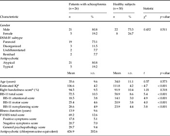
Table 1 describes the characteristics of the 26 patients with schizophrenia and 30 healthy subjects participating in the study. All participants underwent the French version of the Structured Clinical Interview for the Fourth Edition of the Diagnostic and Statistical Manual of Mental Disorders (DSM-IV), Axis I disorders, patient or non-patient version (First et al. Reference First, Spitzer, Gibbon and Williams1996). These interviews confirmed that patients met DSM-IV criteria for schizophrenia and that healthy subjects were free of current or past psychiatric diagnoses. All patients were hospitalized for an acute phase of the disorder and were receiving antipsychotic medication. No participant had a history of medical or neurological disorder that might affect brain function. All participants were right-handed, as assessed by the Edinburgh Handedness Inventory (handedness score ⩾0.7) (Oldfield, Reference Oldfield1971). Approval by the local Biomedical Research Ethics Committee and written informed consent from participants were obtained. Data of 21 patients and 21 healthy subjects of the current study were already used in a prior investigation comparing these two groups with regard to the maps of brain activation elicited by response inhibition (Kaladjian et al. Reference Kaladjian, Jeanningros, Azorin, Grimault, Anton and Mazzola-Pomietto2007).
Demographic and clinical characteristics of patients with schizophrenia and healthy subjects

BIS-11, Barratt Impulsivity Scale – version 11; PANSS, Positive and Negative Syndrome Scale (Kay et al. Reference Kay, Fiszbein and Opler1987).
a Estimated from the French National Adult Reading Test (Mackinnon et al. Reference Mackinnon, Ritchie and Mulligan1999).
b Assessed using the Edinburgh handedness inventory.
Procedures
Within 2 days following the fMRI exam, all participants completed the BIS-11, a 30-item self-report measure of impulsivity that has been validated in both non-clinical and clinical populations (Patton et al. Reference Patton, Stanford and Barratt1995). This scale includes subscales assessing attentional impulsivity, motor impulsivity and non-planning impulsivity.
To elicit response inhibition, we used a Go/NoGo task in conjunction with an event-related fMRI design. This task is usually designed with many more Go than NoGo stimuli to establish a pre-potent tendency to respond. However, under conditions in which NoGo stimuli are rare, brain activation associated with inhibition of the pre-potent response is entailed by that related to processing of novelty and conflict (Braver et al. Reference Braver, Barch, Gray, Molfese and Snyder2001; Nieuwenhuis et al. Reference Nieuwenhuis, Yeung, van den Wildenberg and Ridderinkhof2003). An alternative approach to establish a pre-potent tendency to respond consists of introducing a warning phase to signal the impending presentation of a stimulus, regardless of its type (Go or NoGo) (Konishi et al. Reference Konishi, Nakajima, Uchida, Sekihara and Miyashita1998; Liddle et al. Reference Liddle, Kiehl and Smith2001; Watanabe et al. Reference Watanabe, Sugiura, Sato, Sato, Maeda, Matsue, Fukuda and Kawashima2002; Kaladjian et al. Reference Kaladjian, Jeanningros, Azorin, Grimault, Anton and Mazzola-Pomietto2007; Karch et al. Reference Karch, Jager, Karamatskos, Graz, Stammel, Flatz, Lutz, Holtschmidt-Taschner, Genius, Leicht, Pogarell, Born, Moller, Hegerl, Reiser, Soyka and Mulert2008; Mazzola-Pomietto et al. Reference Mazzola-Pomietto, Kaladjian, Azorin, Anton and Jeanningros2009). The warning phase facilitates speeded responding to Go stimuli and increases the difficulty of withholding responses to NoGo stimuli. This approach makes it possible to match the Go and NoGo trials for frequency and thus to perform unbiased comparison of the brain activations associated with these trials.
The present study used the same version of the warned equiprobable Go/NoGo task as that described by Kaladjian et al. (Reference Kaladjian, Jeanningros, Azorin, Grimault, Anton and Mazzola-Pomietto2007). Briefly, the task consisted of two runs. Each run contained 25 NoGo and 25 Go trials, presented in a pseudo-random order. Each trial began with a 5-s warning phase, consisting of a countdown (i.e. a series of circles with decreasing diameters) followed by the presentation of a letter. Subjects were instructed to press a button with their right index finger as quickly as possible upon the presentation of the letter ‘X’ (i.e. the Go stimuli) and to withhold response on the appearance of the letter ‘A’ (i.e. the NoGo stimuli). Errors of omission (EO), errors of commission (EC) and response times to Go trials (RT) were recorded. Perceptual sensitivity (d') and response bias (β) were calculated for each individual by using the method proposed by Stanislaw & Todorov (Reference Stanislaw and Todorov1999) (see http://psy2.ucsd.edu/~kang/sdt/sdt.htm). Participants completed two brief practice runs of the task before scanning.
All images were obtained using a 3-T whole body MRI scanner (Medspec 30/80 Avance, Bruker, Germany) with a circular polarized head coil. Anatomical images were acquired in the axial plane by using a T1-weighted three-dimensional magnetization- prepared rapid gradient-echo sequence [inversion time=800 ms, repetition time (TR)/echo time (TE)=25/5 ms, delay time=300 ms, flip angle=15°, field of view (FOV)=25.6×23.0×18.0 cm, voxel size=1×1.2×1.73 mm]. Functional images were acquired in the same plane by using a T2*-weighted free induction decay echoplanar sequence (36 contiguous interleaved slices, TR/TE=3000/35 ms, flip angle=83°, FOV=19.2×19.2 cm, voxel size=3×3×3 mm).
Statistical analysis
Between-group differences in demographic, behavioural and psychometric data were assessed by using a χ2 test or independent-sample t tests, as appropriate. Relationships between BIS-11 score and task performance measures were assessed by using Pearson's correlation tests. These statistical analyses were performed with the SPSS statistical package (version 10.1; SPSS Inc., USA).
fMRI data were processed using Statistical Parametric Mapping software (SPM5; Wellcome Department of Cognitive Neurology, UK). For each subject, functional images were corrected for slice timing, spatially realigned, normalized to the standard SPM5/Montreal Neurological Institute template and smoothed using a 9-mm Gaussian kernel. Finally, they were high-pass filtered (120-s cut-off period) to remove low-frequency drifts.
The statistical analyses of the fMRI data were performed using the general linear model (Friston et al. Reference Friston, Holmes, Worsley, Poline, Frith and Frakowiack1995) with separate regressors for the warning phase and each type of trial (i.e. correct Go trial, incorrect Go trial, correct NoGo trial, incorrect NoGo trial). Each regressor was modelled by using the standard haemodynamic response function implemented in SPM5. In the first level of analysis, brain activation specifically associated with response inhibition was assessed in each subject by using the contrast ‘correct NoGo trials minus correct Go trials’. In the second level of analysis, the relationship between impulsivity and activation associated with response inhibition was assessed by performing separate voxel-wise correlation analyses in the schizophrenia and the healthy subject groups. This was achieved in each group by entering the first level contrast image of each individual together with his/her BIS-11 score into a regression analysis with his/her estimated IQ and RT as covariates of non-interest. In an attempt to control for the potential effect of the antipsychotic dosage (expressed as chlorpromazine equivalent) in the schizophrenia group, we also performed a regression analysis that was adjusted for this variable. Statistical significance was set at p=0.05 corrected for multiple comparisons at the voxel level using false discovery rate. To further interpret our data, we performed post-hoc analyses using a region of interest approach. The relationship between each of the BIS-11 subscores and the level of activation (effect size extracted in each subject with MarsBaR, http://marsbar.sourceforge.net/) in the cluster showing a significant correlation with the BIS-11 score was examined by using Pearson's correlation tests.
Complementary analyses were performed to identify within-group and between-group differences in activation associated with response inhibition. The first level images were entered into one-sample t tests and an analysis of covariance with IQ and RT as covariates, respectively. As the results of these analyses were largely similar to those reported in our prior report (Kaladjian et al. Reference Kaladjian, Jeanningros, Azorin, Grimault, Anton and Mazzola-Pomietto2007), they are provided as supplementary material (Figs S1 and S2, available online).
Results
Impulsivity score and task performance
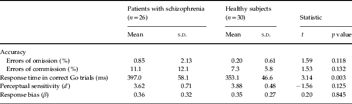
Patients with schizophrenia had significantly increased BIS-11 scores relative to healthy subjects (Table 1). Regarding task performance, there were no significant differences for accuracy (percentage of EO and EC) between the schizophrenia and the healthy subject groups (Table 2). A significant between-group difference was found for RT, with patients being slower than healthy subjects. There were no significant between-group differences for perceptual sensitivity and response bias, indicating that patients and healthy subjects distinguished the Go and NoGo stimuli with a similar ease and developed a similar and strong tendency toward responding. No significant correlations were found between performance measures (RT, percentage of EO and EC) and the BIS-11 score in either healthy subjects or patients (all |r|<0.3, all p>0.15).
Task performance of patients with schizophrenia and healthy subjects

fMRI data
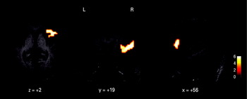
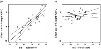
In patients with schizophrenia, regression analysis over the entire brain volume yielded a unique cluster in which the magnitude of activation during response inhibition correlated significantly with the BIS-11 score (Fig. 1). This correlation was positive, indicating that the activation in this cluster increased with increasing impulsivity (Fig. 2 a). It was the strongest within the pars triangularis region of the right VLPFC [Brodmann area (BA) 45] and extended to other parts of the VLPFC (BA 44, BA 47) and to the anterior insula. The result remained largely unchanged when the antipsychotic dosage was entered as a covariate of non-interest in the voxel-wise regression analysis. Post-hoc analyses revealed that the level of activation within this cluster correlated significantly with each of the BIS-11 subscores (motor: r=0.76, p<0.001, non-planning: r=0.68, p<0.001; attentional: r=0.47, p=0.015).
Statistical parametric map resulting from the voxel-wise correlation analysis between the Barratt Impulsivity Scale – version 11 scores and cerebral activation associated with motor response inhibition in patients with schizophrenia (n=26). A unique cluster exhibited a significant correlation. This cluster was localized within the right ventrolateral prefrontal cortex [Talairach coordinates at peak of correlation: x=56, y=19, z=2, Z score=4.51; extent: 171 voxels, Brodmann area (BA) 45 extending to BA 44, BA 47 and anterior insula]. Statistical significance was set at p=0.05 corrected for multiple comparisons at the voxel level using false discovery rate. The cluster is surface-rendered onto slices (axial, coronal and sagittal) of a standard reference brain. The colour scale represents voxel t values. L, left; R, right.

Scatterplots showing the relationship between the level of activation in the right ventrolateral prefrontal cortex (VLPFC) cluster (effect size extracted with MarsBaR; http://marsbar.sourceforge.net/) and the Barratt Impulsivity Scale – version 11 (BIS-11) total score in (a) patients with schizophrenia (n=26) and (b) healthy subjects (n=30). A significant correlation is observed in the patient group (r=0.84, p<0.001) but not in the healthy subject group (r=0.20, p=0.293).

In healthy subjects, the voxel-wise regression analysis did not reveal any significant correlation between brain activation and the BIS-11 score. Additionally, no significant correlations were found in these subjects between the BIS-11 total score (Fig. 2 b) or subscores and the level of activation in the right VLPFC cluster identified in the schizophrenia group (all |r|<0.18; all p>0.3).
Discussion
This study is the first to investigate the relationship between the neural correlates of response inhibition and a psychometric measure of impulsivity in patients with schizophrenia. It reveals a positive correlation between the degree of impulsivity of patients and the magnitude of activation in a cluster localized in the right VLPFC, a key node in the brain network subserving response inhibition (Chamberlain & Sahakian, Reference Chamberlain and Sahakian2007). This cluster largely overlaps the cluster that we have shown as less activated during response inhibition in schizophrenia in a prior investigation comparing subgroups of the current study (Kaladjian et al. Reference Kaladjian, Jeanningros, Azorin, Grimault, Anton and Mazzola-Pomietto2007).
These findings suggest that the amount of VLPFC activation engaged by the patients during motor response inhibition depends on two distinct factors. The first is the diagnosis of schizophrenia, which is associated with a decrease of the average magnitude of activation within this brain region, as evidenced by the comparison of the patient and healthy subject groups. This effect is likely to reflect disease-related structural or neurofunctional alterations that compromise the capacity of this brain region to be activated. The second factor, which appears to have an opposite effect to that of the disease, is the trait impulsivity dimension, as conceived in the BIS-11. The direction of change in VLPFC activation, when impulsivity increases, may provide clues for the understanding of the functionality of this brain region in relation to impulsivity in schizophrenia. Based on the assumption that, at a low task demand, greater activation of a brain region in patients with schizophrenia may reflect a lower ability to engage a cognitive function (Callicott et al. Reference Callicott, Bertolino, Mattay, Langheim, Duyn, Coppola, Goldberg and Weinberger2000; Manoach, Reference Manoach2003; Potkin et al. Reference Potkin, Turner, Brown, McCarthy, Greve, Glover, Manoach, Belger, Diaz, Wible, Ford, Mathalon, Gollub, Lauriello, O'Leary, van Erp, Toga, Preda and Lim2009), a greater right VLPFC activation associated with higher impulsivity in patients may indicate a lower efficiency of this brain region to engage inhibitory processes. In other words, the more impulsive patients may need to recruit greater resources within the right VLPFC to correctly inhibit. This interpretation is in line with the greater inhibition-related brain activation that is observed in healthy volunteers with high self-reported impulsivity (Dimoska & Johnstone, Reference Dimoska and Johnstone2007; Lansbergen et al. Reference Lansbergen, Bocker, Bekker and Kenemans2007). It is also interesting to note that impulsivity in schizophrenia is associated with altered white matter integrity in the right inferior frontal region (Hoptman et al. Reference Hoptman, Volavka, Johnson, Weiss, Bilder and Lim2002). To summarize, despite patients with schizophrenia taken as a group show a deficit of VLPFC activation, they appear to exhibit impulsivity-related differences in engaging brain resources to adapt to the inhibitory demand. When this demand increases, it is likely that the capacity of the VLPFC is more rapidly overwhelmed in the most impulsive ones, leading to failures of inhibitory control. This may help to explain why dysfunctions in inferior frontal areas have been related to impulsivity or violent behaviours in schizophrenia (Naudts & Hodgins, Reference Naudts and Hodgins2006; Joyal et al. Reference Joyal, Putkonen, Mancini-Marie, Hodgins, Kononen, Boulay, Pihlajamaki, Soininen, Stip, Tiihonen and Aronen2007).
In the healthy subjects of the current study, and by contrast to what we evidenced in patients with schizophrenia, there were no significant correlations between the BIS-11 score and brain activation associated with response inhibition. Some prior neuroimaging studies of response inhibition within the frame of the Go/NoGo task in healthy subjects have found a correlation between the BIS-11 score and brain activation within superior frontal and temporal areas (Horn et al. Reference Horn, Dolan, Elliott, Deakin and Woodruff2003) or the ACC and caudate nucleus (Brown et al. Reference Brown, Manuck, Flory and Hariri2006). Other authors have observed that the activity of the right dorsolateral prefrontal cortex correlates with the BIS-11 motor impulsivity subscore, but not with the other subscores or total score (Asahi et al. Reference Asahi, Okamoto, Okada, Yamawaki and Yokota2004). The discrepancies between these studies remain difficult to explain because all employed a quite similar version of the Go/NoGo task. One possible explanation is that they rely on differences in the ranges of the BIS-11 scores examined. An alternative explanation is that the results may have been confounded by the presence of heterogeneities in the profiles of brain activation during response inhibition, particularly those related to genetic factors. For instance, Passamonti et al. (Reference Passamonti, Fera, Magariello, Cerasa, Gioia, Muglia, Nicoletti, Gallo, Provinciali and Quattrone2006) evidenced that a significant positive correlation between the BIS-11 score and activation of the right VLPFC is present only in subjects who have a genetic risk to develop impulsive behaviours. Further studies are needed to clarify this issue.
In the present study, we did not observe a correlation between task performance measures and the BIS-11 score either in patients or in healthy subjects. These findings mirror those of prior studies in schizophrenia (Enticott et al. Reference Enticott, Ogloff and Bradshaw2008) and non-clinical populations (Horn et al. Reference Horn, Dolan, Elliott, Deakin and Woodruff2003; Asahi et al. Reference Asahi, Okamoto, Okada, Yamawaki and Yokota2004; Brown et al. Reference Brown, Manuck, Flory and Hariri2006; Passamonti et al. Reference Passamonti, Fera, Magariello, Cerasa, Gioia, Muglia, Nicoletti, Gallo, Provinciali and Quattrone2006; Dimoska & Johnstone, Reference Dimoska and Johnstone2007; Lansbergen et al. Reference Lansbergen, Bocker, Bekker and Kenemans2007). The absence of correlation may result from the low extent to which the dimensions of impulsivity as assessed by self-reported questionnaires are related to motor response inhibition. However, such explanation is rather inconsistent with the relationship between impulsivity measures and brain functioning that we and others have evidenced. Alternatively, it should be noted that, in all of these studies, the task was not designed to push subjects to work at the edge of their own inhibitory capacity. At a low task demand, subjects having high impulsivity might be able to inhibit correctly by engaging more brain resources (Dimoska & Johnstone, Reference Dimoska and Johnstone2007; Lansbergen et al. Reference Lansbergen, Bocker, Bekker and Kenemans2007). In such context, it is likely that measures of brain activity represent a more sensitive index of the resources engaged to perform a task than performance measures (Rubia et al. Reference Rubia, Lee, Cleare, Tunstall, Fu, Brammer and McGuire2005 a; Raemaekers et al. Reference Raemaekers, Ramsey, Vink, van den Heuvel and Kahn2006). This may explain why impulsivity scores correlate with the right VLPFC activation but not with performance measures in the current study.
A major limitation of the current study is that it focuses on a unique aspect of impulsivity, which is related to poor response inhibition. One should keep in mind that impulsivity is a multi-faceted construct that incorporates disturbances in several and dissociable cognitive domains, such as, for instance, decreased sensitivity to negative consequences of behaviour, inability to delay gratification, sensation seeking or difficulties with maintaining attention (Evenden, Reference Evenden1999; Moeller et al. Reference Moeller, Barratt, Dougherty, Schmitz and Swann2001). Thus, it is likely that the impulsivity expressed by patients with schizophrenia is not underlain solely by the dysfunction of the right VLPFC, but also of other brain regions subserving a variety of cognitive processes involved in the control of behaviour. Of particular interest is the circuitry involved in emotional modulation of inhibitory control, since abnormalities in the amygdala–orbitofrontal system and impaired emotional processing have been associated with violent behaviour and impulsive aggression in this pathology (Naudts & Hodgins, Reference Naudts and Hodgins2006). Another limitation was that all patients were on psychotropic medication. While it was not appropriate to withdraw treatment, it is possible that our results may be relevant to only medicated patients. However, it should be noted that when the antipsychotic dosage was used as a covariate of non-interest into the voxel-wise regression analysis, the results remain largely unchanged, suggesting a minimal confounding effect of medication. Our results may also hold relevance to only a subgroup of patients with schizophrenia. All patients were in an acute phase of their illness and there was an over-representation of the paranoid subtype and male individuals. Further research is needed to extend our findings to patients in remission, other subtypes of schizophrenia and female gender. Finally, on a cautionary note, magnetic susceptibility artefacts altered the functional signal in the most ventral part of the prefrontal cortex. This probably reduced the sensitivity to detect clusters of regression in this region.
In summary, we investigated the relationship between brain activation associated with response inhibition and trait impulsivity in patients with schizophrenia and healthy subjects. Activation within a cluster of the right VLPFC of the patients was found to correlate positively with their degree of impulsivity. The greater neural response within this region in the more impulsive patients can be interpreted as reflecting less efficiency to process inhibitory control. This result supports the hypothesis that some aspects of impulsivity in schizophrenia are underlain by a right VLPFC dysfunction.
Note
Supplementary material accompanies this paper on the Journal's website (http://journals.cambridge.org/psm).
Acknowledgements
The authors thank Hélène Rescigno, who helped with the behavioural data analysis and literature search.
Declaration of Interest
None.