Introduction
Systemic venous anomalies affecting the superior vena cava (SVC) are rare developmental disorders that occur during embryological development. SVC anomalies are usually asymptomatic but can complicate procedures such as cardiac catheterisation, systemic venous cannulation for extracorporeal membrane oxygenation, and transvenous pacemaker implantation. Reference Ylänen, Poutanen, Savikurki-Heikkilä, Uotila, Korppi and Eerola1 These anomalies are typically diagnosed through echocardiography, CT, MRI, or during failed cardiac or venous catheterisation due to IVC course anomalies. Reference Hussain, Chakravarty, Chaikhouni and Smith2 They are more common in males and may be associated with CHDs, rhythm disturbances, or both. Reference Quraishi, Mufti and Wase3 The most common SVC anomalies include the presence of bilateral SVC and the absence of the right SVC with persistent left SVC. Reference Choi, Anderson and Macartney4 Bilateral SVC absence is extremely rare.
Case
A 7-year-3-month-old girl, with no active complaints or symptoms, was referred to the paediatric cardiology department from the paediatric clinic during the investigation of polycythaemia aetiology. The patient”s general condition was good, and no pathological findings were noted on physical examination. Laboratory results showed haemoglobin levels of 15.9 g/dL, haematocrit of 49%, MCV of 76, and platelet count of 364,000. The patient”s history included an episode of swelling in the chest and shoulder area a year prior, which resolved spontaneously within two days. Due to the lack of medical consultation at that time, it could not be determined whether these symptoms were indicative of superior vena cava syndrome (SVCS). There was no significant family history.
Echocardiography was performed as part of the aetiological work-up for polycythaemia to assess for intracardiac shunts and for anomalous systemic venous pathways that could potentially drain into pulmonary veins/left atrium and thereby contribute to right-to-left shunting and subtle desaturation.
Echocardiography revealed venous flow posterior to the descending aorta in the suprasternal view. A persistent left SVC was initially considered; however, the coronary sinus was not dilated, making a typical persistent LSVC draining to the coronary sinus less likely. Given rare variants (e.g. alternative drainage), further imaging was pursued for definitive delineation. Further suprasternal evaluation showed agenesis of the right SVC, with the right innominate vein draining into a caudally directed venous structure. Subcostal long-axis evaluation revealed a dilated inferior vena cava (IVC), with a venous structure draining into the IVC 7.5 cm distal to the right atrial junction at the renal level.
Thoracic CT angiography was performed to delineate systemic venous anatomy given echocardiographic suspicion of a complex systemic venous return anomaly. The CT angiography revealed agenesis of the azygos vein and both SVC. The bilateral innominate veins were seen to converge at the level of the fourth thoracic vertebra and drain into the accessory hemiazygos vein. This vein continued caudally as the hemiazygos vein, coursing posterior to the aorta, and ultimately joined the left renal vein at the renal level, providing drainage into the IVC. Additionally, the right paravertebral vein was tortuous and drained into the superior pulmonary vein. Thin collateral veins also anastomosed with the inferior pulmonary vein (Figures 1 and 2).
Three-dimensional CT angiography reconstructions of systemic venous return in the patient. ( a ) Anterior view demonstrating absence of both SVC and absence of the azygos vein. The right and left brachiocephalic (innominate) veins converge at approximately the T4 vertebral level and drain into the accessory hemiazygos vein, which continues caudally as the hemiazygos vein. The hemiazygos pathway courses posterior to the descending aorta and joins the left renal vein, providing drainage into the IVC. The IVC–right atrium anastomosis may appear narrowed in some projections due to reconstruction angle/partial-volume effects; however, there was no true anatomic stenosis, no contrast hold-up or delayed opacification, and no clinical evidence of venous congestion. ( b ) Posterior view of the same 3D CT reconstruction, highlighting the collateral venous pathway via the accessory hemiazygos/hemiazygos system into the left renal vein and IVC.

CT angiography of patient (Anterior view). The paravertebral collateral draining into the right superior pulmonary vein appeared small in calibre on CT, which is consistent with the absence of clinically significant desaturation (Acc: accessory).

Due to the patient”s polycythaemia and systemic venous structures draining into the pulmonary veins, pulse oximetry was performed to rule out cyanosis, showing an oxygen saturation of 96%. A detailed history confirmed no prior surgeries or illnesses. Abdominal ultrasound and renal Doppler ultrasound were conducted to investigate potential renal and other pathologies, with no abnormalities detected. The asymptomatic patient was discharged with a plan for periodic follow-up.
Discussion and conclusion
The development of systemic venous circulation begins at the start of the fourth week of embryonic development. Initially, it consists of bilateral anterior cardinal veins and bilateral posterior cardinal veins. The SVC is formed from the proximal part of the right anterior cardinal vein and the left cranial anterior cardinal vein. The azygos system originates from supracardinal veins appearing from the sixth week of gestation. The IVC develops from posterior cardinal veins and subcardinal veins, which are part of the supracardinal veins. Reference Minniti, Visentini and Procacci5,Reference Tacy and Silverman6
In cases of bilateral absence of the SVC, the anomaly most likely results from the embryological regression of both common anterior cardinal veins. As a compensatory adaptation—similar to mechanisms observed in SVCS—alternative venous pathways dilate and become tortuous in order to manage the redirected blood flow from the upper body. In our patient, the bilateral innominate veins drained into the accessory hemiazygos vein, which continued as the hemiazygos vein, ultimately connecting to the left renal vein and IVC. The absence of the azygos system and superior caval veins led to the development of prominent collateral venous channels to maintain effective drainage from the head, neck, and upper extremities. These findings are schematically illustrated in Figure 3.
Schematic illustration of normal systemic venous return (left) and venous circulation pattern in our patient with bilateral absence of the SVC (right). ( a ) In the normal anatomy, venous return from the upper limbs, head, and neck drains into the right atrium via the SVC, while blood from the lower body is carried through the IVC. The azygos and hemiazygos systems also contribute to this drainage pathway. ( b ) In our patient, with complete absence of the SVC and azygos vein, the venous return from the upper body—including the jugular and subclavian veins—drains via bilateral innominate veins into the accessory (Acc) hemiazygos vein, which continues as the hemiazygos vein and connects to the left renal vein. The systemic venous blood ultimately reaches the right atrium through the IVC. The diagram also shows bilateral renal veins and compensatory collateral drainage patterns.

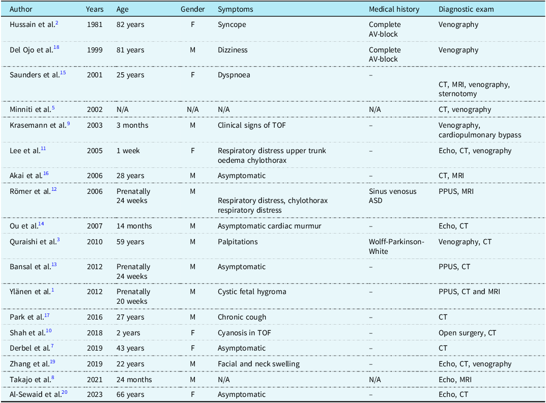
Errors in these processes of merging and regression lead to various combinations of caval anomalies. The failure of regression in both main anterior cardinal veins results in bilateral SVC, while regression of the right common anterior cardinal vein and persistence of the left leads to persistent left SVC. Reference Derbel, Krarti and Ghedira7 Regression of both common anterior cardinal veins results in bilateral SVC agenesis. In this case, other venous structures compensate by expanding and providing drainage through alternate pathways, as observed in this case. Bilateral SVC agenesis is extremely rare, with only a few reported cases to date (Table 1).
Patients with bilateral SVC absence reported in the literature

Takajo et al. in 2021 demonstrated bilateral SVC absence in a 24-month-old patient with abnormal echocardiography findings confirmed by cardiac MRI. In that case, the right jugular and right subclavian veins merged and drained into the azygos vein, while the left jugular and left subclavian veins merged and drained into the hemiazygos vein, with the azygos-hemiazygos veins joining at T9 and draining into the IVC. Reference Takajo, Mahadin and Aggarwal8 In our case, the right jugular and right subclavian veins drained into the accessory hemiazygos via the innominate vein, and the hemiazygos vein merged with the left renal vein at T10 to drain into the IVC.
There have been eight reported cases of bilateral SVC absence in paediatric patients. Krasemann et al. and Shah et al. reported patients with bilateral absence of the SVC in association with tetralogy of Fallot. Reference Krasemann, Kehl, Vogt and Asfour9,Reference Shah, Hiremath, Diwakar and Rema10 In the literature, infants and children more frequently present with symptoms of congestive circulation such as dyspnoea, cyanosis, chylothorax, or upper extremity oedema. Two cases reported in the antenatal period had pleural effusion with postnatal findings of chylothorax and respiratory distress, diagnosed with bilateral SVC absence by CT angiography and cardiac MRI. Reference Lee, Jan, Wang and Chi11,Reference Römer, Opgen-Rhein, Chaoui, Scheer, Czernik and Obladen12 Unlike these cases, our patient was asymptomatic.
Under normal anatomy, the thoracic duct drains into the venous system at the left venous angle (junction of the left internal jugular and subclavian veins), which in turn empties into the left brachiocephalic vein and SVC. In bilateral SVC agenesis, this usual drainage pathway is absent or significantly altered. The body must rely on alternative venous channels for upper-body blood return—for instance, enlarged azygos/hemiazygos veins that carry blood from the head and upper extremities to the IVC or renal veins. Reference Ylänen, Poutanen, Savikurki-Heikkilä, Uotila, Korppi and Eerola1 If these collateral pathways are well-developed, venous return can be rerouted effectively to the right atrium, and lymphatic fluid can drain with little impediment. In fact, many SVC anomalies (e.g. a persistent left SVC draining into the right atrium via the coronary sinus) result in normal haemodynamics and no symptoms and are often discovered incidentally. Reference Ylänen, Poutanen, Savikurki-Heikkilä, Uotila, Korppi and Eerola1 In asymptomatic cases of absent SVC, the thoracic duct likely still empties into a vein that ultimately connects to the IVC (such as the subclavian vein feeding into an enlarged azygos system), allowing lymph to return to the bloodstream without significant backup. Presentation of chylothorax would be an indicator for evaluation of the systemic venous system.
Another patient had a cystic hygroma detected antenatally, which regressed until birth. Postnatal evaluation by echocardiography, CT, and MRI revealed bilateral SVC agenesis, with the patient describing intermittent wheezing and cough. Reference Ylänen, Poutanen, Savikurki-Heikkilä, Uotila, Korppi and Eerola1 In another case, venous return anomaly was suspected in a 24-week obstetric ultrasound, and fetal echocardiography confirmed bilateral SVC absence, with postnatal findings of respiratory distress and facial plethora. Reference Bansal, Strainic and Ashwath13 Only one reported paediatric case was asymptomatic, identified during routine examination due to a cardiac murmur, with the diagnosis confirmed by thoracic CT. Reference Ou, Calcagni and Marini14
In adults or elderly patients, symptoms due to rhythm disturbances are more common. Reference Hussain, Chakravarty, Chaikhouni and Smith2,Reference Quraishi, Mufti and Wase3 Older patients are more likely to be asymptomatic, Reference Derbel, Krarti and Ghedira7,Reference Saunders, Richens and Morris15–Reference Park, Kim and Park17 possibly due to compensatory mechanisms like the ability of venous structures to dilate, becoming more pronounced with age, thereby reducing congestive circulation symptoms.
Unlike reported cases, our patient also had a pulmonary venous return anomaly, with tortuous paravertebral veins draining into the right superior pulmonary vein and thin collateral veins anastomosing with the inferior right pulmonary vein. The paravertebral collateral draining into the right superior pulmonary vein appeared small in calibre on CT, which is consistent with the absence of clinically significant desaturation.
On suprasternal imaging, identification of a venous channel coursing posterior to the descending aorta should raise suspicion for a persistent left SVC. In typical persistent LSVC draining to the right atrium via the coronary sinus, the coronary sinus is dilated; therefore, coronary sinus size should be assessed. If the coronary sinus is not dilated, alternative systemic venous anomalies should be considered and cross-sectional imaging may be required to define anatomy.
In subcostal evaluation, the long-axis view of the IVC should be assessed, and if the IVC is found to be dilated, careful evaluation of venous structures draining into it is necessary. Our patient being asymptomatic differs from the more common finding of symptomatic young patients in the literature. Patients with adequate decompression of head and neck vessels typically remain asymptomatic, as observed in our case. This could indeed lead to underdiagnosis, suggesting that the actual incidence might be higher than reported. In these patients, associated anomalies should be investigated, and genetic consultation should be sought due to the venous anomaly associated with polycythaemia.
This report adds to the limited paediatric literature by describing an asymptomatic school-age presentation discovered during polycythaemia evaluation, with a particularly uncommon systemic venous return pattern including absence of both SVCs and the azygos vein, and accessory hemiazygos/hemiazygos continuation draining to the left renal vein and IVC. In addition, small systemic-to-pulmonary venous collateral connections were identified without clinically significant desaturation. Finally, we highlight echocardiographic “red flags” (a posterior venous channel in the suprasternal view and a dilated IVC with unexpected tributaries) and provide a schematic diagram to facilitate recognition.
Acknowledgements
None.
Financial support
This research received no specific grant from any funding agency, commercial, or not-for-profit sectors.
Competing interests
None.
Ethical standard
Ethics approval statement is not applicable.
Data availability statement
None.