Background
Recently, three-dimensional printing of heart models is being used to plan percutaneous and surgical interventions in patients with CHD. We describe a case where we used a three-dimensional print model to plan a complex percutaneous intervention in a patient with coarctation of the aorta.
Case report
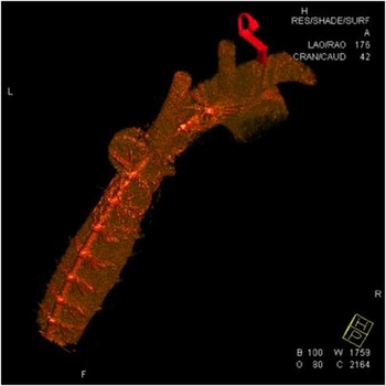
Our patient was a 19-year-old male with a history of coarctation of the aorta. He was diagnosed at 12 years of age after he was referred to us for evaluation of hypertension. He was subsequently taken to the catheterisation laboratory, and underwent successful implantation of a 36-mm Max LD stent (ev3 Endovascular Inc., Plymouth, Minnesota, United States of America) on a 16-mm balloon. He was discharged home the following day, and was subsequently lost to follow-up until he presented again with hypertension 7 years later. Outpatient evaluation revealed upper extremity hypertension with a systolic blood pressure of 148 and a 25 mmHg gradient to the lower extremities by non-invasive blood pressure assessment. He underwent a repeat cardiac catheterisation, which showed a 21- mmHg gradient from the ascending to the descending aorta, circumferential fracture in the mid-portion of the stent, narrowing of the aorta in the area of stent fracture, and a moderate-sized psuedoaneurysm near the take off of the left subclavian artery (Fig 1). We did not have a covered stent in our catheterisation laboratory at the time, and therefore no intervention was performed during this procedure.
Three-dimensional reconstruction of the aorta from rotational angiography performed during repeat catheterisation demonstrating the pseudoaneurysm.

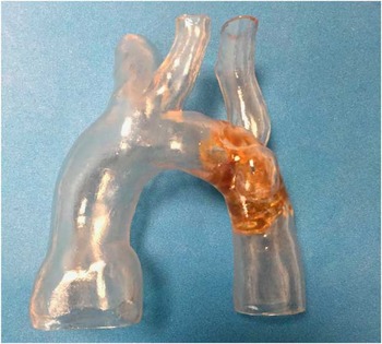
We subsequently applied for and obtained Institutional Review Board approval for compassionate use of a Covered Cheatham Platinum Stent (Numed Inc., Hopkinton, New York, United States of America) to treat the residual coarctation and cover the pseudoaneurysm. We were concerned that implantation of a covered cheatham platinum stent might have to jail the left subclavian artery in order to cover the pseudoaneurysm. A CT angiogram of the chest was performed to image the aortic arch, from which a three-dimensional data set was isolated using Mimics Innovation Suite (Materialise, Plymouth, Michigan, United States of America). From this segmentation, a transparent, rigid, three-dimensional print model of the aorta was made by Materialise using Stereolithography technique with the Watershed material (Fig 2). Simulation of stent implantation in the model showed that, in order to cover the pseudoaneurysm and relieve the residual aortic arch obstruction with a Covered Cheatham Platinum Stent, the left subclavian artery would consequently be jailed. Therefore, in planning the subsequent intervention, we decided to obtain left radial artery access for further imaging of the left subclavian artery and to re-establish patency after implantation of the covered cheatham platinum stent.
Three-dimensional, printed model of the aortic arch made by Materialise. The gold material in the arch represents the previously placed stent.

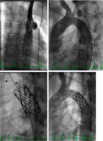
The patient was subsequently brought back to the catheterisation laboratory. He was placed under general anaesthesia, and percutaneous access was obtained in the right femoral vein and artery and the left subclavian artery. A 34-mm-long, covered cheatham platinum stent was implanted with an 18-mm balloon overlying the previously placed stent via a femoral arterial approach. Angiography in the transverse aortic arch demonstrated a well-positioned Covered Cheatham Platinum Stent with no residual aortic arch narrowing, complete exclusion of the pseudoaneurysm, and no antegrade flow into the left subclavian artery. A JR4 catheter was advanced through the left radial artery sheath and positioned at the origin of the left subclavian artery. The stiff end of a 0.014-inch Platinum Plus wire was then used to perforate the ePTFE covering of the Covered Cheatham Platinum Stent. Serial balloon angioplasty was performed to enlarge this opening. Initially, a 3-mm-diameter coronary balloon was used, after which the Platinum Plus wire was removed and re-inserted through the balloon, such that the floppy end of the wire was now in the descending aorta. Subsequent serial angioplasty was performed up to 8-mm diameter. Repeat angiography showed that the covered cheatham platinum stent remained well positioned with no residual narrowing, complete exclusion of the pseudoaneurysm, and patency of the left subclavian artery (Fig 3). There was no residual pressure gradient from the ascending to descending aorta. He tolerated the procedure well and was discharged home the following day. At subsequent outpatient follow-up, there was no residual right arm-to-lower extremity non-invasive blood pressure gradient with a 25-mmHg gradient to the left arm with a strong palpable left radial artery pulse.
Images during implantation of the covered cheatham platinum stent. ( a ) Angiogram of the ascending aorta, demonstrating the pseudoaneuryms. ( b ) Angiogram immediately aftert covered cheatham platinum stent implantation, showing exclusion of the pseudoaneurysm with no flow seen in the left subclavian artery. ( c ) Balloon angioplasty of the covered cheatham platinum stent through the puncture created from the left subclavian artery. ( d ) Final angiogram demonstrating flow in the left subclavian artery and exclusion of the left subclavian artery.

Discussion
Percutaneous stent implantation is now the treatment of choice for coarctation of the aorta in older children and young adults at many centres. Before the Covered Cheatham Platinum Stent recently becoming commercially available in the United States of America, bare metal stents were used. The reported incidence of acute aortic wall injury with bare metal stent implantation is ~1%; however, the incidence of aortic aneurysm during intermediate follow-up is 5%.Reference Forbes, Kim and Du 1 The Covered Cheatham Platinum Stent has been used for treatment of existing aortic wall injury in patients with previous intervention for coarctation of the aorta with a reported 92% success rate for complete coverage of the aortic wall injury during the COAST II trial.Reference Taggart, Minahan and Cabalka 2 Treatment of an aortic arch aneurysm with a Covered Cheatham Platinum Stent that required exclusion of the left subclavian artery and subsequent restoration of flow in a case very similar to ours has been previously reported.Reference Tsai, Hill and Cheatham 3 Exclusion of the left subclavian artery using covered stent grafts for aortic arch repair is reported to be safe, with the potential for symptoms of left subclavian artery steal;Reference Rehders, Petzsch and Ince 4 however, subsequent left-arm claudication requiring surgical translocation of the left subclavian artery to the left carotid artery has been reported.Reference Marcheix, Lamarche and Perrault 5 Given that perforation of the ePTFE for the Covered Cheatham Platinum Stent had been shown to be feasible, we opted to perform this in our patient to minimise the chances of subclavian artery steal or claudication and subsequent need for additional surgical intervention. The Covered Cheatham Platinum Stent is not an open-cell stent design, and therefore the potential to create an opening that led to significant left subclavian artery ostial stenosis did exist. Furthermore, we did have concerns that we could create a more extensive tear in the ePTFE, which could result in endoleak and insufficient covering of the pseudoaneurysm. Fortunately, we were able to achieve complete occlusion of the aortic wall injury. We did note mild ostial stenosis of the left subclavian artery on the final angiogram, and a non-invasive blood pressure discrepancy between the left and right arms during subsequent follow-up; however, our patient has not reported claudication or evidence of left subclavian artery steal.
Real-scale, three-dimensional printed models have been used for surgical planning in CHD and have been shown to be a valuable tool for pre-surgical planning in patients with complex CHD.Reference Garekar, Bharati and Chokhandre 6 , Reference Farooqi, Gonzalez-Lengua and Shenoy 7 , Reference Kiraly, Tofeig and Jha 8 , Reference Valverde, Gomez and Gonzalez 9 In addition, they have shown promise for planning complex percutaneous interventions.Reference Valverde, Gomez and Coserria 10 , Reference Dankowski, Baszko and Sutherland 11 , Reference Olivieri, Krieger and Chen 12 Valverde et al reported using three-dimensional printed models to plan stent implantation for a patient with transverse aortic arch hypoplasia and demonstrated excellent correlation for measurements of the aortic arch between the three-dimensional print model, MRI, and angiography in the catheterisation laboratory.Reference Dankowski, Baszko and Sutherland 11 Another report demonstrated the use of a three-dimensional printed model to determine appropriate catheter positioning for transcatheter mitral valve implantation.
The process of creating a three-dimensional print model of the heart and vascular structures begins with acquiring three-dimensional imaging, usually from an MRI or CT scan; however, more recent reports have demonstrated feasibility for using three-dimensional transthoracic echocardiographic images for creating accurate three-dimensional printed models.Reference Olivieri, Krieger and Loke 13 Once the three-dimensional images have been acquired, segmentation is performed using commercially available software to create a data set from which the three-dimensional printing can be done. Once this is completed, the three-dimensional model is printed. Although the term rapid prototype is often used to describe to process of printing a three-dimensional model, it usually takes several hours to a few days to make the model, depending on the complexity of the structures being printed and the type of material being used for printing.
In our patient, we used the three-dimensional printed model of the aortic arch to simulate stent implantation in the aortic arch in order to determine whether the stent would need to cover the left subclavian artery in order to completely exclude the pseudoaneurysm. We inserted a balloon catheter into the printed model with markings on the balloon to determine the best stent positioning. Once it was determined that the Covered Cheatham Platinum Stent needed to exclude the left subclavian artery to ensure that we completely covered the pseudoaneurysm, we were able to plan the procedure accordingly before bringing the patient to the catheterisation laboratory. Although flexible models used to evaluate vessel wall expansion can be made, we chose a rigid, three-dimensional, printed model of the aortic arch, as our goal was to determine stent positioning.
Conclusion
The use of a three-dimensional printed model of the aortic arch permitted detailed planning of the intervention for the patient before the procedure. This allowed for the intervention to be performed safely, and likely reduced the duration of the procedure, total radiation dose, and contrast administration. Our case shows the feasibility for using three-dimensional printed models for planning and carrying out complex percutaneous interventions in patients with CHD.
Acknowledgements
None.
Financial Support
This research received no specific grant from any funding agency, commercial, or not-for-profit sectors.
Conflicts of Interest
None.