Introduction
In canine oocytes, in vitro maturation (IVM) is characterized by low rates of maturation to metaphase II (MII) (Mahi & Yanagimachi, Reference Mahi and Yanagimachi1976; Yamada et al., Reference Yamada, Shimazu, Kawaji, Nakazawa, Naito and Toyoda1992, Reference Yamada, Shimazu, Kawano, Nakazawa, Naito and Toyoda1993; Nickson et al., Reference Nickson, Boyd, Eckersall, Ferguson, Harvey and Renton1993; Bolamba et al., Reference Bolamba, Borden-Russ and Durrant1998; Hewitt & England, Reference Hewitt and England1998, Reference Hewitt and England1999; Metcalfe, Reference Metcalfe1999; Otoi et al., Reference Otoi, Fujii, Tanaka, Ooka and Suzuki2000a, Reference Otoi, Fujii, Tanaka, Ooka and Suzuki.b, Reference Otoi, Ooka, Murakami, Karja and Suzuki2001, Reference Otoi, Willingham, Shin, Kraemer and Westhusin2002; Saint-Dizier et al., Reference Saint-Dizier, Renard and Chastant-Maillard2001; Songsasen et al., Reference Songsasen, Yu and Leibo2002; Hatoya et al., Reference Hatoya, Sugiyama, Torii, Wijewardana, Kumagai, Sugiura, Kida, Kawate, Tamada, Sawada and Inaba2006). However, the actual reason for the low IVM rates in canines remains unclear. It has been acknowledged that canine oocytes are spontaneously ovulated at the germinal vesicle (GV) stage (Yamada et al., Reference Yamada, Shimazu, Kawaji, Nakazawa, Naito and Toyoda1992; Reynaud et al., Reference Reynaud, Fontbonne, Marseloo, Thoumire, Chebrout, Viaris de Lesegno and Chastant-Maillard2005). After ovulation, they require an extended time in the oviducts to complete the second meiotic maturation (Concannon, Reference Concannon1994). In most mammalian animals, oocyte maturation occurs within ovarian follicles.
A previous study was conducted to assess the in vivo developmental competence of oocytes and embryos (Reynaud et al., Reference Reynaud, Fontbonne, Marseloo, Thoumire, Chebrout, Viaris de Lesegno and Chastant-Maillard2005). However, no canine oocytes at the germinal vesicle breakdown (GVBD) stage were detected in their study. Probably, canine oocytes need such a short time in vivo to develop from GV stage to metaphase I (MI) that oocytes at the GVBD stage have not been observed yet in such studies. Another study used super-ovulated canines to aspirate oocytes from follicles for in vitro maturation (Yamada et al., Reference Yamada, Shimazu, Kawaji, Nakazawa, Naito and Toyoda1992). This group also did not find any oocytes at the GVBD stage in their study. Therefore, these two studies reveal that canine oocytes recovered by follicle aspiration or in the oviduct require a very short time to progress beyond the GVBD stage in the first meiosis. In most other studies, canine oocytes with various nuclear stages (including GVBD stage) can be observed before or after in vitro maturation (De Los Reyes et al., Reference De Los Reyes, De Lange, Miranda, Palominos and Barros2005), when the majority of oocytes for IVM were recovered from the follicles located inside the ovary at the abattoir. It is possible that the configurations of nuclear status of canine GV stage oocytes recovered from follicle-S differ from those of follicle-I.
The objective of this study was to compare the configurations of nuclear envelope, nucleolus, and chromatin in three types of canine GV stage oocytes recovered from between follicle-S and follicle-I.
Materials and Methods
Ovaries and oocytes collection
Ovaries obtained from adult bitches at a local abattoir were collected within 1 h of removal, and transported to the laboratory in a thermal bottle containing prewarmed physiological saline solution at 37°C within 3 h of removal. Follicles were classified into two types based on their location in the ovary: follicle-S if follicles located in the ovarian surface, and follicle-I if follicles located inside the ovary. Cumulus–oocyte complexes (COCs) from follicle-S and follicle-I were collected and the nuclear stages of these were examined by phase contrast microscopy.
Experimental design
Ovaries at pro-estrous and estrous stages with apparent follicles located in the ovarian surface (follicle-S) were selected. In each replicate, canine oocytes from follicle-S were first obtained by follicle aspiration. Subsequently, those from follicle-I from the same ovary were obtained by slicing the ovarian cortex with a scalpel blade. Morphologically degenerated oocytes (such as following zona pellucida rupture) both from follicle-S and follicle-I were discarded. Selected COCs were fixed and stained to determine the nuclear stages by phase contrast microscope.
Assessment of nuclear stage of maturation
After collection, oocytes were denuded, fixed in acetic acid: ethanol:chloroform (3:6:2, v:v:v) for 2 to 3 min prior to fixation with acetic acid:ethanol (1:3, v:v) for 24 to 48 h (Songsasen et al., Reference Songsasen, Yu and Leibo2002; De Los Reyes et al., Reference De Los Reyes, De Lange, Miranda, Palominos and Barros2005; Hatoya et al., Reference Hatoya, Sugiyama, Torii, Wijewardana, Kumagai, Sugiura, Kida, Kawate, Tamada, Sawada and Inaba2006). These oocytes were then stained with 1% (w/v) orcein-acetic (Sigma-Aldrich). Nuclear maturation stages were examined by phase contrast microscopy at ×400 magnification. We classified these nuclear stages as GV, GV breakdown (GVBD), metaphase I (MI), metaphase II (MII), and unidentified, according to the methods described previously (De Los Reyes et al., Reference De Los Reyes, De Lange, Miranda, Palominos and Barros2005).
Statistical analysis
The data for numbers and percentages of oocytes that progressed to various nuclear stages, and the distribution of three types of canine GV stage oocytes were analysed by SPSS 10.0 software. Due to the few numbers of canine oocytes recovered from follicle-S in each replicate, the significant differences in maturational rates between the treatment groups have not been compared in current studies.
Results
The majority of canine oocytes was at the GV stage after isolation both from follicle-S and follicle-I (Table 1). Meanwhile, a small proportion of oocytes had undergone the resumption of the meiosis in both the follicle-S and follicle-I groups.
Per cent of canine oocytes at various nuclear stages recovered from follicle-S and follicle-I.

aFollicle-S, follicles located in the ovarian surface; follicle-I, follicles located inside the ovary.
bDE: degenerated oocytes, GV: germinal vesicle stage, GVBD: germinal vesicle breakdown, MI/TI: metaphase I/telophase I, MII: metaphase II.
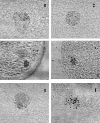
GV stages were classified into three types based on the status of the nuclear envelope and nucleolus: type A, type B, and type C. In follicle-S group, the majority (95.5%) of canine GV stage oocytes was type B, which were characterized by nuclear envelope disappearance prior to nucleolus collapse (Fig. 1). There was no type A canine GV stage oocytes in this group (Table 2). In the follicle-I group, the majority of canine GV stage oocytes was type C. In addition, a small proportion of canine GV stage oocytes from follicle-I (donated type A) were characterized by nuclear envelope disappearance following nucleolus collapse (Fig. 1).
Representative pictures of three types of canine GV stage oocytes (magnification ×400). (a) Type A canine GV stage oocytes, the nuclear envelope was distinct and the nucleolus began to disappear. (b) Type A canine GV stage oocytes, the nuclear envelope was less distinct while the nucleolus completely disappeared. (c) Type B canine GV stage oocytes, the nuclear envelope disappeared and the nucleolus was visible. (d) Type B canine GV stage oocytes, the nuclear envelope has disappeared and the nucleolus has begun to collapse. (e) Type C canine GV stage oocytes, the nuclear envelope has disappeared, while the nucleolus is visible. (f) Type C canine GV stage oocytes, the nuclear envelope has disappeared while the nucleolus has begun to collapse.

Per cent of various types of canine GV stage oocytes recovered from follicle-S and follicle-I.

aCanine oocytes were classified into three types based on the status of their nuclear envelope, nucleolus and chromatin.
Type A, characterized by nuclear envelope disappeared following the nucleolus collapsed completely. Chromatin was clearly seen in type A. Type A was further classified into three stages: A-I, A-II, A-III. A-I indicated that the nuclear envelope was intact and the nucleolus was clear. Chromatin was not observed at this stage; A-II indicated that the nuclear envelope was intact while the nucleolus collapsed. Chromatin was observed clearly at this stage; A-III indicated that the nuclear envelope began to disappear while the nucleolus collapsed completely. Chromatin was observed clearly at this stage.
Type B, characterized by nuclear envelope disappeared prior to the nucleolus collapse completely. Chromatin was not observed in type B. Type B one was further classified into three stages: B-I, B-II, B-III. B-I indicated that nuclear envelope was intact and the nucleolus was clear. Chromatin was not observed at this stage; B-II indicated that the nucleolus was clear while the nuclear envelope disappeared completely. Chromatin was not observed at this stage; B-III indicated that the nuclear envelope disappeared completely while the nucleolus began to collapse. Chromatin was not observed at this stage.
Type C, characterized by nuclear envelope disappeared prior to the nucleolus collapse completely. Chromatin was clearly seen in type C. Type C one was further classified into three stages: C-I, C-II, C-III. C-I indicated that nuclear envelope was intact and the nucleolus was clear. Chromatin was not observed at this stage; C-II indicated that the nucleolus was clear while the nuclear envelope disappeared completely. Chromatin was observed clearly at this stage; C-III indicated that the nuclear envelope disappeared completely while the nucleolus began to collapse. Chromatin was observed clearly at this stage.
Discussion
Unexpectedly, we find that the major types of canine GV stage oocytes from follicle-S (type B) and follicle-I (type C) are different. One possibility is that follicle-I with type B canine GV stage oocytes are predominantly recruited and become dominant follicles. If this assumption holds, type B canine GV stage oocytes may acquire a higher developmental capacity than other types. Therefore, the maturational abilities among three types of canine GV stage oocytes should be evaluated in future studies.
Type B and type C canine GV stage oocytes may be specific as they are characterized by nuclear envelope disappear prior to nucleolus collapse. They differ from those reported in other domestic animals, such as porcine (Christmann et al., Reference Christmann, Jung and Moor1994; Sun et al., Reference Sun, Liu, Yue, Ma and Tan2004), goat (Sui et al., Reference Sui, Liu, Miao, Yuan, Qiao, Luo and Tan2005), and sheep (Liu et al., Reference Liu, Sui, Wang, Yuan, Luo, Xia and Tan2006). As reported in porcine GV stage oocytes, they were characterized by nuclear envelope disappearance following nucleolus collapse (Motlik & Fulka, Reference Motlik and Fulka1976). They are consistent with type A canine GV stage oocytes in this study.
The majority of oocytes obtained from ovaries after collection is still at the GV stage and a few per cent of these have resumed meiosis. This finding is consistent with other reports (Mahi & Yanagimachi, Reference Mahi and Yanagimachi1976; Hewitt et al., Reference Hewitt, Watson and England1995; Bolamba et al., Reference Bolamba, Borden-Russ and Durrant1998). This situation further confirms that a few per cent of canine oocytes from ovaries have competence in resumption of meiosis. For instance, Bolama et al. (Reference Bolamba, Borden-Russ and Durrant1998) reported that 19% to 28% of dog oocytes had undergone GVBD and maximum reported chromosome condensation (GVBD-DK) before culture. Unexpectedly, Yamada et al. (Reference Yamada, Shimazu, Kawaji, Nakazawa, Naito and Toyoda1992) observed that all oocytes after isolation were at GV stage without in vitro culture. It was most likely that maturation rates after recovery associated with the ovaries at various stages of estrous cycles and the majority of oocytes were obtained mainly from ovaries at the non-estrus stage in their study.
In summary, nuclear configurations of three types of canine GV stage oocytes may differ from each other. Distributions of three types of canine GV stage oocytes may associate with the location of the follicle in the ovary.
Acknowledgements
We sincerely thank the workers at the abattoir in Chang Chun city for providing canine ovaries.