Introduction
Nature has evolved an exquisite ability to direct higher order structure using biomolecules, inspiring synthetic analogs. Prominent examples of such bio-templated materials include diatom silica,[Reference Gordon, Losic, Tiffany, Nagy and Sterrenburg1–Reference Vrieling, Beelen, Van Santen and Gieskes4] peptide nanoparticles,[Reference Corra, Shoshan and Wennemers5–Reference Merrill, Yan, Jin, Mu, Chen and Knecht7] and DNA-templated conductive wires.[Reference Braun, Eichen, Sivan and Ben-Yoseph8,Reference Yan, Park, Finkelstein, Reif and Labean9] These examples demonstrate that further potential remains for application-specific optimization of biomolecules, especially in cases such as energetic materials where few natural phenomena exist.
Over the past 20 years, nanoenergetics have been deployed in numerous applications including pyrotechnics, propellants, and initiators.[Reference Glavier, Taton, Ducéré, Baijot, Pinon, Calais, Estève, Djafari Rouhani and Rossi10–Reference Zhou, Torabi, Lu, Shen and Zhang12] In this regard, aluminum nanoparticles (nAl) have attracted attention due to their intrinsically high energy density, large surface area, abundance, and cost.[Reference Zhou, Torabi, Lu, Shen and Zhang12–Reference Dreizin14] Inorganic oxidizers in the form of perchlorate/nitrate salts are added to nanoaluminum to facilitate combustion, increase reaction kinetics, and lower ignition temperature. Metal oxides are combined with aluminum nanoparticles to generate thermite, which releases large amounts of heat upon combustion. However, these mixtures are often unstable and prone to premature degradation or detonation, especially in the case of inorganic salts.[Reference Jian, Feng, Jacob, Egan and Zachariah15,Reference Klapötke, Steemann and Suceska16] Furthermore, poor control over the assembly of these materials at the nanoscale reduces energetic output. To address these issues, metal oxide nanomaterials adapted from Nature have been explored as additives in thermite production. For example, aloe vera gel promoted uniform growth of CuO nanorods that served as oxidizers for aluminum nanoparticles.[Reference Patel and Bhattacharya17] Similarly, DNA hybridization was used to template the assembly of CuO and aluminum particles with complementary DNA sequences.[Reference Calais, Bancaud, Estève and Rossi18,Reference Séverac, Alphonse, Estève, Bancaud and Rossi19] In another example, nAl was coated with a variable number of layers of the iron-sequestering protein ferritin.[Reference Slocik, Crouse, Spowart and Naik20] In all examples, these biothermites demonstrated significant increases in energetic output relative to traditional thermite mixtures.
We previously identified hemozoin, a crystalline form of heme found in malarial parasites, as an excellent oxidizer for nanoaluminum.[Reference Slocik, Drummy, Dickerson, Crouse, Spowart and Naik21] The resulting biothermites possessed high stability and large energy output relative to analogous inorganic oxidizers. Hemozoin is naturally produced by Plasmodium parasites and is a polymer of heme joined via carboxylate-iron bonds.[Reference Coronado, Nadovich and Spadafora22] Plasmodium extracts and digests hemoglobin from the host erythrocyte to generate amino acids and heme. The toxic heme is quickly converted to hemozoin by specialized proteins.[Reference Chugh, Sundararaman, Kumar, Reddy, Siddiqui, Stuart and Malhotra23–Reference Sullivan, Gluzman and Goldberg25] HisRPII and HDP have been shown to nucleate hemozoin in vitro.[Reference Jani, Nagarkatti, Beatty, Angel, Slebodnick, Andersen, Kumar and Rathore24,Reference Sullivan, Gluzman and Goldberg25] HisRPII is a 30 kDa protein with several repeats of a core histidine-rich motif. The imidazole group of histidine residues likely organizes heme molecules through metal coordination and π–π interactions with the porphyrin ring.[Reference Ziegler, Chang and Wright26] HDP is a 24 kDa protein containing only nine histidine residues and is thought to bind heme primarily through a hydrophobic pocket near key histidine residues.[Reference Nakatani, Ishikawa, Aono and Mizutani27] However, the detailed relationship between protein sequence and hemozoin nucleation remains poorly understood.
In our prior report, hemozoin was produced without the addition of a nucleating protein through a high-temperature (70 °C) incubation in concentrated (12 M) acetate buffer.[Reference Slocik, Drummy, Dickerson, Crouse, Spowart and Naik21] Although hemozoin is accessible via synthetic methods,[Reference Jaramillo, Bellemare, Martel, Shio, Contreras, Godbout, Roger, Gaudreault, Gosselin, Bohle and Olivier28] it may be possible to engineer proteins that offer greater control over hemozoin nucleation, allowing for programmable energy yield and reaction kinetics. Additionally, the ability to nucleate hemozoin crystals under physiological conditions (pH 7, 37 °C) could yield organisms that are engineered to produce and assemble nanoenergetic composites in vivo. Inspired by Nature [Reference Chugh, Sundararaman, Kumar, Reddy, Siddiqui, Stuart and Malhotra23–Reference Sullivan, Gluzman and Goldberg25,Reference Lynn, Chandra, Malhotra and Chauhan29] and seminal work in biomolecule-templated inorganic structures,[Reference Nalluri, Shivarova, Kanibolotsky, Zelzer, Gupta, Frederix, Skabara, Gleskova and Ulijn30–Reference Krajina, Proctor, Schoen, Spakowitz and Heilshorn33] we set out to explore the protein sequence–function relationship for hemozoin nucleation. We hypothesized that Nvjp1, a marine sandworm jaw protein,[Reference Broomell, Chase, Laue and Waite34] could seed hemozoin crystals, based on its sequence and amino acid composition similarities with HisRPII as well as its ability to coordinate metal ions. Nvjp1 is the predominant protein isolated from the jaw of Nereis virens and coordinates zinc ions to achieve impressive mechanical properties. Nvjp1 has been extensively studied in our laboratory for material applications in hydrogels and is available in gram scale quantities through recombinant expression.[Reference Chou, Martin-Martinez, Qin, Dennis, Gupta, Naik and Buehler35,Reference Gupta, Becknell, Crosby, Bedford, Wright, Dennis and Naik36] Here, we compare Nvjp1 to HisRPII and demonstrate that Nvjp1 nucleates hemozoin. Scanning electron microscopy (SEM) and x-ray diffraction (XRD) analyses confirm that Nvjp1 seeds hemozoin crystals. We then performed differential thermal analysis (DTA) to measure the energetic output of biothermite made from protein-nucleated hemozoin and compare it to a synthetic standard. Ultimately, biothermite produced with hemozoin nucleated by Nvjp1 had superior heat flow and heat of combustion than comparable thermite from HisRPII-nucleated hemozoin.
Results and discussion
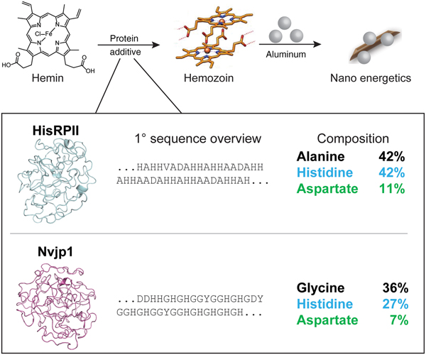
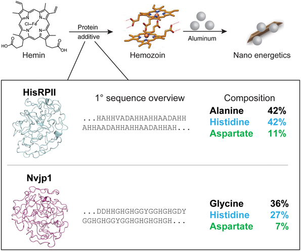
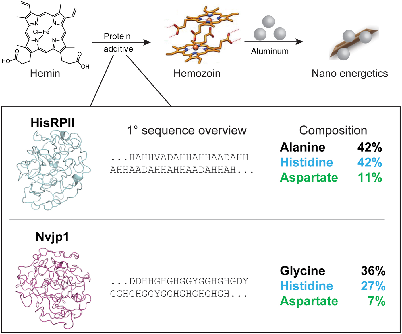
Like HisRPII, Nvjp1 features multiple histidine-rich repeats interspersed with aspartic acid (Fig. 1). While HisRPII has a high alanine content (42% of sequence), Nvjp1 is enriched in glycine (36%). As expected from the primary sequences, computational models generated using ROBETTA software[Reference Kim, Chivian and Baker37] predicted highly disordered folded ensembles for both proteins. We analyzed the influence of hemin on the structures of HisRPII and Nvjp1 via circular dichroism spectroscopy in pH 6 phosphate buffer (Fig. S1). HisRPII had a minimum near 200 nm suggestive of a disordered fold in agreement with prior results.[Reference Lynn, Chandra, Malhotra and Chauhan29] Addition of hemin reduced the intensity of the minimum but did not otherwise suggest significant structural changes to the protein. The low signal observed for HisRPII with hemin was likely due to binding of insoluble hemin as a small pellet was observed after centrifugation of the sample. A negligible signal was detected from Nvjp1 in the presence and absence of hemin. This is likely due to the poor solubility of Nvjp1 in aqueous buffers above pH 5, leading to the lack of a measurable CD signal. Attempts to measure CD spectra in pH 5.0 sodium acetate were complicated by high background from the buffer,[Reference Kelly, Jess and Price38] precluding conclusive results.
Overview of hemozoin synthesis, ROBETTA computational models, and sequence composition of HisRPII and Nvjp1.
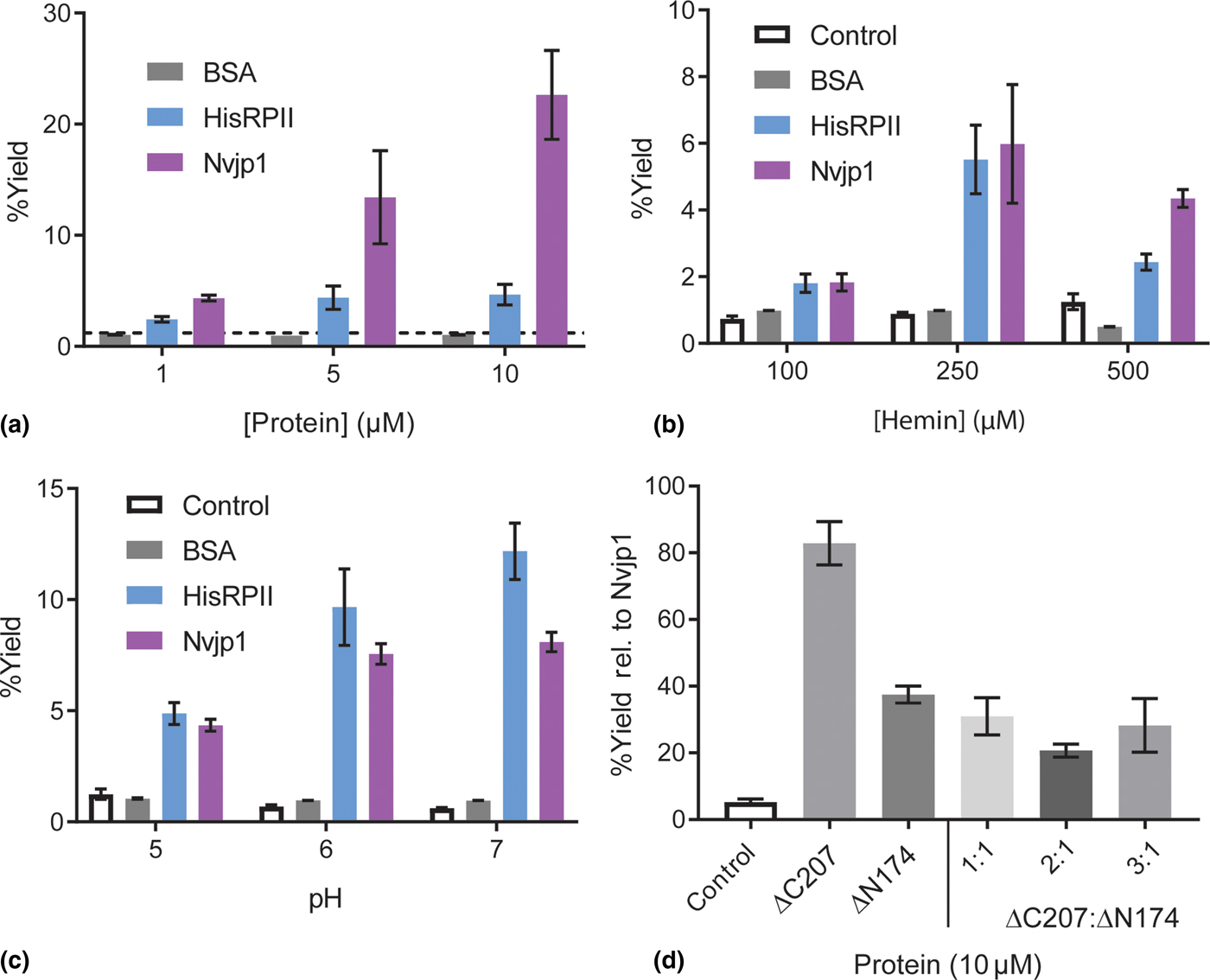
Recombinant HisRPII and Nvjp1 were evaluated for their ability to nucleate hemozoin from commercially available hemin based on established protocols.[Reference Jani, Nagarkatti, Beatty, Angel, Slebodnick, Andersen, Kumar and Rathore24–Reference Sullivan, Gluzman and Goldberg25,Reference Lynn, Chandra, Malhotra and Chauhan29] The effects of protein concentration, hemin concentration, and pH on the yield of hemozoin were explored (Fig. 2). In acidic buffer (pH 5.0) with a large excess of hemin (250 µM), both proteins promoted the production of crystals [Fig. 2(a)]. Reactions containing Nvjp1 consistently produced equal or higher amounts of hemozoin compared with HisRPII (up to ~4-fold more at 10 µM), with a dose-dependent correlation between Nvjp1 concentrations and levels of hemozoin formation observed. The concentration of HisRPII protein had little effect on the yield of hemozoin. Hemozoin formation was specific to the presence of histidine-rich proteins as bovine serum albumin (BSA) did not enhance hemozoin production compared to the buffer in agreement with published results.[Reference Sullivan, Gluzman and Goldberg25] Similarly, no significant amount of hemozoin was detected in control reactions lacking protein.
Hemozoin formation as a function of (a) protein concentration, (b) hemin concentration, (c) and pH. The dashed line in (a) represents the value of control reactions lacking protein. (d) Percent activity of Nvjp1 fragments ΔC207 and ΔN174 and variable ratios relative to full-length Nvjp1. Initial hemin concentration for reactions in (d) was 250 µM. Ratios are of ΔC207: ΔN174, with 1:1 being 10 µM of each fragment. Unless otherwise indicated, protein concentration was 1 µM and hemin concentration was 500 µM in 0.5 M sodium acetate pH 5.0. All samples were incubated at 37 °C with mixing for 24 h. Error bars are standard error of measurement for at least n = 3 samples.
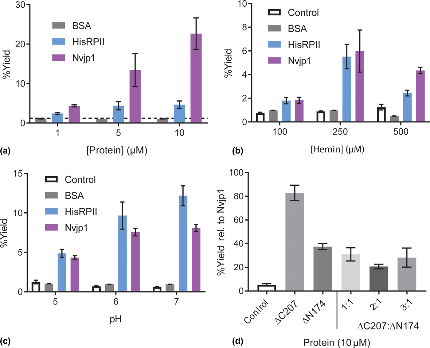
Hemin crystallization reactions were set up using 100, 250, and 500 µM of hemin to identify optimal initial concentrations. The hemozoin produced was directly proportional to initial hemin levels [Fig. 2(b)]. Linear increases in the amount of hemozoin formed were detected up to 250 µM. At 500 µM, significant aggregation of hemin was observed and likely contributed to the reduced yield. Hemin is known to aggregate in acidic media and slow release of free heme into the solution is a key step toward hemozoin formation.[Reference Egan, Mavuso and Ncokazi39]
The effect of pH on hemozoin formation was examined to optimize the reaction conditions [Fig. 2(c)]. In Plasmodium, hemozoin is produced in the acidic food vacuole.[Reference Coronado, Nadovich and Spadafora22] Crystallization of hemin in vitro has also been shown to occur only at pH ≤5.0.[Reference Lynn, Chandra, Malhotra and Chauhan29,Reference Egan, Mavuso and Ncokazi39] The pH range 5.0–7.0 was chosen based on known solubility limits of HisRPII and Nvjp1. Interestingly, a small increase in yield was measured for HisRPII-catalyzed reactions at pH >5. In contrast, the efficiency of Nvjp1 was not significantly altered throughout the pH range explored. At pH 7, both proteins still produced hemozoin at levels similar to that observed at pH 6, supporting the possibility of recombinant hemozoin production in vivo.
While both HisRPII and Nvjp1 possess histidine-rich sequences, the impact of sequence length on hemozoin formation has not been elucidated. Dendrimers of nonamer peptides derived from HisRPII were capable of nucleating hemozoin.[Reference Ziegler, Chang and Wright26] Inspired by this work, we sought to separate full-length Nvjp1 into two unique domains to determine if there was a differential contribution of hemozoin nucleation capacity. Examination of patterns in the sequence of Nvjp1 led us to design and express two fragments: ΔC207 and ΔN174, the amino terminal 174 and the carboxy terminal 207 amino acids of Nvjp1, respectively (see Supplementary Information). Both Nvjp1 fragments contain the same number of histidines; however, the amino acid content of the ΔC207 fragment is more homogeneous than that of the ΔN174 fragment and contains more well-defined repeats. Due to these differences, we hypothesized that the ΔC207 fragment would be superior at promoting hemozoin crystallization. We measured the formation of hemozoin for each fragment at 10 µM in acetate buffer with 250 µM hemin [Fig. 2(d)]. Despite having less than half as many amino acids as full-length Nvjp1, the ΔC207 fragment retained 80% of full-length activity. In contrast, ΔN174 was considerably less effective (40% relative to Nvjp1) at nucleating hemozoin. Interestingly, combining ΔC207 with ΔN174 in equimolar amounts or in stoichiometric excess did not improve the yield of hemozoin relative to ΔN174 alone. These results could indicate that ΔN174 has a higher affinity for heme than ΔC207, yet is unable to efficiently promote hemozoin formation. In addition, the fragments do not appear to reconstitute full-length protein when combined in solution, suggesting the need to exist together as a single polypeptide to display full activity. This observation is supported by previous work in dendrimers containing peptides derived from HisRPII.[Reference Ziegler, Chang and Wright26]
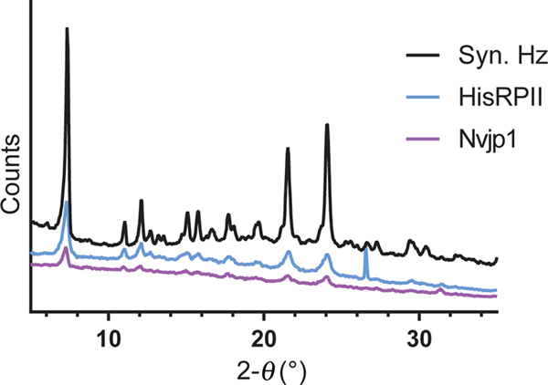
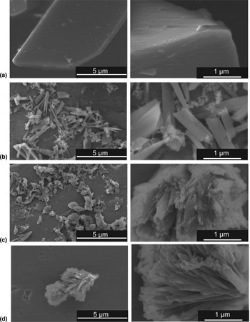
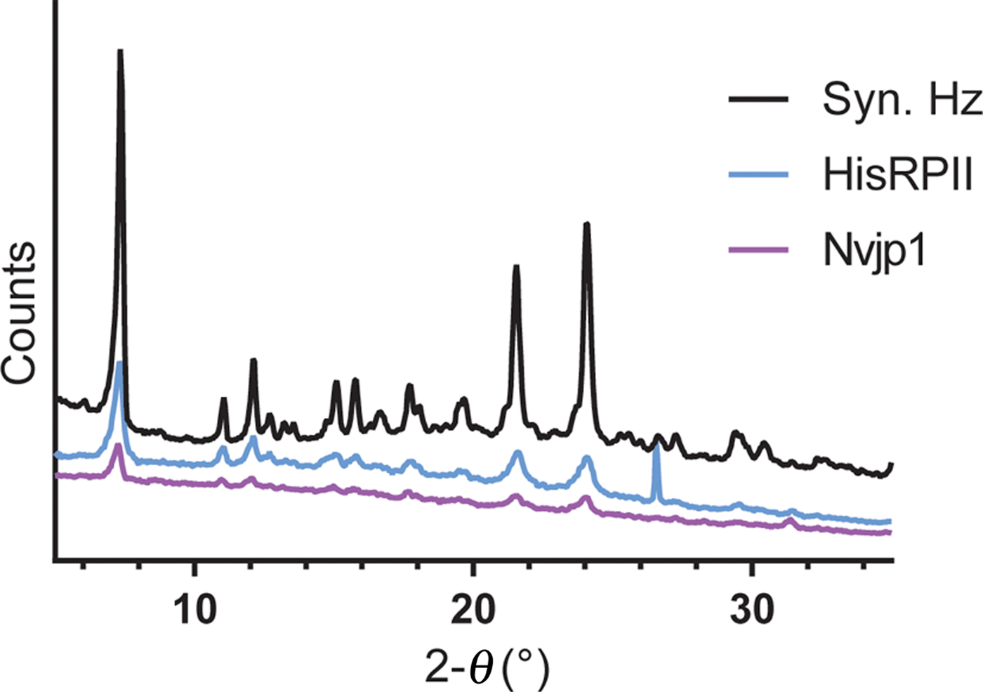
To confirm the identity of the protein-synthesized hemozoin, XRD was performed (Fig. 3). Major peaks identified in HisRPII and Nvjp1-nucleated hemozoin matched literature spectra[Reference Egan, Mavuso and Ncokazi39] with minor hemin impurities present. Furthermore, SEM of HisRPII and Nvjp1-nucleated hemozoin revealed needle-like structures characteristic of malarial hemozoin, which are distinct from those of hemin (Fig. 4). Crystals initiated from proteins were smaller than those of synthetic origin (300–500 nm versus 1 µm in length) though heterogeneous sizes were observed in all cases. We noted that crystals were difficult to disperse and often associated into large clusters spanning several micrometers.
XRD spectra of synthetic and protein-nucleated hemozoin.
SEM of (a) hemin, (b) synthetic hemozoin, and hemozoin nucleated by (c) HisRPII, and (d) Nvjp1.

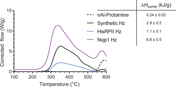
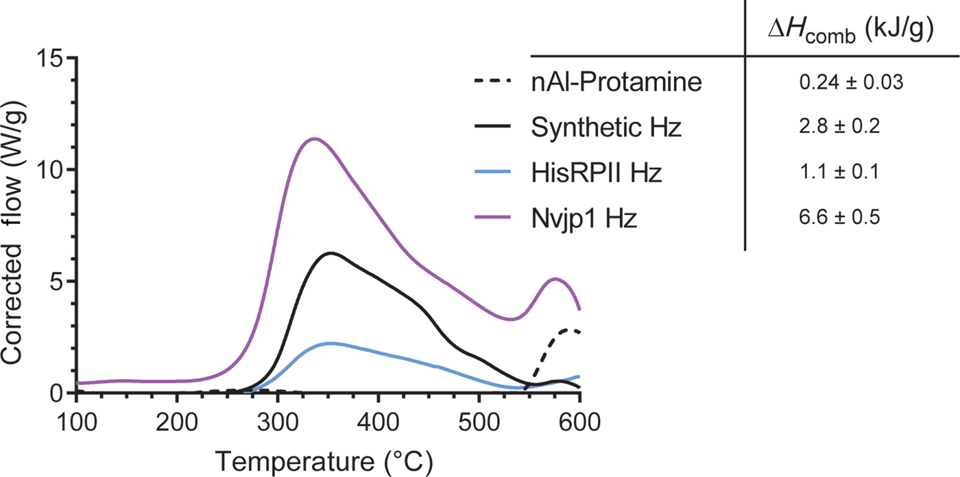
To determine the energetic output of protein-nucleated hemozoin, we assembled hemozoin nucleated by HisRPII or Nvjp1 with protamine-coated nAl at a 1:1 hemozoin:nAl-protamine ratio as previously reported.[Reference Slocik, Drummy, Dickerson, Crouse, Spowart and Naik21] The biothermites were characterized by DTA and thermogravimetric analysis (TGA) under an argon atmosphere (Fig. 5). As anticipated, unassembled aluminum-protamine did not produce measurable exothermic events in the absence of oxidizers by DTA; however, nAl was partially oxidized by minor amounts of atmospheric oxygen as determined by a mass gain in TGA due to the formation of aluminum oxide (Fig. S2). In contrast, nAl assembled with hemozoin nucleated with or without proteins led to large and broad exothermic peaks beginning at ~300 °C and achieved maximum output at 350 °C (Fig. 5). These peaks are consistent and characteristic of previously reported materials using chemically synthesized hemozoin.[Reference Slocik, Drummy, Dickerson, Crouse, Spowart and Naik21] The heat profile suggests that hemozoin is mainly acting as a metal oxide via a thermite-type reaction and less as a molecular oxidizing agent during combustion. By TGA, we observed a mass loss for hemozoin associated with decomposition/volatilization of the carbon/nitrogen-rich porphyrin ring followed by a gain in mass due to the formation of Al2O3. In parallel, we determined the heats of combustion (ΔHcomb) for each sample by integrating the area under the curve of heat flow versus time (Fig. S3). It should be noted that the energy outputs of nAl assembled with chemically synthesized or protein-nucleated hemozoin are artificially lower than previously reported values based on integration over a smaller temperature range. Biothermite assembled with hemozoin from Nvjp1 reactions had substantially higher heat flow and heat of combustion when compared with the synthetic standard or HisRPII. However, biothermite assembled with HisRPII-nucleated hemozoin had considerably lower heat flow than chemically synthesized hemozoin-based thermite (1.1 versus 2.8 kJ/g). The differences in energy output could likely be due to varying degrees of hemozoin purity among proteins, the presence of different amounts of protein trapped on hemozoin crystal surfaces, and inhomogeneity of crystals. In all, biothermite containing the Nvjp1-nucleated hemozoin yielded the best overall energetic performance; however, more energetic testing is needed with larger samples.
DTA of synthetic and protein-nucleated hemozoin assembled with nAl-protamine. The table shows heats of combustion for each thermite mixture. Heat flow was measured under argon at a temperature ramp of 20 °C/min.
Conclusions
Our data demonstrate that a marine sandworm jaw protein functions in an alternate and non-natural biologic process to promote hemozoin formation. Despite its aquatic origins and fewer histidine residues (102 versus 121), comparable nucleation by Nvjp1 was observed relative to HisRPII, a known malarial protein that nucleates hemozoin. It is interesting to note that some degree of order or spacing of histidine residues is important for nucleation as polyhistidine does not initiate hemozoin formation.[Reference Sullivan, Gluzman and Goldberg25] The presence of a non-specific protein is also insufficient to seed hemozoin crystals as shown by the negligible improvement of BSA over buffer in hemozoin synthesis.[Reference Sullivan, Gluzman and Goldberg25] In comparing the primary sequences of HisRPII and Nvjp1, the high glycine content of Nvjp1 may alter the dynamic folded ensemble[Reference D'aquino, Gómez, Hilser, Lee, Amzel and Freire40] and contribute to the measured differences in hemozoin crystallization relative to HisRPII. Crystals obtained from protein-containing syntheses were confirmed to be hemozoin by XRD and appeared to be moderately smaller than synthetic standards (300–500 versus ~1000 nm) with no observable differences in morphology observed between the protein-nucleated hemozoins. Experiments varying the pH of reaction conditions showed that hemozoin could be formed at neutral pH. This result is in contrast with the requirement of acidic pH in synthetic hemozoin formation and in vivo.[Reference Coronado, Nadovich and Spadafora22–Reference Jani, Nagarkatti, Beatty, Angel, Slebodnick, Andersen, Kumar and Rathore24,Reference Egan, Mavuso and Ncokazi39] It is possible that under neutral or basic pH, the protein promotes protonation of a hemin carboxylate to generate the requisite inter-heme hydrogen bond. In addition, we demonstrated that proteins can nucleate hemozoin at physiologic pH without compromising the yield. This result could enable recombinant heme crystallization in vivo, setting the stage for sustained biothermite production in bacteria. Such technology would enhance the scalability of hemozoin production and advance the design of related nanoenergetic materials.
Currently, our goal is to identify peptides and proteins that can efficiently nucleate hemozoin to produce energetic materials when combined with aluminum nanoparticles. Although Nvjp1 is capable of seeding hemozoin crystals near physiologic pH, its poor solubility in neutral media limits its promise for downstream applications. It is interesting to note that a fragment corresponding to removal of the carboxy-terminal half of Nvjp1 retained 80% of the full-length activity, underscoring the potential for further sequence minimization. Future engineering efforts combining primary sequences from HisRPII, and Nvjp1 may yield improvements in hemozoin nucleation with finer control over crystal size, enhanced nanoenergetics, and improved solubility. Ultimately, this would accelerate the development of recombinant biothermite production in vivo and enhance current and future nanoenergetic applications.
Experimental methods
All reagents were purchased from Sigma-Aldrich (St. Louis, MO) and used without further purification. All solutions were made using 18 mΩ water obtained from a Barnstead GenPure system (Dubuque, IA). A pET15b plasmid encoding HisRPII was a generous gift from the Goldberg Lab (Washington University in St. Louis). Nvjp1 was cloned as previously described.[Reference Gupta, Becknell, Crosby, Bedford, Wright, Dennis and Naik36] Fresh hemin stocks were prepared in 1.4 M NaOH. Synthetic hemozoin was prepared according to a published protocol.[Reference Slocik, Drummy, Dickerson, Crouse, Spowart and Naik21]
Protein expression and purification
The plasmid encoding HisRPII was transformed and expressed in BL21(DE3) cells and purified as previously described.[Reference Sullivan, Gluzman and Goldberg25] Briefly, the pellet from induced cultured cells was resuspended in 50 mM Tris pH 8.0 500 mM NaCl 30 mM imidazole. After sonication, lysates were clarified by centrifugation and the supernatant was loaded onto an IMAC column containing cobalt. HisRPII was eluted in 500 mM imidazole and dialyzed against 0.1 M NaOAc pH 5.0.
Nvjp1 was expressed and purified following a literature protocol.[Reference Gupta, Becknell, Crosby, Bedford, Wright, Dennis and Naik36] Cultured and induced cells were pelleted, resuspended in 50 mM Tris-HCl pH 8.0, 150 mM NaCl, 100 µg/ml lysozyme and disrupted by sonication. After centrifugation, the insoluble pellet containing inclusion bodies was washed twice with 50 mM Tris-HCl, pH 8.0, 150 mM NaCl, 0.01% Triton X-100 followed by 50 mM Tris-HCl, pH 8.0, 150 mM NaCl. The pellet was resuspended in water and the pH was adjusted to 3 with 1 M HCl followed by centrifugation. NaCl was then added to the supernatant to a final concentration of 100 mM and the pH was adjusted to 8.0 with 1 M Tris pH 9 followed by centrifugation. The pellet was dissolved in 6 mM HCl and dialyzed overnight against water. After lyophilization, Nvjp-1 was reconstituted to a final concentration of 60 mg/ml in water.
Circular dichroism spectroscopy
CD spectra of HisRPII and Nvjp1 were performed on a Jasco J-815 spectrometer in 2 mm quartz cuvettes. Samples consisted of 10 µM protein in 20 mM sodium phosphate buffer pH 6.0 with or without 50 µM hemin. Scans were collected from 190 to 260 nm with 1 nm increments and baseline corrected for the blank buffer with and without hemin.
Hemozoin synthesis
Hemozoin was produced following a modified published protocol.[Reference Sullivan, Gluzman and Goldberg25] Proteins were diluted in 0.5 M NaOAc pH 5.0 and hemin was added in a final volume of 1 mL. Unless otherwise indicated, final reaction concentrations were 1 µM protein and 500 µM hemin. The mixture was vortexed and incubated with shaking at 37 °C for 24 h. The reactions were pelleted by centrifugation at 15,000 g and the supernatant decanted. The pellet was washed with 0.1 M NaHCO3 2.5% SDS pH 9.1 and subsequently with water until the supernatant was colorless. For quantification, hemozoin was decomposed in 0.1 M NaOH and heme absorbance measured on a UV-Vis plate reader (SpectraMax M3, Molecular Devices) at 385 nm using an extinction coefficient of 58,400/M/cm. We note that day-to-day variability was observed and data shown are a representative experiment.
Assembly of nAl-hemozoin thermite
Aluminum nanoparticles (10 mg, Novacentrix, Inc.) were suspended in 5 mL of deionized water and sonicated with an ultrasonic bath for 5 min. Two milligrams of protamine (Sigma) was added and incubated for 30 min at room temperature. The nAl-protamine suspension was centrifuged at 15,000× g for 5 min, washed with water to remove excess protamine, and centrifuged again. A total of three wash cycles were performed. The nAl-protamine suspension was then mixed 1:1 with hemozoin in 1 mL of water, sonicated for 5 min, and lyophilized to obtain an assembled thermite.
Hemozoin characterization
SEM images were obtained on an FEI Quanta SEM microscope operating at 4 kV and a working distance of 10 mm. Hemozoin was lyophilized and dried onto metal stubs using methanol. XRD data were collected via drop-casting sample on a glass substrate with a scan speed of 3°/min. The scans were from 5° to 90° with a 10 mm length limited. All scans were performed using the 1D continuous scan mode of the Hy-Pix 3000 detector. DTA was performed in an SDTQ600 TGA. Samples (~2–5 mg) were loaded into tared alumina crucibles with an empty crucible serving as a reference. All data were collected in dynamic mode under argon (100 mL/min) from ambient temperature to 800 °C heated at a rate of 20 °C/min. Heats of combustion in kJ/g were obtained by plotting corrected heat flow versus time in seconds and integrating the area under the curve.
Supplementary material
The supplementary material for this article can be found at https://doi.org/10.1557/mrc.2019.46
Acknowledgments
The authors thank Anthony Pelton for assistance in XRD data acquisition and team members of AFRL and UES for thoughtful discussion. P.B.D. is an adjunct faculty at Wright State University, Dayton, Ohio. This work was supported by the Applied Research for the Advancement of Science and Technology Priorities Program on Synthetic Biology for Military Environments funded by the Office of the Secretary of Defense.