INTRODUCTION
There has been a dramatic rise in the incidence of fascioliasis in recent years, and a spread of the disease into areas that were previously fluke free (Mitchell, Reference Mitchell2002; Pritchard et al. Reference Pritchard, Forbes, Williams, Salimi-Bejestani and Daniel2005). This, coupled with increasing instances of drug resistance to triclabendazole (TCBZ), the current drug of choice against the disease (Fairweather, Reference Fairweather2005; Alvarez-Sanchez et al. Reference Alvarez-Sanchez, Mainar-Jaime, Perez-Garcia and Rojo-Vasquez2006), poses a serious threat to animal health and the sustainability of the agri-food industry. In order to optimize drug treatment and to reduce drug resistance, clarification of the mechanism of TCBZ action is urgently required. Much is known about the absorption, plasma kinetics, tissue distribution, metabolism and elimination of TCBZ in the host (e.g. Hennessy et al. Reference Hennessy, Lacey, Steel and Prichard1987; Virkel et al. Reference Virkel, Lifschitz, Sallovitz, Pis and Lanusse2006; Mestorino et al. Reference Mestorino, Formentini, Lucas, Fernandez, Modamio, Hernández and Errecalde2008). The ability of the fluke to metabolize the drug is also recognized (Mottier et al. Reference Mottier, Virkel, Solana, Alvarez, Salles and Lanusse2004; Robinson et al. Reference Robinson, Lawson, Trudgett, Hoey and Fairweather2004). Data on the mode of action against the presumed target protein (tubulin) in the fluke has been reviewed by Fairweather (Reference Fairweather2005) and the relative activities of different metabolites against the fluke discussed by Halferty et al. (Reference Halferty, Brennan, Hanna, Edgar, Meaney, McConville, Trudgett, Hoey and Fairweather2008). This paper addresses another aspect of drug pharmacokinetics, namely, the entry of TCBZ into the fluke. This is an important aspect of the drug/parasite relationship (Alvarez et al. Reference Alvarez, Mottier and Lanusse2007). For an anthelmintic to have a beneficial effect, it must reach and maintain therapeutic levels within the target parasite. Helminth parasites may take up drug via oral ingestion, trans-tegumental/trans-cuticular diffusion or a combination of both these routes (Thompson and Geary, Reference Thompson, Geary, Marr and Muller1995). As F. hepatica is a blood–feeder, it may be presumed that oral uptake predominates, and the high binding of TCBZ and its metabolites to plasma proteins (>99%) further consolidates this assumption (Mohammed Ali et al. Reference Mohammed Ali, Bogan, Marriner and Richards1986). Another fasciolicide, clorsulon, has been shown to enter F. hepatica predominantly via the oral route, causing extensive disruption to the tegument and gut; it binds to red blood cell carbonic anhydrase (Meaney et al. Reference Meaney, Haughey, Brennan and Fairweather2005a, Reference Meaney, Haughey, Brennan and Fairweatherb). However, the accumulated literature suggests that trans-tegumental diffusion predominates as the major route of drug entry into trematode (Alvarez et al. Reference Alvarez, Imeperiale, Sánchez, Murno and Lanusse2000, Reference Alvarez, Mottier, Sánchez and Lanusse2001, Reference Alvarez, Mottier and Lanusse2004, Reference Alvarez, Mottier and Lanusse2007; Mottier et al. Reference Mottier, Alvarez, Ceballos and Lanusse2006a) and cestode parasites (Mottier et al. Reference Mottier, Alvarez, Pis and Lanusse2003, Reference Mottier, Alvarez, Ceballos and Lanusse2006a) and that trans-cuticular uptake dominates in nematode parasites (Ho et al. Reference Ho, Geary, Barsuhn, Sims and Thompson1990; Sims et al. Reference Sims, Ho, Geary, Thomas, Day, Barsuhn and Thompson1996; Cross et al. Reference Cross, Renz and Trees1998).
A series of experiments has been carried out in vitro to determine the relative susceptibilities of the tegument and gut to TCBZ action. In order to evaluate the contribution of oral uptake, ligatures were secured around the oral cone of the flukes, closing off the pharynx and thus preventing oral ingestion of drug. Trans-tegumental uptake was inhibited by allowing drug to bind to an excess of the plasma protein, bovine serum albumin (BSA), added to the incubation medium. Furthermore, non-ligatured flukes were incubated in TCBZ.SO in the presence of red blood cells (RBCs) to clarify that oral feeding does occur in vitro and therefore that the drug can enter the fluke during incubation. Changes to the tegumental and gastrodermal surfaces following the various drug treatments were evaluated by scanning electron microscopy, and compared to ligatured, drug-treated and control specimens in order to ascertain the susceptibility of each interface to anthelmintic action.
MATERIALS AND METHODS
Isolate of Fasciola hepatica
This study was carried out using the Cullompton isolate of Fasciola hepatica. It was obtained in 1998 as a field isolate from an abattoir in Cullompton, Devon, UK. It has been shown to be susceptible to albendazole (Buchanan et al. Reference Buchanan, Fairweather, Brennan, Trudgett and Hoey2003), clorsulon (Meaney et al. Reference Meaney, Fairweather, Brennan, McDowell and Forbes2003, Reference Meaney, Fairweather, Brennan and Forbes2004), triclabendazole (Robinson et al. Reference Robinson, Trudgett, Hoey and Fairweather2002; McCoy et al. Reference McCoy, Fairweather, Brennan, Kenny, Ellison and Forbes2005; Halferty et al. Reference Halferty, Brennan, Hanna, Edgar, Meaney, McConville, Trudgett, Hoey and Fairweather2008) and nitroxynil (McKinstry et al. Reference McKinstry, Fairweather, Brennan and Forbes2003, Reference McKinstry, Brennan, Halferty, Forbes and Fairweather2007). Adult male Sprague-Dawley rats were each orally infected with 20 metacercarial cysts under light ether anaesthesia via a stomach tube.
In vitro drug treatment
Adult flukes were recovered from the bile ducts of the experimentally infected Sprague-Dawley rats under sterile conditions in a laminar flow cabinet. Flukes were washed several times in warm (37°C) sterile NCTC 135 culture medium containing antibiotics (penicillin 50 IU/ml; streptomycin 50 μg/ml). Some flukes were ligatured by tying surgical thread just behind the oral sucker to prevent oral entry of TCBZ. This technique has been used successfully in previous experiments on F. hepatica (Meaney et al. Reference Meaney, Haughey, Brennan and Fairweather2005a, Reference Meaney, Haughey, Brennan and Fairweatherb; Mottier et al. Reference Mottier, Alvarez, Ceballos and Lanusse2006a). Use of scanning electron microscopy has shown that the ligature causes only limited abrasion to the tegumental surface in the immediate vicinity of the ligature, but this does not allow penetration of an Evan's Blue dye (0·01%, w/v) (Fairweather et al. Reference Fairweather, Holmes and Threadgold1983; Meaney et al. Reference Meaney, Haughey, Brennan and Fairweather2005a; Mottier et al. Reference Mottier, Alvarez, Ceballos and Lanusse2006a). Some flukes were left non-ligatured. All flukes were transferred to fresh NCTC 135 culture medium containing triclabendazole sulphoxide (TCBZ.SO) at a concentration of 15 μg/ml and incubated at 37°C for 24 h. Ligatured and non-ligatured control flukes were incubated in NCTC 135 culture medium at 37°C for 24 h. Controls at 0 h were also prepared. TCBZ.SO was used for the experiments as it is believed to be the principal metabolite of TCBZ and its concentration was chosen to correspond to maximum blood levels in vivo following a therapeutic dose of 10 mg/kg body weight (Hennessy et al. Reference Hennessy, Lacey, Steel and Prichard1987).
In separate experiments, ligatured and non-ligatured flukes were washed several times in warm (37°C) sterile NCTC 135 culture medium containing antibiotics (penicillin 50 IU/ml; streptomycin 50 μg/ml). Bovine serum albumin (BSA) (30 mg/ml) was added to fresh NCTC 135 culture medium containing TCBZ.SO at a concentration of 15 μg/ml and the solution left to stand for 5 min to allow the TCBZ.SO to bind. Ligatured and non-ligatured flukes were then incubated in the drug medium at 37°C for 24 h. Ligatured and non-ligatured control flukes were incubated in NCTC 135 culture medium containing BSA (30 mg/ml) at 37°C for 24 h. The concentration of BSA used was similar to that used by Mottier et al. (Reference Mottier, Alvarez, Ceballos and Lanusse2006a) and equates to a physiological concentration of albumin in sheep plasma.
A further experiment involved the use of RBCs to verify that feeding by the flukes does occur in vitro and that drug can be taken up orally. Whole blood was collected from adult male Sprague-Dawley rats at necropsy using heparinized tubes to prevent clotting. Blood was centrifuged at 3354 g for 10 min and the supernatant discarded. From the remaining red blood cell fraction, 1·5 ml of RBCs was spread onto the bottom of sterile wells in a 6-well multi-well plate. Four ml NCTC 135 culture medium containing TCBZ.SO at a concentration of 15 μg/ml was added to each of the wells. Flukes displayed full gut contents on recovery from the bile ducts of the rats. Flukes were then washed in several changes of warm (37°C) sterile NCTC 135 culture medium and allowed to regurgitate their gut contents. One fluke was then added to each well in the 6-well multi-well plate and allowed to graze on the RBCs for 24 h at 37°C. At the end of the experiment, gut contents were present in all flukes, demonstrating that feeding had occurred. A control experiment was carried out in which flukes were allowed to graze for 24 h on RBCs in the absence of TCBZ.SO. A minimum of 6 intact flukes were fixed and processed for scanning electron microscopy (SEM) for each treatment and a minimum of 3 flukes per treatment used for the preparation of gut sections for SEM.
Preparation of gut sections for scanning electron microscopy
Following flat-fixing, the oral cone and tail regions were removed from flukes using a razor blade. The remaining midbody region was further dissected into longitudinal sections 1–2 mm thick. The specimens were further prepared as described in the scanning electron microscope (SEM) protocol. It should be noted that the sections were mounted on their side so that internal structures faced upwards.
Tissue preparation for scanning electron microscopy
Initially, the flukes were lightly flat-fixed for 1 h at room temperature in 4% (w/v) aqueous glutaraldehyde and subsequently free–fixed at 4°C in a 3:1 mixture of 4% (w/v) aqueous glutaraldehyde and 1% osmium tetroxide overnight. Flukes were then rinsed in 70% (v/v) ethanol and dehydrated in an ascending series of ethanols. Following this, the flukes were dried in hexamethyldisilazane, mounted (with the ventral or dorsal surface facing upwards) on aluminium stubs and sputter-coated with gold-palladium. The flukes were viewed using a FEI Quanta 200 scanning electron microscope operating at an accelerating voltage of 10 keV.
RESULTS
Changes to the tegumental surface
Non-ligatured and ligatured flukes treated with TCBZ.SO (15 μg/ml)
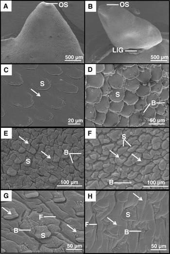
There was no apparent difference in disruption to the tegument between the ligatured and non-ligatured flukes and so the results will be discussed together. Disruption was more severe on the dorsal than ventral surface and the micrographs presented (Figs 1 and 2) are from that surface. Some flattening of the surface was common in the central area of the oral cone region (Fig. 1C), due to swelling of the tegument, but the latter was especially evident along the lateral margins of the fluke (Fig. 1A–D). As a result, the spines appeared submerged, lying almost flat against the surface (Fig. 1C). In the region of the oral cone anterior to the ligature, some blebs were seen in association with the spines, but the blebbing was not severe (Fig. 1D).
Scanning electron micrographs (SEMs) of the dorsal surface of adult non-ligatured (A,C,E,G) and ligatured (B,D,F,H) Fasciola hepatica treated in vitro with TCBZ.SO (15 μg/ml). (A) Non-ligatured fluke. Low-power micrograph showing the oral cone region. OS, oral sucker. (B) Ligatured fluke. SEM showing the ligature (LIG) behind the oral sucker (OS). (C) Non-ligatured fluke, oral cone. High-power SEM shows swelling of the tegument between the spines (S), giving a ‘flattened’ appearance (arrow) to the tegument. (D) Ligatured fluke, oral cone. There is some localized blebbing (B) on the tegumental surface. S, spine. (E) Non-ligatured fluke, anterior midbody region. The tegument between and covering the spines is swollen (arrows) and there is some blebbing (B) on the tegumental surface. S, spine. (F) Ligatured fluke, anterior midbody region. The tegument covering and between the spines (S) is very swollen so that the spines appear submerged (arrows) and there is some blebbing (B) on the tegumental surface. (G) Non-ligatured fluke, anterior midbody region. The tegument is swollen (arrows) and furrowed (F). Blebs (B) are associated with the inter-spine tegument. S, spine. (H) Ligatured fluke, anterior midbody region. The tegument is swollen (arrows) and furrowed (F), so that the spines (S) appear submerged and patches of blebs (B) adorn the tegumental surface.

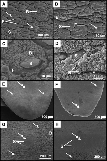
Scanning electron micrographs (SEMs) of the dorsal surface of adult non-ligatured (A,C,E,G) and ligatured (B,D,F,H) Fasciola hepatica treated in vitro with TCBZ.SO (15 μg/ml). (A) Non-ligatured fluke, posterior midbody region. There is widespread swelling (arrows), furrowing (F) and blebbing (B) of the tegument. S, spine. (B) Ligatured fluke, posterior midbody region. Blebs (B) are scattered over the tegumental surface. Swelling (arrow) and furrowing (F) of the tegument are also evident. S, spine. (C) Non-ligatured fluke, posterior midbody region. Many blebs (B) are present on the surface of the spines (S) and on the inter-spine tegument, and the tegument appears furrowed (F). (D) Ligatured fluke, posterior midbody region. The tegument and spines (S) are covered with a carpet of blebs (B). The tegument appears furrowed (F) and swollen. (E) Non-ligatured fluke, tail region. The tegument is severely swollen (arrows) and, in some areas, the surface has a flattened appearance (double arrow). (F) Ligatured fluke, tail region. Low-power SEM showing an area of swelling (arrow), also flattening of the tegument (double arrow). (G) Non-ligatured fluke, tail region. High-power SEM showing swelling (arrows) of the tegument. In some areas the tegument has a flattened appearance (double arrow) where the spines (S) appear sunken. A patch of blebs (B) is also evident. (H) Ligatured fluke, tail region. High-power SEM showing swelling (arrow) and flattening (double arrow) of the tegument where the spines (S) appear sunken.

In the anterior midbody region, the tegument between and covering the spines was very swollen, and the spines appeared submerged by the swollen tegument surrounding them; this was most pronounced along the lateral margins (Fig. 1E–H). The tegument also appeared furrowed with patches of blebs between the spines (Fig. 1G and H). In the posterior midbody region, the tegument was extremely swollen, to the extent that the spines were barely visible (Fig. 2A–D). The tegument was thrown into deep furrows (Fig. 2A and B). Blebbing on the surface was very extensive (Fig. 2A–D) and the disruption observed generally was more severe along the lateral margins of the flukes. In the tail region, there were distinct areas of swelling and flattening of the tegument. The spines appeared submerged by the swollen tegument surrounding them (Fig. 2E–H). Isolated patches of blebs were observed in this region (Fig. 2G).
Non-ligatured and ligatured flukes treated with TCBZ.SO (15 μg/ml)+BSA (30 mg/ml)
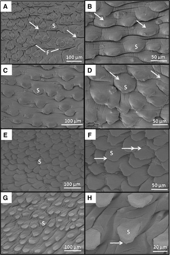
Changes observed were greater on the dorsal than ventral surface and so the micrographs presented (Fig. 3A–D) are from that surface. The micrographs are taken from the posterior midbody region of flukes, as this was consistently the most affected area. There was no apparent difference in disruption between the ligatured and non-ligatured flukes. Some swelling and furrowing of the inter-spinal tegument and the tegumental covering of the spines themselves was observed on non-ligatured flukes treated with TCBZ.SO+BSA (Fig. 3A and B). Ligatured flukes treated with TCBZ.SO+BSA also showed limited swelling of the tegument, both between and covering the spines (Fig. 3C and D).
Scanning electron micrographs (SEMs) of the dorsal surface of the posterior midbody region of adult non-ligatured and ligatured Fasciola hepatica treated in vitro with TCBZ.SO (15 μg/ml)+BSA (30 mg/ml) (A–D) and BSA (30 mg/ml) (E–F), also 24 h controls (G–H). (A) Non-ligatured fluke. Low-power SEM showing some swelling (arrows) and furrowing (F) of the tegument. S, spine. (B) Non-ligatured fluke. High-power SEM showing some swelling of the tegument (arrows) covering and between the spines (S). (C) Ligatured fluke. Low-power SEM showing minimal swelling of the tegument covering and between the spines (S). (D) Ligatured fluke. High-power SEM showing limited swelling of the tegument (arrows) covering and between the spines (S). (E) Non-ligatured, BSA control fluke. Low-power SEM of the tegumental surface. S, spine. (F) Non-ligatured, BSA control fluke. High-power SEM showing very limited swelling of the tegument (double arrow) between the spines (S). Spinelets (arrow) are visible at the tips of the spines. (G) Non-ligatured control fluke. Low-power SEM showing tegument covered with spines (S). (H) Non-ligatured control fluke. High-power SEM showing spines (S) with intact spinelets (arrow).

Non-ligatured and ligatured flukes treated with BSA (30 mg/ml)
In the posterior midbody region of non-ligatured flukes incubated in BSA, there was minimal swelling of the tegument (Fig. 3E and F). The spines were clearly visible, projecting free from the surface. The surface morphology of ligatured flukes incubated in BSA for 24 h was similar.
0 h and 24 h non-ligatured and ligatured controls
The tegumental surface in the posterior midbody region of non-ligatured flukes incubated in NCTC culture medium for 24 h remained normal (Fig. 3G and H). The general surface morphology of 24 h ligatured controls and the 0 h controls matched the images presented by Bennett (Reference Bennett1975) and Fairweather et al. (Reference Fairweather, Threadgold, Hanna and Dalton1999).
Non-ligatured flukes treated with TCBZ.SO (15 μg/ml) in the presence of RBCs
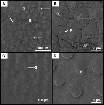
Changes observed were more severe on the dorsal surface of the flukes, with greatest disruption occurring along the lateral margins in the posterior midbody region: it is these changes that will be illustrated. The tegument between and covering the spines was severely swollen, so that the spines appeared submerged (Fig. 4A and B). The tegument was thrown into a number of furrows (Fig. 4A and B) and extensive patches of large and small blebs adorned the tegumental surface (Fig. 4A and B).
Scanning electron micrographs (SEMs) of the dorsal surface of the posterior midbody region of adult non-ligatured Fasciola hepatica treated in vitro with TCBZ.SO (15 μg/ml) in the presence of RBCs (A–B) and 24 h controls incubated in the presence of RBC's (C–D). (A) Low-power SEM showing some swelling (arrows) around the spines (S) and furrowing (F) of the tegument. Blebbing (B) is visible on the surface of the tegument. (B) High-power SEM showing swelling of the tegument (arrow), both that covering and that between the spines (S). The tegument has been thrown into a number of furrows (F) and patches of large and small blebs adorn the tegumental surface (B). (C) Low-power SEM of the tegumental surface. S, spine. (D) High-power SEM of the tegument. S, spine.

Non-ligatured control flukes incubated in the presence of RBCs
In the posterior midbody region of flukes exposed to RBCs for 24 h, the tegumental architecture remained normal with spines projecting free from the tegumental surface (Fig. 4C and D).
Changes to the gut surface
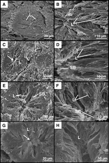
The results are presented in Fig. 5A–H and Fig. 6. In the normal gut caeca, the luminal surface is covered by a regular series of broad, sheet-like lamellae (for images, see Figs 7 and 8 in Meaney et al. Reference Meaney, Haughey, Brennan and Fairweather2005a). The lamellae project into the gut lumen from the epithelial lining. Following examination of all flukes in the present study, whether ligatured or non-ligatured, treated with TCBZ.SO or TCBZ.SO+BSA, the gut surface retained a normal morphology (Fig. 5A–H). In some of the specimens, small, rounded bodies were observed attached to the surface of the lamellae (Fig. 5D–F): they have been described variously as spherical bodies, blisters or secretory bodies (Threadgold, Reference Threadgold1978; Fujino et al. Reference Fujino, Uni, Ishii and Takada1987).
Scanning electron micrographs (SEMs) of gut sections of adult non-ligatured and ligatured Fasciola hepatica treated in vitro with TCBZ.SO (15 μg/ml) (A–D) and TCBZ.SO (15 μg/ml)+BSA (30 mg/ml) (E–H). (A) Non-ligatured fluke. The gut lamellae (L) exhibit a normal morphology. (B) Non-ligatured fluke. The lamellae (L) are normal and extend into the lumen of the gut. (C) Ligatured fluke. Low-power SEM of a section through two gut caeca. The gut lamellae (L) appear normal and extend towards the central lumen (arrow). (D) Ligatured fluke. High-power SEM showing the normal structure of the gut lamellae (L). Spherical bodies (SB) are present on the surface of the lamellae. (E) Non-ligatured fluke. Low-power SEM of a transverse section through the gut, showing the arrangement of the gut lamellae (L). Spherical bodies (SB) are present on the surface of the lamellae. (F) Non-ligatured fluke. High-power SEM showing the sheets of lamellae (L). Spherical bodies (SB) are present on the surface of the lamellae. (G) Ligatured fluke. Low-power SEM showing the normal organization of the gut lamellae (L), which extend towards the central lumen (arrow). (H) Ligatured fluke. High-power SEM showing that the gut lamellae (L) have a normal morphology.

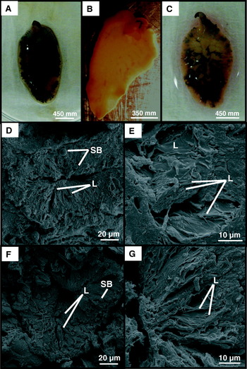
Images of intact flukes showing the gut contents (A–C); also, scanning electron micrographs (SEMs) of gut sections of adult non-ligatured Fasciola hepatica treated in vitro with TCBZ.SO (15 μg/ml) in the presence of RBC's (D–E) and 24 h controls incubated in the presence of RBCs (F–G). (A) Fluke with full gut contents immediately following removal from the bile duct of an experimentally infected rat. (B) Fluke with empty gut following regurgitation of gut contents. (C) Fluke with full gut contents following 24 h incubation in TCBZ.SO (15 μg/ml) in the presence of RBCs. (D) Fluke treated with TCBZ.SO (15 μg/ml) in the presence of RBCs. Low-power SEM of a transverse section through the gut, showing the arrangement of the gut lamellae (L). Spherical bodies (SB) are present on the surface of the lamellae. (E) Fluke treated with TCBZ.SO (15 μg/ml) in the presence of RBCs. High-power SEM showing stacked sheets of lamellae (L). (F) RBC control fluke. Low-power SEM showing the normal organization of the gut lamellae (L). Some spherical bodies (SB) are visible. (G) RBC control fluke. High-power SEM showing the sheet-like gut lamellae (L), which have a normal morphology.

On incubation of flukes in TCBZ.SO in the presence of RBCs, oral feeding was confirmed in vitro. On isolation from the rat host, the flukes had full gut contents (Fig. 6A). They were then allowed to void their gut contents prior to incubation (Fig. 6B). After a 24 h incubation in TCBZ.SO in the presence of RBCs, the gut was seen to be almost full, indicating that feeding had taken place during the incubation period (Fig. 6C). The gastrodermal surface remained normal at the end of the incubation in TCBZ.SO with RBCs and in the 24 h RBC controls (Fig. 6D–G).
Summary of results
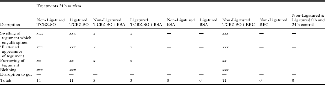
The results of the various treatments are summarized in Table 1.
Summary of SEM results
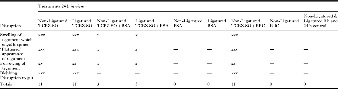
—, no noticeable disruption; x, mild disruption; xx, severe disruption; xxx, extremely severe disruption; TCBZ.SO, triclabendazole sulphoxide; BSA, bovine serum albumin; RBC, red blood cell.
DISCUSSION
The aim of this study was to determine the predominant route by which TCBZ.SO enters the liver fluke, by isolating either the oral or the trans-tegumental route of uptake. TCBZ.SO was used in this study as it is believed to be the major metabolite responsible for the flukicidal activity of the drug (Fairweather, Reference Fairweather2005). Disruption to the tegumental and gastrodermal surfaces was evaluated by scanning electron microscopy. The results demonstrated clearly that the tegument was consistently the more disrupted at the two surfaces (see summary of results in Table 1).
Comparable drug changes to the tegument were observed in the non-ligatured and ligatured TCBZ.SO-treated flukes. This would suggest that restricting the oral uptake of drug via the ligature does not affect the ability of the drug to enter the fluke and exert its anthelmintic effect. The result is consistent with that of a previous absorption kinetics study which showed that the concentrations of TCBZ.SO within ligatured and non-ligatured F. hepatica were not statistically different after incubation in this compound (Mottier et al. Reference Mottier, Alvarez, Ceballos and Lanusse2006a). There was no difference in disruption between TCBZ.SO-treated ligatured and non-ligatured flukes and those flukes treated with the drug in the presence of RBCs (Table 1). Even though oral ingestion was stimulated by the presence of blood cells, the gut retained a normal morphology, whilst the tegument was severely affected. On addition of BSA to the incubation medium, there was a very notable decline in the level of morphological disruption to the tegument (Table 1). Again, this is consistent with a previous observation that, when BSA was added to the incubation medium, uptake of TCBZ.SO was reduced significantly by 85% in both the ligatured and non-ligatured flukes, leaving only ~15% of the TCBZ.SO free to pass into the fluke (Mottier et al. Reference Mottier, Alvarez, Ceballos and Lanusse2006a). This corresponds with the lower level of disruption to the flukes when exposed to TCBZ.SO plus BSA that was observed in the present study. Throughout all these experiments, the gross morphology of the gut remained unaffected by TCBZ.SO action. The combined results emphasize the importance of the tegument as the principal route of entry for TCBZ.SO into the fluke.
The non-ligatured, TCBZ.SO-treated flukes acted as the primary experiment, against which the results of the subsequent experiments could be compared. The addition of a ligature around the oral cone, closing off the pharynx, did not reduce the overall level of disruption caused by TCBZ.SO (Table 1), suggesting that trans-tegumental uptake of TCBZ into the liver fluke is predominant. SEM revealed that the dorsal surface of non-ligatured and ligatured flukes was more severely impacted by drug action than the ventral surface. Severity of drug effect also increased posteriorly along the fluke, an observation noted in previous studies with TCBZ.SO (Smeal and Hall, Reference Smeal and Hall1983; Stitt and Fairweather, Reference Stitt and Fairweather1993; Meaney et al. Reference Meaney, Fairweather, Brennan, Ramasamy and Subramanian2002, Reference Meaney, Allister, McKinstry, McLaughlin, Brennan, Forbes and Fairweather2006). Disruption took the form of swelling of the tegument, both that covering and that between the spines, which was so severe in some midbody areas that the tegument appeared flattened. Swelling along the lateral margins of the flukes was very extreme, causing the spines to appear submerged and the tegument to be thrown into many furrows. Blebbing of the tegument was also widely observed, especially in the posterior midbody region of the flukes. Blebbing is a common stress reaction mounted in response to anthelmintic treatment, where secretory bodies are rapidly transported to, and released from, the apical plasma membrane in an effort to replace damaged membrane and maintain the integrity of the tegumental surface (Stitt and Fairweather, Reference Stitt and Fairweather1993; McKinstry et al. Reference McKinstry, Fairweather, Brennan and Forbes2003; Meaney et al. Reference Meaney, Fairweather, Brennan, McDowell and Forbes2003; McConville et al. Reference McConville, Brennan, McCoy, Castillo, Hernandez-Campos, Ibarra and Fairweather2006).
Due to the haematophagous nature of the liver fluke, and because triclabendazole and its metabolites bind so strongly (>99%) to plasma proteins (Mohammed Ali et al. Reference Mohammed Ali, Bogan, Marriner and Richards1986; Mottier et al. Reference Mottier, Alvarez, Ceballos and Lanusse2006a), it would be reasonable to expect that the drug would be taken in orally and that the gastrodermal tissue would be adversely affected by drug treatment. However, in the present investigation no disruption was visible to the gastrodermis following treatment with TCBZ.SO in either ligatured or non-ligatured flukes. The gut lamellae retained their normal morphology and there was no indication that gut functioning would be impaired. A further experiment was devised to stimulate oral feeding in vitro, in which flukes were exposed to TCBZ.SO in the presence of RBCs. Flukes exhibited almost full gut contents following 24 h incubation when they were allowed to graze freely on blood cells. Again, the gut architecture remained normal, suggesting that – even though the fluke had taken up blood and TCBZ.SO from the incubation medium – the gut is not sensitive to the anthelmintic effect of the drug. The tegument of flukes treated with TCBZ.SO and RBCs, however, was severely affected, again pointing to the tegument as the principal interface for drug uptake. This is in stark contrast to the situation with the flukicide clorsulon. Clorsulon is known to bind to red blood cell carbonic anhydrase (Schulman et al. Reference Schulman, Valentino, Cifelli, Lang and Ostlind1979), so it is likely that the drug is taken in orally by the fluke. Clorsulon was found to cause extensive disruption to the gut, primarily, and also to the tegument of F. hepatica when flukes were non-ligatured, and when they were allowed to feed on clorsulon-bound red blood cells. Furthermore, the gut remained normal when the oral route was isolated by means of a ligature (Meaney et al. Reference Meaney, Haughey, Brennan and Fairweather2005a, Reference Meaney, Haughey, Brennan and Fairweatherb). Observations on nitroxynil-treated flukes suggest that the oral route of uptake may predominate for this flukicide (McKinstry et al. Reference McKinstry, Brennan, Halferty, Forbes and Fairweather2007). The present results also contrast sharply with those of experiments involving albendazole and its sulphoxide metabolite: the gut was equally disrupted whether the fluke was ligatured or not (Haughey, Reference Haughey2008). The difference between the two benzimidazoles may lie in the greater lipophilicity of albendazole compared with TCBZ (Alvarez et al. Reference Alvarez, Mottier and Lanusse2004; Mottier et al. Reference Mottier, Virkel, Solana, Alvarez, Salles and Lanusse2004).
Although clorsulon has been shown to be taken up orally by F. hepatica (and nitroxynil, too), much of the literature suggests that trans-tegumental and/or trans-cuticular uptake may play a more important role in drug entry into trematode, cestode and nematode parasites. Traditionally, the complex nematode cuticle was believed to act as a barrier to external molecules such as anthelmintics, although there has been much evidence to the contrary. For example, closantel, another flukicidal drug which is known to bind strongly to plasma proteins, has been detected within ligatured Haemonchus contortus (Rothwell and Sangster, Reference Rothwell and Sangster1997). Geary et al. (Reference Geary, Sims, Thomas, Vanover, Davis, Winterrowd, Klein, Ho and Thompson1993) have also demonstrated the importance of drug uptake across the nematode cuticle. Ivermectin induces a chemical ligation in H. contortus, paralysing the feeding muscles in the nematode and, as a result, preventing oral uptake of this drug, so any drug measured in the worm could only have entered it via the cuticle. Similarly, ivermectin induces a chemical ligation in the filarial nematode, Onchocerca ochengi, further demonstrating the importance of the large surface area of the cuticle that is available for drug absorption (Cross et al. Reference Cross, Renz and Trees1998). The trans-cuticular route also predominates for levamisole uptake in Ascaris suum (Verhoeven et al. Reference Verhoeven, Willemsens, Van den Bossche and Van den Bossche1980).
The present results point to trans-tegumental uptake as the major route of entry of TCBZ into the liver fluke. In this, they support previous pharmacological data on the route of uptake of triclabendazole (Mottier et al. Reference Mottier, Alvarez, Ceballos and Lanusse2006a). The large concentrations of TCBZ metabolites in bile would provide the opportunity for considerable chemical contact with the fluke (Hennessy et al. Reference Hennessy, Lacey, Steel and Prichard1987) and the amplification of the available surface area by the invaginations of the apical plasma membrane would assist in the process. Understanding the route of entry of TCBZ has relevance to determining the mechanism of resistance, as uptake is reduced in triclabendazole-resistant flukes (Alvarez et al. Reference Alvarez, Solana, Mottier, Virkel, Fairweather and Lanusse2005; Mottier et al. Reference Mottier, Alvarez, Fairweather and Lanusse2006b). The mechanism appears to be specific to TCBZ, as the uptake of albendazole is equal in triclabendazole-susceptible and-resistant flukes (Mottier et al. Reference Mottier, Alvarez, Fairweather and Lanusse2006b). Oral uptake may play a (very) minor role in drug entry, as both juvenile and adult flukes will ingest blood laden with drug metabolites. The current work focussed on SEM analysis of gross surface changes to the tegument and gut. A parallel transmission electron microscope (TEM) study has been carried out to examine internal, fine-structural changes to the two tissues and the results of that study will be presented in a separate communication. Transmission electron microscopy may more accurately indicate the functional state of a tissue and reveal changes that are not reflected at the surface.
This work was supported by a grant from the European Union (DELIVER grant, no. FOOD-CT-200X-023025).