INTRODUCTION
Infections caused by protozoa of the genus Leishmania are a serious health problem worldwide that cause morbidity and mortality, principally in tropical countries with low socio-economic status (World Health Organization, 2010). Based on the clinical presentations, American tegumentary leishmaniasis (ATL) is normally classified as localized cutaneous leishmaniasis (LCL), mucosal leishmaniasis (ML), disseminated leishmaniasis (DL) and diffuse cutaneous leishmaniasis (DCL). In Brazil, Leishmania (Viannia) braziliensis is the most common species and it is frequently associated with cases of LCL, ML and DL (Silveira et al. Reference Silveira, Müller, De Souza, Laison, Gomes, Laurenti and Corbett2008). LCL is characterized by a single or multiple ulcers with elevated borders and a reddish central crust. ML primarily affects the nasal mucosa, but the oral mucosa can also be affected. With disease progression cartilages can be compromised as well, resulting in facial disfigurement (Goto and Lindoso, Reference Goto and Lindoso2012). DL is characterized by multiple pleomorphic acneiform, ulcerated and papular cutaneous lesions in 2 or more non-contiguous areas of the body (Carvalho et al. Reference Carvalho, Barral, Costa, Bittencourt and Marsden1994).
No effective vaccines for preventing leishmaniasis have been developed to date. Disease control is based on preventive measures, and treatment of affected individuals mainly with pentavalent antimonials, the first choice drugs for the treatment of these diseases in the world for over 70 years (Santos et al. Reference Santos, Coutinho, Madeira, Bottino, Vieira, Nascimento, Bernadino, Bourguignon, Corte-Real, Pinho, Rodrigues and Castro2008). The use of antimony has a number of disadvantages such as a parenteral administration route, which requires hospitalization of the patient and raises the treatment costs, and the side effects, which include cardiac, renal, hepatic and pancreatic alterations (Santos et al. Reference Santos, Coutinho, Madeira, Bottino, Vieira, Nascimento, Bernadino, Bourguignon, Corte-Real, Pinho, Rodrigues and Castro2008; Frézard et al. Reference Frézard, Demicheli and Ribeiro2009; Sundar and Chakravarty, Reference Sundar and Chackavarty2010; Astelbauer and Walochnik, Reference Astelbauer and Walochnik2011). Moreover, failures in the treatment of leishmaniasis with antimonials have become commonplace (Decuypere et al. Reference Decuypere, Rijal, Yardley, De Doncker, Laurent, Khanal, Chappuis and Dujardin2005; Dube et al. Reference Dube, Singh, Sundar and Singh2005; Polonio and Efferth, Reference Polonio and Efferth2008). In Brazil, treatment failure may reach 29% (Tuon et al. Reference Tuon, Amato, Graf, Siqueira, Nicodemo and Neto2008), reaching up to 70% when only ML, whose aetiological agent is generally L. (V.) braziliensis, is analysed (Goto and Lindoso, Reference Goto and Lindoso2010). Because of the current therapy problems, the search for new low-toxicity and low-cost effective drugs for treating different clinical forms of leishmaniasis is necessary.
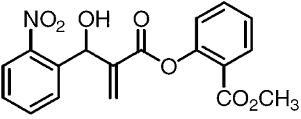
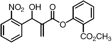
Morita-Baylis-Hillman adducts (MBHA) are compounds that are easily prepared in a high yield one-step synthetic reaction involving activated alkenes and aldehydes, ketones, among others, by nucleophilic catalysis (Basavaiah et al. Reference Basavaiah, Rao and Reddy2007). Since 2006, our group has been describing the synthesis and biological evaluation of several MBHA. These molecules presented anti-parasitic activity against amastigotes and promastigotes of Leishmania (De Souza et al. 2007; Barbosa et al. Reference Barbosa, Junior, Silva, Lopes, Figueiredo, Sousa, Batista, Silva, Silva, Oliveira and Vasconcellos2009; Silva et al. Reference Silva, Assis, Junior, Andrade, Cunha, Oliveira and Vasconcellos2011). We have recently described the synthesis of methyl 2-{2-[hydroxy(2-nitrophenyl)methyl])acryloyloxy} benzoate (HNAB) (Fig. 1) and 6 similar adducts. Among the compounds evaluated for anti-leishmanial activity, HNAB, which was designed based on the medicinal chemistry strategy of molecular hybridization between the methyl salicylate and an adduct that was derived from ortho-nitrobenzaldehyde, was the most active against Leishmania (Leishmania) amazonensis and L. (L.) infantum/chagasi (Barbosa et al. Reference Barbosa, Sousa, Amorim, Rodrigues, Assis, Caldas, Oliveira and Vasconcellos2011). It should be noted that the chemical structure of HNAB has similarity to the chalcones, and the anti-parasitic activity of a number of chalcone-like structure compounds has been described in the literature (Boeck et al. Reference Boeck, Falcão, Leal, Yunes, Filho, Torres-Santos and Rossi-Bergmann2006).
Chemical structure of Morita-Baylis-Hillman adduct methyl 2-{2-[hydroxy(2-nitrophenyl)methyl])acryloyloxy} benzoate (HNAB).
The aim of this work was to investigate the anti-leishmanial activity of HNAB in vitro against promastigotes and amastigotes of L. (V.) braziliensis isolates. The immunomodulatory properties of the drug on infected macrophages were also investigated.
MATERIALS AND METHODS
Reagents and commercial drugs
The following reagents and drugs were used: fetal bovine serum (FBS), RPMI-1640 medium, streptomycin and penicillin (Cultilab, SP, Brazil); pentavalent antimony meglumine antimoniate (Aventis Pharma™, SP, Brazil); Schneider's medium, DMSO and potassium antimonyl tartrate trihydrate (Sigma-Aldrich™, St Louis, USA); thioglycollate (Himedia, Mumbai, India); recombinant murine interferon (IFN)-γ (rIFN-γ) (Peprotech™, Rocky Hill, USA); alkaline phosphatase-conjugated streptavidin, recombinant murine tumor necrosis factor (TNF)-α, recombinant murine interleukin (IL)-6, recombinant murine IL-10, anti-TNF-α, anti-IL-6 and anti-IL-10 monoclonal antibodies (eBioscience™, CA, USA); sodium nitrite (Vetec Química Fina, RJ, Brazil); haematological stain Panótico rápido (Laborclin, PR, Brazil).
Morita-Baylis-Hillman adduct
Morita-Baylis-Hillman adduct methyl 2-{2-[hydroxy(2-nitrophenyl)methyl])acryloyloxy} benzoate (HNAB) was synthesized according to the protocol described by Barbosa et al. (Reference Barbosa, Sousa, Amorim, Rodrigues, Assis, Caldas, Oliveira and Vasconcellos2011). The drug was diluted in DMSO to prepare the stock solution (20 mg/ml). For each experiment the stock solution was diluted in culture medium in order to prepare the working solution (0·1% DMSO).
Parasites
Three isolates of Leishmania (Viannia) braziliensis obtained from humans with different clinical forms of ATL were used in this study: L. (V.) braziliensis MHOM/BR/2011/JMTS (isolated from a patient with localized cutaneous leishmaniasis), L. (V.) braziliensis MHOM/BR/2010/JCNS (isolated from a patient with mucosal leishmaniasis) and L. (V.) braziliensis MHOM/BR/2011/AF (isolated from a patient with disseminated cutaneous leishmaniasis). The species was identified by use of PCR techniques (Bruijn and Barker, Reference Bruijn and Barker1992), in the Laboratório Biologia de Leishmania (Instituto de Ciências Biológicas, Universidade Federal de Minas Gerais, Belo Horizonte, Brazil). The reference strain L. (V.) braziliensis MHOM/BR/1975/M2903 was also used in the promastigote sensitivity assays. Promastigote cultures were maintained at 25 °C in test tubes with biphasic medium consisting of rabbit blood agar (Novy-MacNeal-Nicolle) and Schneider's medium supplemented with 20% of fetal bovine serum, streptomycin (100 μg/ml) and penicillin (100 U.I./ml). The cultures were used in testing up to a maximum of 20 passages in vitro.
Animals
Male and female young BALB/c mice were obtained from the Laboratório de Imunopatologia Keizo Asami (Universidade Federal de Pernambuco, Recife, Brazil) and maintained at constant room temperature (21 ± 2 °C), on a 12/12 h light-dark cycle, with free access to food pellets and water in the Professor Thomas George Animal House Facility (Centro de Biotecnologia, Universidade Federal da Paraíba, João Pessoa, Brazil). All experiments were performed using 8 to 12-week-old animals and the protocols were previously approved by Animal Research Ethical Committee of the Universidade Federal da Paraíba (process number 208/2007).
Promastigote sensitivity assay
The sensitivity of promastigotes to HNAB, trivalent (potassium antimonyl tartrate trihydrate) and pentavalent antimony (meglumine antimoniate) was evaluated as described by Barbosa et al. (Reference Barbosa, Sousa, Amorim, Rodrigues, Assis, Caldas, Oliveira and Vasconcellos2011). Each experiment was performed in duplicate and repeated at least 3 times. The 50% inhibitory concentration (IC50) was calculated by Probit analysis (SPSS 8.0 for Windows).
Isolation of BALB/c peritoneal macrophages
The method used was similar to that described by Oliveira et al. (Reference Oliveira, Tafuri, Afonso, Oliveira, Nicoli, Vieira, Scott, Melo and Vieira2005). Briefly, 5 days before the experiments, the animals were injected intraperitoneally with 1 ml of thioglycollate (3%). The mice were euthanized by cervical dislocation and the macrophages were obtained by washing the peritoneal cavity with 10·0 ml of PBS supplemented with FBS (3%). The suspension was centrifuged for 10 min at 111 g. The cell density in the suspension was adjusted in RPMI-1640 medium.
Cytotoxicity and determination of selectivity index
The drug cytotoxicity to peritoneal macrophages was evaluated by the 3-(4,5-dimethyl-2-thiazole)-2,5-diphenyltetrazolium bromide (MTT) colourimetric assay as described by Silva et al. (Reference Silva, Assis, Junior, Andrade, Cunha, Oliveira and Vasconcellos2011). The viability of macrophages incubated in the presence of different concentrations of HNAB was determined by comparing to culture control (macrophages cultivated in medium only). The concentration that caused a 50% reduction in cell viability (CC50) was calculated by Probit analysis (SPSS 8.0 for Windows). The selectivity index (SI) was calculated by dividing the CC50 by IC50 that was previously determined for promastigote forms of L. (V.) braziliensis (Dos Santos et al. Reference Dos Santos, Júnior, Rosa, Torquato, Bassi, Ribeiro, Júnior, Bessera, Fontes, Da Silva and Piuvezam2011). Each experiment was performed in duplicate and repeated at least 3 times.
Treatment of macrophages with Morita-Baylis-Hillman adduct
Treatment of infected macrophages was performed as described by Oliveira et al. (Reference Oliveira, Tafuri, Afonso, Oliveira, Nicoli, Vieira, Scott, Melo and Vieira2005), with modifications. Briefly, peritoneal macrophages were adhered onto glass cover-slips by incubating the cells in 24-well culture plates (5 × 10⁵ cells/ml) in RPMI medium for 2 h at 37 °C and 5% CO2 atmosphere. After this period, stationary-phase promastigotes were added at a ratio of 10 parasites per 1 macrophage. After 3 h, each well was washed 3 times with pre-warmed RPMI-1640 medium to remove non-ingested promastigotes and fresh medium with or without different concentrations of HNAB was added. In parallel, meglumine antimoniate was used as reference drug. The cells were cultured for additional 24 or 72 h and after this time the percentage of infected macrophages and the number of amastigotes per infected macrophage was determined by optical microscopy examination of 300 macrophages in stained cover-slips. These values were multiplied to determine the infection index as described by Campos et al. (Reference Campos, Gomes, De Souza, Laison, Corbett and Silveira2008). The infection index values were used to determine the 50% effective concentration (EC50) of the drugs. The EC50 was calculated by Probit analysis (SPSS 8.0 for Windows). Each experiment was performed in duplicate and repeated at least 3 times.
Cytokine production
The supernatants of macrophage cultures were harvested at 24 and 72 h post- Leishmania infection and stored at −20 °C for TNF-α, IL-6 and IL-10 determination. The quantification was done by sandwich ELISA, according to the manufacturer´s instructions (eBioscience™, EUA). Standard curves were generated by using murine recombinant TNF-α, IL-6 and IL-10. The results obtained were expressed in pg/ml.
Nitric oxide production
Nitric oxide (NO) production was estimated by determining nitrite concentration in the supernatant of the culture of infected macrophages, using the Griess method (Green et al. Reference Green, Wagner, Glogowski, Skipper, Wishnok and Tannenbaum1982). This method consists of a colourimetric reaction in which the supernatant is incubated with an equal volume of freshly prepared Griess reagent (1% sulfanilamide, 0·1% dihydrochloride of N-(1 naphytyl)-ethylenediamine, 2·5% ortho-phosphoric acid) for 10 min at room temperature. The absorbance at 545 nm was determined using a spectrophotometer (Thermoplate, TP-Reader). The nitrite levels of each sample were determined by extrapolation from a previously determined standard curve. As a positive control, the supernatant of the infected macrophages incubated with rIFN-γ (100 U/ml) was used (Oliveira et al. 2005). The results obtained were expressed in μM.
Statistical analysis
The data obtained were presented as mean values ± standard errors of the mean (s.e.m.). Student's t-test was used to evaluate the significance of individual data and one-way analysis of variance (ANOVA) was used to compare the differences between groups. All analyses were done using GraphPad Prism software version 5.0 for Windows (GraphPad Software, San Diego, CA, USA). Values of P ⩽ 0·05 were considered as significant.
RESULTS
Activity of Morita-Baylis-Hillman adduct against promastigotes forms
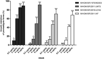
In order to evaluate the sensitivity of promastigotes of the reference strain and 3 isolates of L. (V.) braziliensis to HNAB, the percentage of growth inhibition was analysed in the presence of different concentrations of the drug (Fig. 2). Anti-promastigote activity against the reference strain of L. (V.) braziliensis was observed at 0·5 μg/ml. The growth inhibition of the isolate JMTS was observed at 2 μg/ml. On the other hand, JCNS and AF had their growth inhibited at 4 μg/ml. The IC50 values of HNAB ranged from 3·26 μg/ml (±1·61) to 7·77 μg/ml (±1·25) (Table 1). In parallel, the meglumine antimoniate (SbV) and potassium antimonyl tartrate (SbIII) IC50 were also calculated. The results showed that the anti-promastigote activity of HNAB was similar to that of SbIII. However, the anti-promastigote activity of the adduct against the reference strain was higher than that of the SbIII (P ⩽ 0·001). As expected, only at high concentrations was the SbV able to inhibit the growth of promastigotes. The IC50 values of SbV ranged from 3847 μg/ml (±751·7) to 8693 μg/ml (±136·2).
Growth inhibition of promastigote forms of the reference strain and different isolates of Leishmania (Viannia) braziliensis in the presence of different concentrations of HNAB. Parasites were cultivated for 96 h in Schneider medium at 25 °C. The graph represents the mean±s.e.m of at least 3 independent experiments performed in duplicate. *ND, not detected. Student's t-test was performed to compare the inhibition observed in the presence of different concentrations of HNAB to control (parasite growth in medium alone). * P ⩽ 0·05 and *** P ⩽ 0·001.

Concentrations of HNAB, trivalent antimony (SbIII) and pentavalent antimony (SbV) able to inhibit growth of promastigote forms of the reference strain and different isolates of Leishmania (Viannia) braziliensis by 50% (IC50)

Cytotoxicity and selectivity index of Morita-Baylis-Hillman adduct
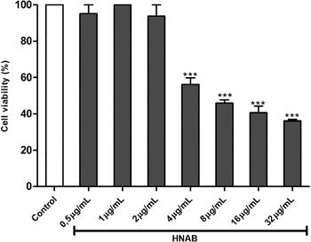
When the cytotoxicity of HNAB was analysed, it was observed that up to a concentration of 2 μg/ml the macrophage viability was not altered (Fig. 3). However, there was a significant reduction in the percentage of viable cells when they were incubated in the presence of 4 μg/ml of the adduct. The CC50 obtained was 18·09 μg/ml. The selectivity index (SI) showed that HNAB was approximately 3·6 times more toxic to L. (V.) braziliensis promastigotes than murine macrophages. The SI obtained using the reference strain was 5·55 (MHOM/BR/1975/M2903) and for the isolates it was 3·37 (MHOM/BR/2011/JMTS), 3·13 (MHOM/BR/2010/JCNS) and 2·33 (MHOM/BR/2011/AF). Similar results were obtained when the trypan blue exclusion assay was performed, even when the analysis was performed after a 72-h macrophage incubation in the presence of HNAB (data not shown).
Cytotoxicity of HNAB for BALB/c peritoneal macrophages. The macrophages were incubated for 24 h at 37 °C, 5% CO2, in the presence and absence of different concentrations of HNAB. Thereafter, cell viability was evaluated by the MTT reduction method. The graph represents the mean±s.e.m. of at least 3 independent experiments performed in duplicate. Student's t-test was performed to compare the cell viability in the presence of different concentrations of adduct to control (medium alone). *** P ⩽ 0·001.

Activity of Morita-Baylis-Hillman adduct against intracellular amastigotes
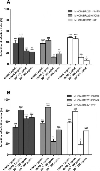
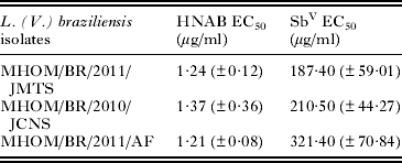
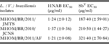
The activity of HNAB against intracellular amastigotes of the different isolates of L. (V.) braziliensis was evaluated using the in vitro model of macrophage infection (Fig. 4). In parallel, the activity of the reference drug meglumine antimoniate (SbV) was evaluated. After a 24-h incubation of infected macrophages with HNAB, it was observed that concentrations of 1 μg/ml and 2 μg/ml were able to reduce the infection index of all isolates analysed. The reductions ranged from 37·47% to 48·19% (Fig. 4A) and were higher than those observed in the presence of 100 μg/ml or 200 μg/ml of SbV. After 72 h, the incubation of infected macrophages with the adduct resulted in reductions of infection index that were even higher than that observed at 24 h (Fig. 4B). The data obtained after 72 h of treatment with HNAB or SbV were used to calculate the EC50 (Table 2). The EC50 analysis showed that the concentration of adduct required to reduce the infection rate of different isolates of L (V.) braziliensis by 50% was on average 189 times lower than that of SbV, after a 72-h treatment (P ⩽ 0·0127). The isolates analysed were equally sensitive to HNAB. However, the isolate obtained from a patient with disseminated cutaneous leishmaniasis, L. (V.) braziliensis MHOM/BR/2011/AF, was less sensitive to SbV than the others.
Reduction of infection index of macrophages infected with different isolates of Leishmania (Viannia) braziliensis and treated with different concentrations of HNAB and pentavalent antimony (SbV). Infected macrophages were treated with HNAB (1 μg/ml or 2 μg/ml) or SbV (100 μg/ml or 200 μg/ml) and incubated for 24 h (A) or 72 h (B) at 37 °C, 5% CO2. The graph represents the mean±s.e.m. of 3 independent experiments performed in duplicate. Student's t-test was performed to compare the infection index of treated macrophages to the infection index of control (untreated). * P ⩽ 0·05, ** P ⩽ 0·01 and *** P ⩽ 0·001.

Concentrations of HNAB and pentavalent antimony (SbV) able to reduce the infection index of macrophages infected with different isolates of Leishmania (Viannia) braziliensis by 50% (EC50)
Effect of Morita-Baylis-Hillman adduct on TNF-α, IL-6 and IL-10 production
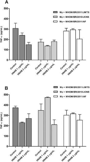
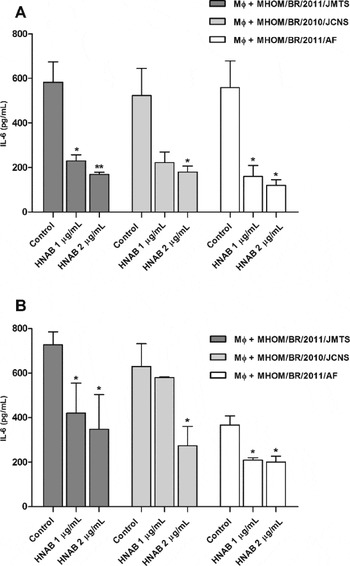
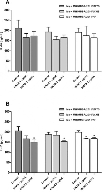
The production of TNF-α, IL-6 and IL-10 by infected macrophages treated with HNAB was evaluated. TNF-α production was not altered by HNAB treatment (Fig. 5). However, it decreased IL-6 (Fig. 6) and IL-10 (Fig. 7) production. The reduction in IL-6 production was observed after 24 and 72 h of treatment, but IL-10 production decreased only after 72 h of treatment.
Effect of HNAB on TNF-α production by infected macrophages. TNF-α concentration was evaluated in the supernatant of macrophages infected with different isolates of Leishmania (Viannia) braziliensis that were treated or not (control) with HNAB (1 μg/ml or 2 μg/ml) after 24 h (A) or 72 h (B) of incubation at 37 °C, 5% CO2. The graph represents the mean±s.e.m. of 3 independent experiments performed in duplicate. Student's t-test was performed to compare the production of TNF-α by adduct-treated macrophages to that of control (untreated) macrophages.

Effect of HNAB on IL-6 production by infected macrophages. IL-6 concentration was evaluated in the supernatant of macrophages infected with different isolates of Leishmania (Viannia) braziliensis that were treated or not (control) with HNAB (1 μg/ml or 2 μg/ml) after 24 h (A) or 72 h (B) of incubation at 37 °C, 5% CO2. The graph represents the mean±s.e.m. of 3 independent experiments performed in duplicate. Student's t-test was performed to compare the production of IL-6 by adduct-treated macrophages to that of control (untreated) macrophages. * P ⩽ 0·05 and ** P ⩽ 0·01.

Effect of HNAB on IL-10 production by infected macrophages. IL-10 concentration was evaluated in the supernatant of macrophages infected with different isolates of Leishmania (Viannia) braziliensis that were treated or not (control) with HNAB (1 μg/ml or 2 μg/ml) after 24 h (A) or 72 h (B) of incubation at 37 °C, 5% CO2. The graph represents the mean±s.e.m. of 3 independent experiments performed in duplicate. Student's t-test was performed to compare the production of IL-10 by adduct-treated macrophages to that of control (untreated) macrophages. * P ⩽ 0·05.

Effect of the Morita-Baylis-Hillman adduct on nitric oxide production
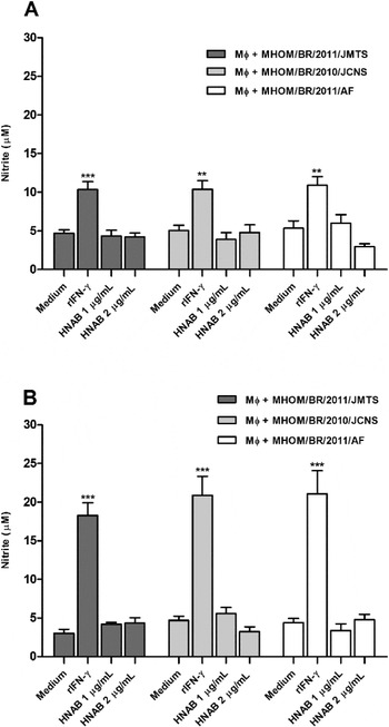
In order to evaluate the involvement of nitric oxide production in the reduction of the infection index observed after treatment of the infected macrophages with HNAB, nitrite levels in the supernatant of the cultures were measured (Fig. 8). The results showed that, independent of time and concentration evaluated, nitric oxide production by the infected cells was not altered. However, as expected, macrophages that were exposed to 100 U/ml of IFN-γ exhibited an increased production of nitric oxide. These results revealed that the reduction of the infection index observed when macrophages infected with different isolates of L. (V.) braziliensis were treated with HNAB was independent of NO production.
Effect of HNAB on nitric oxide (NO) production by infected macrophages. Nitrite concentration was evaluated in the supernatant of macrophages infected with different isolates of Leishmania (Viannia) braziliensis that were treated or not (control) with rIFN-γ (100 U/ml) or with HNAB (1 μg/ml or 2 μg/ml) after 24 h (A) or 72 h (B) of incubation at 37 °C, 5% CO2. The graph represents the mean±s.e.m. of 3 independent experiments performed in duplicate. Student's t-test was performed to compare the production of nitric oxide by rIFN-γ or HNAB-treated macrophages to that of control (untreated) macrophages. ** P ⩽ 0·01 and *** P ⩽ 0·001.

DISCUSSION
In the present study the activity of the Morita-Baylis-Hilman adduct, methyl 2-{2-[hydroxy(2-nitrophenyl)methyl])acryloyloxy} benzoate (HNAB), against L. (V.) braziliensis parasites was investigated. The anti-promastigote activity of HNAB, as well as the activity of the other 6 adducts, has been previously demonstrated for L. (L.) amazonensis and L. (L.) chagasi (Barbosa et al. Reference Barbosa, Sousa, Amorim, Rodrigues, Assis, Caldas, Oliveira and Vasconcellos2011). The results obtained in the present study showed that HNAB was also effective in inhibiting the growth of promastigote forms of L. (V.) braziliensis isolated from patients with different clinical manifestations of tegumentary leishmaniasis.
The choice of HNAB for further pharmacological studies was based on the fact that it exhibited higher anti-leishmanial activity than other MBHAs previously studied (Barbosa et al. Reference Barbosa, Sousa, Amorim, Rodrigues, Assis, Caldas, Oliveira and Vasconcellos2011). In general, adducts that were derived from ortho-nitro benzaldehyde in our laboratory are more active against promastigote forms of Leishmania than other isomers (meta and para). Furthermore, they showed a higher selectivity index (Silva et al. Reference Silva, Assis, Junior, Andrade, Cunha, Oliveira and Vasconcellos2011). We have recently described that in a congeneric series of 12-aryl nitro-compounds investigated by an electrochemical (cyclic voltammetry) method, the ortho-nitro derivatives had the lowest ionization potential (De Paiva et al. 2012). This observation suggests that the mechanism of action via oxidative stress may play a role in the anti-promastigote activity of the adduct studied.
The anti-promastigote activity of HNAB for the reference strain and different isolates of L. (V.) braziliensis was compared with the activity displayed by trivalent antimony (SbIII) and pentavalent antimony (SbV). Studies have shown that SbV is more active against amastigote than promastigote forms of Leishmania (Ephros et al. Reference Ephros, Bitnun, Shaked, Waldman and Zilberstein1999; Vermeersch et al. Reference Vermeersch, Da Luz, Toté, Timmermans, Cos and Maes2009). However, the activity of SbIII is similar in both (Ephros et al. Reference Ephros, Bitnun, Shaked, Waldman and Zilberstein1999). The IC50 values obtained in the present study demonstrated that the efficacy of HNAB in inhibiting the growth of promastigote forms of the different isolates of L. (V.) braziliensis was similar to that of SbIII. However, against the reference strain it was more active than SbIII. As expected, SbV was only able to inhibit the growth of the promastigotes at high concentrations.
Chemotherapy of leishmaniasis is usually aimed at clinical cure of the lesions and containment of the parasites, either by destroying them directly or inducing an immune response in the host to control the infection (Croft et al. Reference Croft, Sundar and Fairlamb2006). Since in the vertebrate host Leishmania parasites are found within macrophages and in the amastigote form, it is essential to assess the activity of HNAB on the infected host cell. For this analysis, macrophages were infected with the different isolates of L. (V.) braziliensis and then treated with the adduct. Concentrations which showed no cytotoxicity to macrophages were used. The results show that HNAB was effective in reducing the infection rate of macrophages infected with the different isolates. In fact, it was more active than SbV. This fact was demonstrated by the EC50 values, as the concentration of the adduct required to reduce the infection rate of different isolates of L (V.) braziliensis by 50% were, on average, 189 times lower than that of SbV, after a 72-h treatment. Furthermore, HNAB was equally effective in reducing the infection rate of the different isolates. On the other hand, SbV was less effective in reducing the infection rate of the isolate L. (V.) braziliensis MHOM/BR/2011/AF, obtained from a patient with disseminated cutaneous leishmaniasis. It is noteworthy that pentavalent antimonials are considered pro-drugs which are converted inside the macrophages (Gourbal et al. Reference Gourbal, Sonuc, Bhattacharjee, Legare, Sundar, Oullette, Rosen and Mukhopadhyay2004) or amastigotes (Denton et al. Reference Denton, McGregor and Coombs2004) to the trivalent antimonial, a more active form against the amastigote forms of Leishmania. We may therefore hypothetically state that HNAB is also more active against amastigotes of L. (V.) braziliensis than SbIII.
Comparing the results obtained with intracellular amastigotes to those obtained with promastigotes, the former were more sensitive to HNAB (P ⩽ 0·0045) than the latter. The low ionization potential of HNAB can contribute to the generation of free radicals once inside the macrophages or the intracellular parasites (De Paiva et al. Reference De Paiva, Souza, Lima-Junior, Silva, Filho, De Vasconcelos, De Abreu, Goulart and Vasconcellos2012). These free radicals in turn may exert a direct effect on the intracellular amastigotes, resulting in the death of the parasites. However, the anti-amastigote activity observed may also be due to an indirect action of HNAB by stimulating macrophages to contain the infection.
In order to investigate the mechanism by which HNAB was able to reduce the infection rate of macrophages, its potential immunomodulatory effect was investigated, by analysing production of selected cytokines such as TNF-α, IL-6, IL-10 and of nitric oxide by the infected and treated macrophages.
The immune response to Leishmania infection is mainly cellular and the elimination of intracellular parasites depends ultimately on the microbicidal activity of macrophages (Bogdan and Röllinghoff, Reference Bogdan and Röllinghoff1998). Experimental models of L. (L.) major infection of inbred mice showed that resistance to infection is associated with the development of Th1 lymphocytes which produce IFN-y, a cytokine that activates anti-leishmanial activity of macrophages (Sacks and Noben-Trauth, Reference Sacks and Noben-Trauth2002). Moreover, the susceptibility has been associated with the development of Th2 lymphocytes which secrete cytokines such as IL-4, IL-10 and transforming growth factor (TGF)-β to inhibit macrophage activation resulting in proliferation of intracellular parasites (Sacks and Noben-Trauth, Reference Sacks and Noben-Trauth2002).
Superoxides (O2−) and nitric oxide (NO) are important antimicrobial agents produced by macrophages that contribute to the elimination of Leishmania parasites (Gantt et al. Reference Gantt, Goldman, Miller, McCormick, Miller, Jeronimo, Nascimento, Britigan and Wilson2001). The inhibition of production of one of these two agents by murine and human macrophages increases the intracellular growth of L. (L.) chagasi (Gantt et al. Reference Gantt, Goldman, Miller, McCormick, Miller, Jeronimo, Nascimento, Britigan and Wilson2001). Although the process of phagocytosis can stimulate the production of superoxides, nitric oxide is synthesized by the inducible nitric oxide synthase (iNOS) only when macrophages are exposed to stimuli such as IFN-γ and TNF-α (Liew et al. Reference Liew, Li and Millot1990; Bogdan, Reference Bogdan2001; Vila-del Sol et al. Reference Vila-del Sol, Díaz-Munöz and Fresno2007).
TNF-α is a pro-inflammatory cytokine produced by various cell types, including macrophages (Baud and Karin, Reference Baud and Karin2001). This cytokine, by binding to its receptor, induces the activation of several intracellular signalling pathways such as NF-kB signalling, which in turn modulates the transcription of several genes related to the immune response, among which is the iNOS gene (Baud and Karin, Reference Baud and Karin2001). In the present study, analysis of TNF-α in the supernatant of macrophages that were infected with the different isolates of L. (V.) braziliensis and treated with HNAB, revealed that this treatment had no effect on the production of this cytokine. Thus, the decrease observed in infection rate of macrophages was not correlated with the production of TNF-α.
IL-6 is a cytokine produced by a variety of cells, including macrophages, dendritic cells and Th2 cells (Rincón et al. Reference Rincón, Anguita, Nakamura, Firkrig and Flavell1997). This cytokine contributes indirectly, by induction of IL-4, to the differentiation of Th2 cells (Rincón et al. Reference Rincón, Anguita, Nakamura, Firkrig and Flavell1997). On the other hand, it inhibits the differentiation of Th1 cells (Diehl et al. Reference Diehl, Anguita, Hoffmeyer, Zapton, Ihle, Fikrig and Rincón2000). Using an in vitro infection model, it was found that IL-6 can inhibit the activation of macrophages for killing of L. (L.) amazonensis by TNF-α and IFN-γ (Hatzigeorgiou et al. 1993). In the present study it was observed that the treatment of macrophages infected with different isolates of L. (V.) braziliensis with HNAB resulted in reduction of IL-6 release. Therefore, the decrease in IL-6 may facilitate the activation of these macrophages and therefore the containment of infection.
IL-10, an important regulator of the immune response, can be produced by macrophages, dendritic cells, B lymphocytes and different subpopulations of T cells (reviewed by Couper et al. Reference Couper, Blount and Riley2008). Using a model of in vitro infection, it was observed that the addition of anti-IL-10 increases the leishmanicidal activity of macrophages stimulated by IFN-γ or IL-7, indicating that endogenous IL-10 produced by macrophages can act in an autocrine mode as a deactivating factor (Vieth et al. Reference Vieth, Will, Schröppel, Röllinghoff and Gessner1994). The antagonistic effect of IL-10 on IFN-γ induction of microbicidal activity in human macrophages infected with L. (L.) major or L. (L.) infantum has been demonstrated (Vouldoukis et al. Reference Vouldoukis, Bécherel, Riveros-Moreno, Arock, Da Silva, Debré, Mazier and Mossalayi1997). IL-10, by inducing the activity of arginase, an enzyme that competes for the substrate with the iNOS, negatively regulates NO production by macrophages (Bogdan, Reference Bogdan2001). We observed in the present work that HNAB was able to reduce the production of IL-10 by the infected macrophages. This reduction may facilitate the activation of the macrophages and thus the control of infection. In a recent study, the in vitro leishmanicidal activity of a plant extract and its isolated compound against L. (V.) braziliensis was directly linked to increases in NO and decreases in IL-10 production by the infected macrophages (Dos Santos et al. Reference Dos Santos, Júnior, Rosa, Torquato, Bassi, Ribeiro, Júnior, Bessera, Fontes, Da Silva and Piuvezam2011).
It can be concluded that the immunomodulatory activity of HNAB, in promoting a reduction in the levels of IL-6 and IL-10, may favour the activation of macrophages by TNF-α produced by the macrophages (although TNF-α production in treated and non-treated macrophages was not different), resulting in a decrease in the infection rate. However, the anti-amastigote activity of the adduct was independent of nitric oxide production. This work revealed the potential of HNAB in the treatment of human infections caused by L. (V.) braziliensis and demonstrates the need to perform more profound mechanistic studies with this drug.
ACKNOWLEDGEMENTS
We thank Dr Maria Norma Melo (UFMG, Belo Horizonte, Brazil) for generously providing the reference strain L. (V.) braziliensis MHOM/BR/1975/M2903 and Rosângela Gomes Cordeiro, José Crispin Duarte and Renata Márcia Costa Vasconcelos for their technical support.
FINANCIAL SUPPORT
This work was supported in part by CNPq (Conselho Nacional de Desenvolvimento Científico e Tecnológico) and CAPES (Coordenação de Aperfeiçoamento de Pessoal de Nível Superior).