Introduction
Recent studies have revealed that epigenetic modifications, such as DNA methylation and histone modifications, play important roles in the regulation of chromatin structure and gene expression (Shahbazian et al., Reference Shahbazian and Grunstein2007). Two families of enzymes, histone acetyltransferases (HATs) and histone deacetyltransferases (HDACs) regulate histone acetylation.
The genomic state of oocytes is altered globally during oocyte growth and post fertilization (Panero et al., Reference Panero, Trelles, Rodano, Montserrat, Iglesias, Lewkowicz and Iribarren2006; Zuccotti et al., Reference Zuccotti, Piccinelli, Giorgi Rossi, Garagna and Redi1995). A recent study reported that H4K5, H4K8, H4K12, H4K16, H3K9, and H3K14 are acetylated in mouse oocytes at the germinal vesicle (GV) stage. However, with the restart of meiosis, deacetylation occurs at all sites except for H4K8, and this state is maintained until the end of the second meiotic metaphase (MII) (Kim et al., Reference Kim, Ogura, Nagata and Aoki2002).
It has been shown that acetylation of core histones is important in regulating gene expression by producing conformational changes in chromatin structure that allow DNA to be accessible to transcription factors (Long et al., Reference Long, Lu, Kuang, Yan, Liang, Yu, Chai, Yan and Lyu2013). In addition, histone acetylation can serve as an epigenetic marker by retaining this information in the mitotic phase, and propagating it from one cell to the next generation (Jenuwein and Allis, Reference Jenuwein and Allis2001). Specifically, it was suggested that H4K12 is a critical epigenetic marker for active genes (Kruhlak et al., Reference Kruhlak, Hendzel, Fischle, Bertos, Hameed, Yang, Verdin and Bazett-Jones2001; Jin et al., Reference Jin, Zhu, Guo, Li, Zhang, Zhang, Xing, Xuan, Luo, Yin and Kang2016).
The goal of this study was to determine the exact pattern of H4K12 acetylation in mouse oocytes during meiosis and after parthenogenetic activation (PA). We found that hyperacetylation decreased just after GV, was weakly detected during germinal vesicle break down (GVBD), and no signals were detected in the MII phase. However, H4K12 acetylation reappeared at the 1-cell embryos at 4 h after PA. This study reveals the dynamic alterations of H4K12 acetylation during meiotic maturation and after PA, and provides the theoretical foundation for further research into the function of acetylation during the reproductive process.
Materials and methods
Collection and culture of mouse oocyte
To obtain fully grown oocytes arrested at prophase I of meiosis, 3-week-old KunMing (KM) female mice were superovulated with 10 IU pregnant mare’s serum gonadotrophin (PMSG). The ovaries were removed from the mice at 48 h post-PMSG and transferred to M2 medium (Sigma) as described previously (Sobajima et al., Reference Sobajima and Kohmoto1993). The GV, GVBD, and MII-stage oocytes were obtained at 0 h, 4 h, and 12 h of culturing at 38.5°C and 5% CO2 in air, at maximum humidity.
Parthenogenetic activation
The in vivo matured mouse oocytes (MII oocytes) were activated by PA. These detailed procedures have been described previously (Zhang et al., Reference Zhang, Jin, Zhu, Guo, Li, Zhang, Xing, Xuan, Luo, Luo, Wang, Cui, Li, Cui, Yin and Kang2017). Briefly, the MII mouse oocytes were parthenogenetically activated by incubating in activation medium, which consisted of Ca2+-free potassium-supplemented simplex optimised medium–bovine serum albumin (KSOM–BSA) supplemented with 10 mM SrCl2 and 5 μg/ml cytochalasin B, at 38.5°C and 5% CO2 in air, at maximum humidity for 6 h.
Quantitative real-time PCR
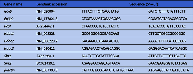
Quantitative real-time PCR (qPCR) was used to measure the relative expression of histone acetyltransferase (Gcn5), E1A binding protein (Ep300), 3-oxoadipyl-CoA thiolase (Pcaf), histone deacetylase 1 (Hdac1), histone deacetylase 2 (Hdac2), histone deacetylase 3 (Hdac3), sirtuin 1 (Sirt1), and sirtuin 2 (Sirt2) mRNA in GV, MII, and post PA oocytes. Total RNA was extracted from oocytes using the RNeasy Micro Kit (Qiagen), according to the manufacturer’s protocol. Reverse transcription and qPCR were performed using the ReverTra Ace qPCR RT KitQ-PCR and SYBR Premix Ex Taq (TaKaRa) in a reaction volume of 20 μl and the ABI StepOne system (Applied Biosystems). Each experiment was repeated four times. Primer sequences are listed in Table 1.
Primers used in this study

Immunofluorescence assay
Immunofluorescence was performed as described (Huang et al., Reference Huang, Wang, Qi, Wang, Tong and Zhang2011). Briefly, oocytes at the GV, GVBD, MII stages and embryos were fixed in 4% (w/v) paraformaldehyde at 4°C overnight and then permeabilized with 0.5% (v:v) Triton X-100 for 30 min. After blocking in phosphate-buffered saline with 1% bovine serum albumin (PB1) for 1 h at room temperature, all samples were incubated with the primary antibodies against AcH4K12 (ab46983, Abcam) for 1 h at 37°C. After three washes in PB1 of 5 min each, oocytes were incubated with rabbit anti-mouse IgG secondary antibody (Cowin Biotech) for 1 h. After three washes, all samples were counterstained with 4′,6-diamidino-2-phenylindole (DAPI) (Vector Laboratories) to visualize the DNA. To confirm, the antibody we used in the experiment was specific for Lys-12 of histone H4, control experiments were carried in accordance with the same experimental protocol without using the primary antibody. Finally, fluorescence was detected using a confocal laser scanning system.
Results and discussion
H4K12 acetylation changes during meiosis
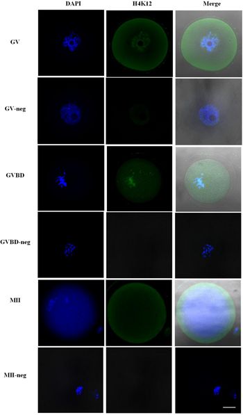
The global H4K12 acetylation status of histones during mouse oocyte maturation is shown in Fig. 1. There was an obvious GV in the middle of the GV oocyte, and H4K12 was hyperacetylated in the nucleus. However, after 4 h of maturation, the GV oocytes reached the GVBD stage, at which time the GV broke down, and H4K12 acetylation was lower than that at the GV stage. In the MII stage, the oocytes extruded the first polar body (PB1), and no fluorescence signal was detected. From the results, H4K12 is deacetylated during meiosis in mouse oocytes. Our results were consistent with the finding of Aoki’s group that H4K12 is deacetylated when oocytes enter the first meiotic phase, temporarily acetylated around the time of completion of the first meiosis, and then deacetylated again when the oocytes enter the second meiotic phase (Akiyama et al., Reference Akiyama, Kim, Nagata and Aoki2004).
Immunostaining of global histone acetylation status during meiotic maturation of oocytes. GV, GVBD and MII indicate oocytes at the germinal vesicle, germinal vesicle breakdown and metaphase II stages that were stained with antibodies against AcH4K12. While GV-neg, GVBD-neg and MII-neg indicate control oocytes at the same stage, but with only the secondary antibody, and without antibodies against AcH4K12, which is shown as green. Each sample was counterstained with 4′,6-diamidino-2-phenylindole (DAPI) to visualize DNA (blue). Scale bar, 20 μm.

H4K12 acetylation distribution in oocytes after 4-hour-PA
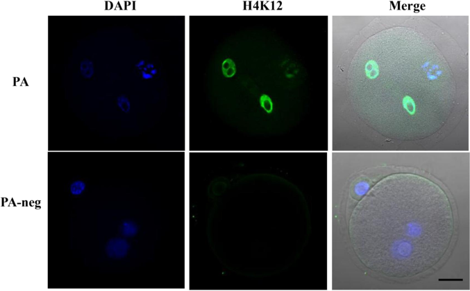
The MII oocytes were then subjected to PA using SrCl2 and cytochalasin B, and the H4K12 acetylation distribution is shown in Fig. 2. The oocytes had formed two pronuclei after 4 h of PA, shown on the left-hand side of Fig. 2. Our results also showed that H4K12 acetylation reappeared after PA. There have been a few studies on the changes in acetylation after PA until now. However, some research on the changes in acetylation after fertilization has been published, showing that acetylation of histones occurs in both male and female pronuclei (Kageyama et al., Reference Kageyama, Liu, Kaneko, Ooga, Nagata and Aoki2007).
Immunostaining of global histone acetylation status of oocytes after parthenogenetic activation (PA). Oocytes after PA were stained with or without (PA-neg) antibodies against AcH4K12, which is shown as green; and DAPI to visualize DNA (blue). Scale bar, 20 μm.

Relative mRNA expression of several representative HDACs and HATs
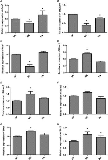
As histone acetylation levels are closely related to levels of HDACs and HATs, we selected three HAT genes, Gcn5, Ep300, and Pcaf, and five HDAC genes, Hdac1, Hdac2, Hdac3, Sirt1, and Sirt2, and measured their gene expression levels in the oocytes at GV and MII stages, and at 4 h after PA. The results are shown in Fig. 3. Expression levels of Gcn5, Ep300, and Pcaf all decreased significantly in MII compared with GV, and had an increasing trend from MII to PA [mean ± standard error of the mean (SEM) relative expression of Gcn5: 0.70 ± 0.10 at metaphase MII and 1.27 ± 0.31 at PA; Ep300: 0.41 ± 0.09 at MII and 0.80 ± 0.04 at PA; and Pcaf: 0.35 ± 0.12 at MII and 1.12 ± 0.06 at PA; P < 0.05]. All five selected HDAC genes showed a significant increase in MII compared with that in the GV, while the expression levels in PA declined and were similar to those in the GV (mean ± SEM relative expression of Hdac1: 1.41 ± 0.10 at metaphase MII and 0.79 ± 0.05 at PA; Hdac2: 1.50 ± 0.19 at MII and 1.21 ± 0.03 at PA; Hdac3: 1.19 ± 0.04 at MII and 0.87 ± 0.11 at PA; Sirt1: 1.32 ± 0.02 at MII and 1.11 ± 0.09 at PA; Sirt2: 1.50 ± 0.21 at MII and 1.31 ± 0.11 at PA; P < 0.05). The same genes were selected in a previous study to investigate the changes in the expression levels in GV, MI, and MII oocytes from control and diabetic mice (Ding et al., Reference Ding, Pan, Huang, Wang, Shen, Xu, Zhang, Liu, He, Yang, Qi and Wang2012). The difference between the control and diabetic mice oocytes was reported, but the changing pattern of these genes in control oocytes was not described, which we have done in this study. The expression levels of these genes matched the immunostaining results.
Relative mRNA expression of Gcn5 (A), Ep300 (B), Pcaf (C), Hdac1 (D), Hdac2 (E), Hdac3 (F), Sirt1 (G) and Sirt2 (H) in mouse oocytes at the germinal vesicle (GV), metaphase II (MII), and after parthenogenetic activation (PA) stages (*P < 0.05).

Financial support
This work was supported by the National Natural Science Foundation of China (31802063), the Youth Foundation of Hebei Educational Committee (2018166) and Scientific Research Foundation for the Introduction of Talent in Hebei Agricultural University (ZD201718).
Conflicts of interest
The authors declare no conflicts of interest.
Ethical standards
All experiments were performed in accordance with the guide for the Care and Use of Laboratory Animals prepared by the Institutional Animal Care and Use Committee of Hebei Agricultural University, China.