Introduction
Healthy People 2020, a Center for Disease Control initiative, set a goal of achieving 57.8% of women entering pregnancy with a normal weight by 2020. 1 Unfortunately, data from the USA show that 61.9% of reproductive aged women in 2016 had a body mass index (BMI) ≥ 25.0 kg/m2. Reference Berge, Fertig, Tate, Trofholz and Neumark-Sztainer2 Maternal overweight and obesity are associated with poor offspring outcomes during pregnancy, delivery, and postnatally. During pregnancy, women who are overweight and obese are more likely to be diagnosed with gestational diabetes, pre-eclampsia, and other complications of pregnancy. Reference Fuchs, Senat and Rey3 At delivery, the same group of women tend to have higher rates of C-section and higher risk of other delivery complications. Reference Ballesta-Castillejos, Gomez-Salgado, Rodriguez-Almagro, Ortiz-Esquinas and Hernandez-Martinez4 Moreover, offspring outcomes are affected by maternal weight status, including infant complications at birth such as hypoglycemia, macrosomia, and still birth. Reference Ruager-Martin, Hyde and Modi5 As infants born to obese mothers transition into adulthood, they tend to have higher risk of metabolic perturbations. Reference Bider-Canfield, Martinez and Wang6–Reference Lu, Zhang and Li8
Many studies based on the Developmental Origins of Health and Disease hypothesis Reference Barker9 have identified the long-lasting effects of maternal factors on offspring health. One crucial physiological system that appears to be developmentally programmed by maternal factors is the fetal autonomic nervous system (ANS). Reference Gustafson, Popescu, Reissland and Kisilevsky10 Heart rate variability (HRV), defined as the beat to beat variation in heart rate, is one reliable and valuable proxy of ANS development in the fetus. Reference Schneider, Bode and Schmidt11 In utero measurement of fetal HRV has allowed for insights into how maternal behaviors and characteristics relate to neurodevelopment before birth and moved the field toward interventions for improving long-term offspring health outcomes. For example, maternal physical activity results in higher offspring HRV at 36 weeks gestational age, a finding that remains at 1-month postnatal age. Reference May, Glaros, Yeh, Clapp and Gustafson12
While there is a known relationship between maternal weight and infant outcomes, the link between fetal ANS development and maternal weight throughout pregnancy is largely understudied. In a healthy pregnancy, fetal heart rate decreases and variability increases as gestation progresses, indicating maturation and integration of the ANS. Reference Gustafson, Popescu, Reissland and Kisilevsky10 We hypothesize maternal weight will be inversely related to fetal ANS development, thereby elucidating a possible mechanism initiated in utero leading to poor developmental outcomes among infants born to obese mothers. Therefore, the goal of the current analysis was to evaluate the influence of maternal weight during early (enrollment) and late (36 weeks) pregnancy on fetal HRV.
Methods
This study was a secondary analysis of a longitudinal, randomized, double-blind, placebo-controlled trial at the University of Kansas Medical Center that investigated the effect of docosahexaenoic acid (DHA) supplementation during pregnancy on fetal autonomic development. Eligible participants were generally healthy pregnant women living in the Kansas City area. Participants received 600 mg DHA per day or placebo throughout their pregnancy. Details of the parent study intervention and primary outcomes have been published previously. Reference Gustafson, Carlson and Colombo13
Maternal–fetal magnetocardiograms were recorded using an 83-channel dedicated fetal biomagnetometer (CTF MEG International Services LP, Coquitlam, BC, Canada) at 36 weeks gestation (n = 46). Data were collected in two blocks of continuous 18-min recording (for a total of 36 min) using a 300 Hz sampling rate. Maternal and fetal cardiac components were separated using independent components analysis. Reference Gustafson, Carlson and Colombo13 Fetal HRV was represented by the standard deviation of sinus beat-to-beat (NN) intervals (SDNN). Maternal weight was measured at enrollment (12–20 weeks) and 36 weeks gestation.
The primary aim of this research was to investigate the relationship between fetal HRV and maternal weight during pregnancy. Continuous variables were expressed as mean ± SD and categorical variables reported as proportion of the sample. Fetal HRV was calculated and treated as a continuous measure. Relationship between fetal HRV and maternal weight at enrollment and 36 weeks was modeled using unadjusted ordinary least squares (OLS) regression models. Adjusted OLS models included (1) a model adjusted for categorical DHA group assignment (DHA group vs. control) and (2) a model to assess for interaction between maternal weight at 36 weeks and DHA group assignment. We also constructed OLS models to investigate relationship between self-reported gestational weight gain (GWG) and fetal HRV. Assumptions for all models were assessed through residual analyses including visualization of Q–Q plots, residual histograms, and scale-location plots. Statistical analyses were performed using R (version 3.6.1; R Foundation, Vienna, Austria). Statistical significance was set at P ≤ 0.05.
Results
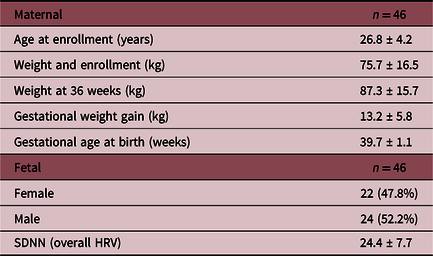
Data were available for 46 generally healthy pregnant women who had both biomagnetometry and weight data at 36 weeks gestation. Mean maternal age was 26.8 ± 4.2 years, weight at enrollment was 75.7 ± 16.5 kg, and weight at 36 weeks was 87.3 ± 15.7 kg. Maternal and fetal characteristics are described in Table 1.
Maternal and fetal characteristics

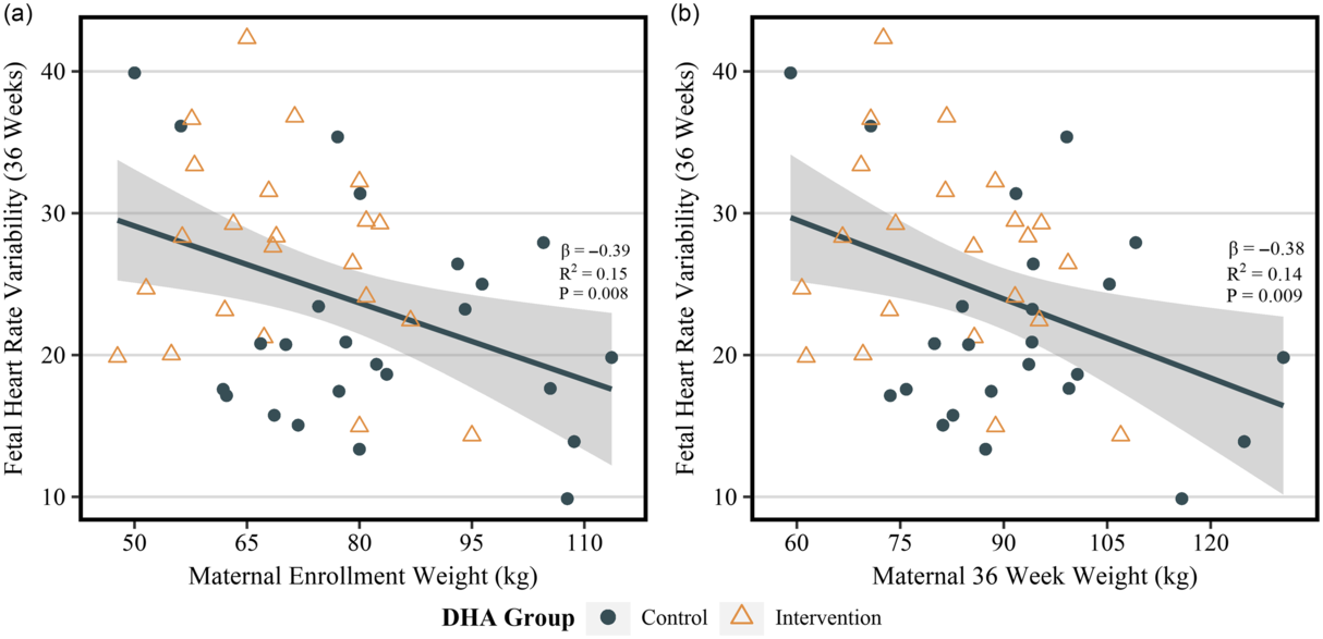
Maternal weight at enrollment and at 36 weeks were inversely related to fetal HRV (β = −0.39, P = 0.008 and β = −0.38, P = 0.009, respectively), illustrated in Fig. 1, even when controlled for random group assignment (P = 0.049, P = 0.04, respectively). There was no interaction between maternal weight at either time point and DHA group assignment (a variable already determined to be significantly associated with fetal HRV Reference Gustafson, Carlson and Colombo13 ), indicating the inverse linear relationship between maternal weight and fetal HRV was the same in both participants randomly assigned to the intervention and control. Self-reported GWG was not related to fetal HRV (P = 0.71). We controlled for the influence of known confounders such as maternal smoking status and fetal sex, and none were related to fetal HRV in this sample.
Unadjusted OLS models for fetal HRV and maternal weight at enrollment (A) and 36 weeks (B).

Discussion
The goal of this study was to investigate the association between maternal weight in early and late pregnancy and fetal ANS development, as indexed by overall HRV at 36 weeks gestational age. Our results show that maternal weight is a significant predictor of fetal overall HRV; specifically, higher maternal weight predicts lower fetal HRV.
The HRV metric used in this analysis, SDNN, is a reliable time domain metric of fetal overall HRV, reflecting both sympathetic and parasympathetic input. Reference Gustafson, Popescu, Reissland and Kisilevsky10 In a typically developing and healthy fetus, overall HRV increases with gestational age, becoming more complex in the period between 30 and 32 weeks, due in part to increasing influence of the vagus and the emergence of characteristic fetal neurobehavioral states. Reference Schneider, Bode and Schmidt11 The ontogenetic functional development of the fetus is largely mediated by the ANS; therefore, HRV serves as a proxy for central-autonomic neural integration.
Although our results only imply a relationship between weight and fetal HRV, it is important to note that weight class determined via BMI is an important variable to consider in future research. Maternal overweight and obesity are predictors of numerous maternal and offspring outcomes including cardiometabolic disease risk. Reference Kawasaki, Arata and Miyazaki14–Reference Osmond and Barker16 However, the point of origination of the link between maternal and offspring health has been difficult to determine. Our findings suggest the link begins early in pregnancy and persists throughout gestation. Likewise, other investigators have reported a link between gestational diabetes mellitus Reference Fehlert, Willmann and Fritsche17 and maternal obesity Reference Mat Husin, Schleger and Bauer18 and fetal HRV, reporting lower fetal SDNN when compared to both normoglycemic mothers and normal weight mothers. Our findings are consistent with these reports and will require further study to understand the causal mechanisms.
These results are from a secondary analysis of an existing dataset designed to detect the impact of maternal omega 3 (DHA) consumption on fetal autonomic development. As such, the present analysis was not powered to detect a relationship between maternal weight and fetal HRV. Within our sample, we saw large variation in HRV which could be explained by a variety of fetal and/or maternal variables worth exploring in the future. A significant strength of this study is that maternal weight was measured by trained study personnel during visits, and not self-reported. However, we were not able to collect measured pre-pregnancy weight and height, and therefore were not able to look at the relationship between pre-pregnancy BMI and fetal ANS outcomes. Pre-pregnancy BMI is a topic that warrants examinations in future studies of fetal ANS.
In conclusion, our analysis suggests maternal weight is a factor that may constrain fetal autonomic development, even early in pregnancy. Maternal factors during pregnancy, and possibly preconception, lay the groundwork for fetal autonomic development and should be considered when designing interventions to promote the health and well-being of mothers and their offspring. Our findings suggest further study is needed to better understand how maternal obesity influences fetal autonomic development and how this may predict long-term neurodevelopmental outcomes.
Acknowledgements
The authors would like to acknowledge Hoglund Biomedical Imaging Center which has been supported by a generous gift from Forrest and Sally Hoglund and funding from the National Institutes of Health including U54 HD090216: Kansas Intellectual and Developmental Disabilities Research Center. The content is solely the responsibility of the authors and does not necessarily represent the official views of the funding agencies.
Financial support
This study was supported by The Eunice Kennedy Shriver National Institute of Child Health and Development (R21 HD059019) (KMG) and in part received core support from the Kansas Intellectual Development and Disabilities Research Center (P30 NICHD HD 002528). The investigational product was generously donated by DSM Nutritional Products. This trial was registered at clinicaltrials.gov, Identifier: NCT01007110.
Conflict of interest
None.
Ethical standards
The authors assert that all procedures contributing to this work comply with the ethical standards of the relevant national guidelines on human experimentation and with the Helsinki Declaration of 1975, as revised in 2008, and have been approved by the institutional committees at the University of Kansas Medical Center.