Sudden cardiac arrest is an unexpected, often highly publicised, harrowing event that occurs in seemingly healthy young individuals. The true incidence for sudden cardiac arrest in the young remains unclear. Some studies have estimated the incidence as high as one in 3100 in National Collegiate Athletic Association African-American athletes or as low as one in 300,000.Reference Harmon, Asif, Klossner and Drenzer 1 – Reference Schoenbaum, Denchev, Vitiello and Kaltman 7 Although the exact number is unknown, it can be appreciated that the survival rate of sudden cardiac arrest is low.Reference Maron, Friedman and Kligfield 3 – Reference Meyer, Stubbs Bm Fahrenbruch, Maeda, Harmon, Eisenber and Drenzer 5 It was previously thought that elite athletes were at a greater risk for sudden cardiac arrest compared with individuals who do not participate in regular organised sport; however, more recently, it has been shown that non-athletes are also at risk for sudden cardiac arrest.Reference Maron, Friedman and Kligfield 3 , Reference Chandra, Bastianenen and Papadakis 8 – Reference Vetter, Elia and Erickson 11 Pre-athletic screening protocols that include a history and physical examination are widely used throughout the United States of America. The addition of an electrocardiogram to pre-athletic screening for improved detection of individuals at risk of sudden cardiac arrest is controversial in the United States of America because of the large population of young athletes (estimated 60 million), a high false-positive interpretation rate, and the stress and cost to families associated with an abnormal electrocardiogram result.Reference Maron, Friedman and Kligfield 3 , Reference Maron, Thompson and Ackerman 4 This is in contrast with the International Olympic Committee & European Society of Cardiology who recommend that pre-athletic screening include an electrocardiogram.Reference Chandra, Bastianenen, Papadakis and Sharma 12 , Reference Ljunggvist, Jenoure and Engebretsen 13 In 2013, Seattle Criteria were developed by an expert consensus panel with a goal of improving the specificity for electrocardiogram identification of cardiac conditions associated with sudden cardiac arrest.Reference Drezner, Ackerman and Anderson 14 The purpose of this study was to compare the number of abnormal electrocardiogram interpretations using standard methods and Seattle Criteria. Positive electrocardiograms would trigger additional cardiac testing and evaluation in order to determine the true- and false-positive frequency of electrocardiograms in an Arizona middle-school population.
Materials and methods
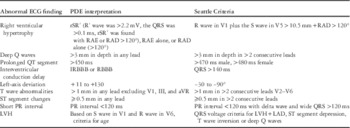
A retrospective analysis was conducted using electrocardiograms obtained between 2011 and 2014 on middle-school students aged 11–13 years who underwent pre-athletic screening as part of a community volunteer-based programme (Andraheart, www.andraheart.org). It consisted of an American Heart Association-endorsed questionnaire, which included a 14-point cardiac-specific history and physical exam. A 12-lead Physio Control Lifepak 15 model (Physio-Control, Inc., Redmond, Washington, United States of America) electrocardiogram was obtained and interpreted by a paediatric electrophysiologist. Students found to have a positive screen or abnormal standard electrocardiogram completed an additional work-up with a paediatric cardiologist, which included an additional electrocardiogram, a two-dimensional echocardiogram, or both. Echocardiograms were performed using a Philips IE33 ultrasound system (Philips Healthcare, Andover, Massachusetts, United States of America). All electrocardiograms that were referred for additional evaluation were re-interpreted by a paediatric electrophysiologist using Seattle Criteria in a blinded manner (Table 1).Reference Drezner, Ackerman and Anderson 14
Abnormal electrocardiogram (ECG) criteria by standard paediatric electrophysiologist (PDE) examination and Seattle Criteria guidelines.

aVR=augmented vector right; IRBBB=incomplete right bundle branch block; LAD=left-axis deviation; LVH=left ventricular hypertrophy; RAD=right axis deviation; RAE=right atrial enlargement; RBBB=right bundle branch block
Modified from Drezner et alReference Drezner, Ackerman and Anderson 14
Institutional Review Board approval was obtained before the initiation of this study. Written consent was obtained from a guardian for each of the patients evaluated during this study.
Data organisation analysis and graphic presentation were performed using Excel (Microsoft, Redmond, Washington, United States of America).
Results
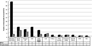
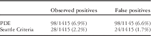
A total of 1424 students between the ages of 11 and 13 were evaluated before sports participation from 2011 to 2014. Of the 1424 students screened, 107 had an abnormal screening electrocardiogram and were referred for additional follow-up; 77 patients underwent an echocardiogram, and 24 patients received repeat electrocardiograms. No referrals for additional testing were made on the basis of pre-athletic screening history or physical examination alone. A total of 29 electrocardiograms were found to be positive based on Seattle Criteria following blinded electrocardiogram re-interpretation by a paediatric electrophysiologist; six patients were lost to follow-up. Letters were sent to these families, but, unfortunately, no contact was made; one of these patients was found to be both positive on paediatric electrophysiologist and Seattle Criteria interpretation, but no additional testing could be completed. Moreover, three electrocardiograms when re-interpreted by a paediatric electrophysiologist were read as normal and were excluded from the study. Therefore, a total of 1415 patients were included in the present study; 98 of 1415 (6.9%) electrocardiogram abnormalities were identified by standard paediatric electrophysiologist interpretation, whereas Seattle Criteria identified only 28 of 1415 (2%) abnormal electrocardiograms (Fig 1). Formal cardiology evaluation confirmed cardiac phenotypes associated with sudden cardiac arrest in four of 1415 students (0.3%): long QT syndrome (n=2), Wolff–Parkinson–White (n=1), and pulmonary hypertension (n=1). Several incidental findings were isolated, including right bundle branch block (n=1), mitral valve prolapse (n=1), and patent foramen ovale (n=1). All students with at-risk phenotypes for sudden cardiac arrest were identified by both paediatric electrophysiologist and Seattle Criteria electrocardiogram interpretation and not by history or physical examination. Overall, the false-positive electrocardiogram interpretation rate was 6.6% for paediatric electrophysiologist-interpreted electrocardiograms; this decreased to 1.7% when Seattle Criteria was applied, but true-positive electrocardiogram number remained equal (Table 2). Utilising current Centers for Medicare and Medicaid Services pricing schedules in Arizona for new patient visit (99203) and global non-congenital echocardiogram (93306) of $115 and $235 (CMS.gov), the downstream fees for follow-up of a positive screen using standard paediatric electrophysiologist versus Seattle Criteria was $24 versus $7/individual screened. 15
Bar graph representing the number of abnormal electrocardiogram (ECG) findings when interpreted by standard paediatric electrophysiologist (PDE) examination compared with the guidelines set forth by Seattle Criteria. IRBBB=incomplete right bundle branch block; IVCD=interventricular conduction delay; LAD=left-axis deviation; LVH=left ventricular hypertrophy; PAC=premature atrial contraction; RAD=right axis deviation; RAE=right atrial enlargement; RBBB=right bundle branch block; RVH=right ventricular hypertrophy; WPW=Wolff–Parkinson–White.

Observed positive versus false-positive findings based on electrocardiogram interpretation.

PDE=paediatric electrophysiologist
Comparison of the observed positive and false-positive findings based on standard paediatric electrophysiologist and Seattle Criteria interpretations
Discussion
Sudden cardiac arrest is an important cause of death in young athletes, accounting for up to 75% of fatalities during sport. The survival rate of sudden cardiac arrest has been documented as <15%.Reference Meyer, Stubbs Bm Fahrenbruch, Maeda, Harmon, Eisenber and Drenzer 5 Some studies have shown a decrease in sudden cardiac arrest with the addition of screening electrocardiograms for competitive athletes.Reference Corrado, Pelliccia and Bjornstad 9 , Reference Chandra, Bastianenen, Papadakis and Sharma 12 , Reference Ljunggvist, Jenoure and Engebretsen 13 . Previously, it was believed that competitive athletes had an increased risk for sudden cardiac arrest compared with their non-athletic counterparts; however, recent reports have shown similar risks in non-athletes and suggest that cardiac screening should also include children not involved in organised sport.Reference Maron, Friedman and Kligfield 3 , Reference Chandra, Bastianenen, Papadakis and Sharma 12 The International Olympic Committee and the European Society of Cardiology recommend inclusion of electrocardiograms in pre-athletic screening, but the benefits of large-scale electrocardiogram use remain controversial.Reference Chandra, Bastianenen, Papadakis and Sharma 12 , Reference Ljunggvist, Jenoure and Engebretsen 13
In the United States of America, the large number of youth athletes, combined with the high number of false-positive electrocardiograms that result from standard interpretation, would result in high costs and an overwhelming amount of work for current practitioners; thus, electrocardiograms are not endorsed as part of pre-athletic screening by the American Heart Association.Reference Curtis and Bourji 16 In response to these obstacles, a summit of experts in sports cardiology and sports medicine convened to develop the Seattle Criteria for electrocardiogram interpretation for pre-athletic screening. These criteria were envisioned to streamline interpretations of electrocardiograms for clinicians and cut down false-positive readings and unnecessary cardiology follow-up.Reference Drezner, Ackerman and Anderson 14 The summit itself recognised such limitations to the Seattle Criteria, including the inability of an electrocardiogram to detect all conditions predisposing to sudden cardiac arrest and being designed for athletes aged 14–35; however, criteria were formulated with the specific intention to increase electrocardiogram specificity for sudden cardiac arrest-associated cardiac phenotypes, while also being easy to learn and implement by physicians.
In the 2014 statement on electrocardiogram screening by the American College of Cardiology and American Heart Association, the ethics and logic of screening wide populations for abnormalities were plainly summarised. The authors considered the potential utility for mass screenings of the general population, rather than the athlete-specific population, and addressed important factors including the low prevalence of cardiovascular diseases related to sudden cardiac arrest in young populations, the low risk of sudden cardiac arrest in patients who have these diseases, a potential screening population of over 60 million young people, and the imperfection of the electrocardiogram as a screening tool. Although this statement ultimately recommends against including electrocardiogram screening for the United States of America population under 35, it does acknowledge a lack of efficacy in the current American Heart Association-endorsed pre-athletic screening based upon history and examination alone.Reference Meyer, Stubbs Bm Fahrenbruch, Maeda, Harmon, Eisenber and Drenzer 5
This statement also discussed the efforts of the Seattle Criteria to increase specificity of electrocardiogram screening, but recommended a prospective application of this approach to a large, general population under 35 years of age. To date, several studies have demonstrated the efficacy of the Seattle Criteria in elite athletes and young adults.Reference Berge, Gjesdal, Anderson, Solberg and Steine 17 – Reference Brosnan, La Gerche and Kalman 20 Our study is the first to look at applying the Seattle Criteria to a representative population of peri-adolescent students, less than 14 years of age, in Arizona. The results demonstrate that Seattle Criteria electrocardiogram interpretation decreases the rate of false-positive results without adverse effects on identifying true-positive electrocardiograms. In our middle-school-aged population, 6.6% of students had false-positive electrocardiograms by standard paediatric electrophysiologist interpretation; the majority of “abnormal” interpretations were right ventricular hypertrophy (rSR′ or right atrial deviation). In contrast, applying the more restrictive Seattle Criteria decreased the rate of “abnormal” electrocardiograms to 1.7% without an adverse impact on identifying students at risk for sudden cardiac arrest.
Study limitations included the project’s retrospective application of the Seattle Criteria and that all patients did not undergo an echocardiogram. Study subject demographics including ethnicity and gender were not available, but general demographic characteristics for the school district where the programme was conducted had a distribution of 56.9% Caucasian, 36.9% Hispanic, and 1.2% black. 21 It is not known whether differences in ethnic backgrounds impact electrocardiogram results; however, in the present study, the most common sudden cardiac arrest substrate identified was long QT Syndrome, which accounted for 50% (n=2) of the total true-positive electrocardiograms. Interestingly, no study subjects were found to harbour a structural heart condition associated with sudden cardiac arrest, such as hypertrophic cardiomyopathy. In addition, we did not evaluate for participation in physical activity and cannot comment on its effects.
Although the number of false-positive results are reduced with more stringent electrocardiogram guidelines, it is inevitable that not all adolescents with phenotypes concerning for sudden cardiac arrest can be identified by history and electrocardiogram studies. As the guidelines become stricter, it is possible that the number of individuals with true disease – that is, false negatives – will increase. It is recognised that electrocardiograms and follow-up echocardiography will likely fail to detect some forms of heart disease associated with sudden cardiac death, including anomalous coronary artery, regional cardiomyopathy, or forms of exercise-induced arrhythmia that require stress testing. On the other hand, inclusion of a screening electrocardiogram will likely identify more individuals at risk for sudden cardiac arrest that would be otherwise missed by careful history and physical examination alone. In addition, in this study, the true-positive rate remained the same whether standard electrocardiogram interpretation or Seattle Criteria were used. This is reassuring and demonstrates that Seattle Criteria detected all identified at-risk individuals in this cohort.
Although questions remain regarding inclusion of electrocardiograms in screening protocols for America’s youth, our study demonstrates that the number of positive electrocardiograms in an adolescent population that would require cardiology referral can be dramatically decreased by applying the Seattle Criteria. Compared with standard electrocardiogram interpretation by a paediatric electrophysiologist, this approach may decrease costs and minimise undue stress on families associated with an abnormal test result for youth screening programmes. Utilising current Centers for Medicare and Medicaid Services pricing for a paediatric cardiology visit and echocardiography for individuals with abnormal screening history and electrocardiogram, downstream costs associated with screening with standard paediatric electrocardiogram or Seattle Criteria were projected at $24 versus $7, respectively.
In conclusion, this study demonstrates that application of the Seattle Criteria for electrocardiogram interpretation significantly decreases the rate of false-positive results in a peri-adolescent Arizona population compared with standard electrocardiogram interpretation. This was completed without omitting any true-positive electrocardiogram findings. For paediatric screening programmes that include an electrocardiogram, the use of the Seattle Criteria may decrease the costs and stress associated with unnecessary paediatric cardiology referrals.
Acknowledgement
None.
Financial Support
This research received no specific grant from any funding agency, commercial, or not-for-profit sectors.
Conflicts of Interest
None.