Introduction
Tissue engineering has emerged in recent decades for treating injuries or disorders of human body tissues that are difficult to be cured by conventional methods.[Reference Griffith and Naughton1] In tissue engineering, scaffold-based strategies are popular as tissue-engineered scaffolds with well-designed properties can provide an appropriate structural and mechanical platform for supporting cell growth and tissue regeneration.[Reference Webber, Khan, Sydlik, Tang and Langer2] Nanofibrous scaffolds are of high interest owing to their resembling structure to the natural extracellular matrix (ECM), hence offering biomimetic structural cues for directing cell attachment and cell function.[Reference Stevens and George3] Electrospinning, a simple yet effective technique for fabricating nanofibers, has been widely employed and investigated for the fabrication of nanofibrous tissue-engineered scaffolds.[Reference Jiang, Carbone, Lo and Laurencin4–Reference Peng, Jin, Li, Li, Srinivasan, Ramakrishna and Chen6] However, conventional electrospun scaffolds lack desirable functionalities for desirable therapeutic effects. Through specifically adapted electrospinning, nanofibrous scaffolds can be tailored for the delivery of various cargo substances, ranging from small molecular drugs or biomacromolecules to even living mammalian cells,[Reference Zhao, Wang and Wang7] which bring the possibility to endow scaffolds with different functions. For example, electrospun scaffolds have been investigated for the delivery of small molecular anticancer drugs such as doxorubicin hydrochloride (DOX), showing potential in cancer treatment.[Reference Xu, Chen, Ma, Wang and Jing8] Electrospun scaffolds with controlled release of biomacromolecules such as growth factors have been also shown to enhance bioactivity and assist tissue regeneration.[Reference Zhao, Lu and Wang9] Different electrospinning methods such as blend electrospinning and emulsion electrospinning as well as different types of biodegradable polymers have been employed for preparing nanofibrous drug release systems. The encapsulation efficiency and release profile of the cargo molecules can be modulated by the electrospinning technique applied and the fiber composition.[Reference Wei, Hu, Li, Chen and Chen10–Reference Tiwari, Hwang, Oh, Maharjan, Chun, Kim, Joshi, Park and Kim12] As such, the excellent versatility of electrospinning motivated us to develop multifunctional scaffolds with dual release of small molecular drugs and biomacromolecules. One potential envisaged application is in cancer postoperative care where the implantation of multifunctional scaffolds onto the resected sites could inhibit cancer recurrence and simultaneously assist in situ tissue regeneration via the local delivery of anticancer drugs and biomacromolecules, respectively. However, the successful development of multifunctional scaffolds with dual release of small molecular drugs and biomacromolecules meeting the specific therapeutic requirements remains challenging, as it requires high-efficiency encapsulation of cargo molecules and good control over their release behavior in a spatiotemporal manner. It is therefore of great importance to provide a systematic study on the release behavior of the different cargo molecules as well as on influencing factors modulating their release profiles, which should provide valuable insight into the design of multifunctional scaffolds with desirable combinational therapeutic effects.
In this study, multifunctional scaffolds were fabricated by dual-source dual-power (DSDP) electrospinning for the dual release of a small molecular drug, DOX, and a popular model protein for assessing the release behavior of biomacromolecules,[Reference Li, Su, Liu, Tan, Mo and Ramakrishna13] bovine serum albumin (BSA). The two molecules are separately encapsulated in nanofibers formulated using a biodegradable polymer, poly(lactic-co-glycolic acid) (PLGA) with different monomer ratios. The scaffolds fabricated by different electrospinning techniques (either blend electrospinning or emulsion electrospinning) and consisting of PLGA with different LA:GA ratios having similar physiochemical properties but different degradation behaviors were prepared for controlling the release of different cargo molecules. The morphology and properties of the resulting DOX- and BSA-incorporated scaffolds were characterized by different techniques. The influence of scaffold properties on the encapsulation and release behavior of the molecules were systematically investigated. Overall, bicomponent nanofibrous scaffolds with high encapsulation efficiency of DOX and BSA as well as sustained release profiles were successfully developed, paving the way for combinational therapeutic effects.
Materials and methods
Medical-grade PLGA with a LA:GA molar ratio of 75:25 (PLGA 75/25; inherent viscosity: 0.6–0.8 dL/g) and PLGA with a LA:GA molar ratio of 50:50 (PLGA 50/50; inherent viscosity: 0.6–0.8 dL/g) were purchased from Lakeshore Biomaterials Inc., USA and used for fabrication of electrospun scaffolds owing to their desirable biocompatibility, biodegradability, and appropriate mechanical properties for soft tissue engineering.[Reference Nair and Laurencin14] All chemicals and reagents were purchased from Sigma-Aldrich, USA and directly used without further purification.
Electrospinning
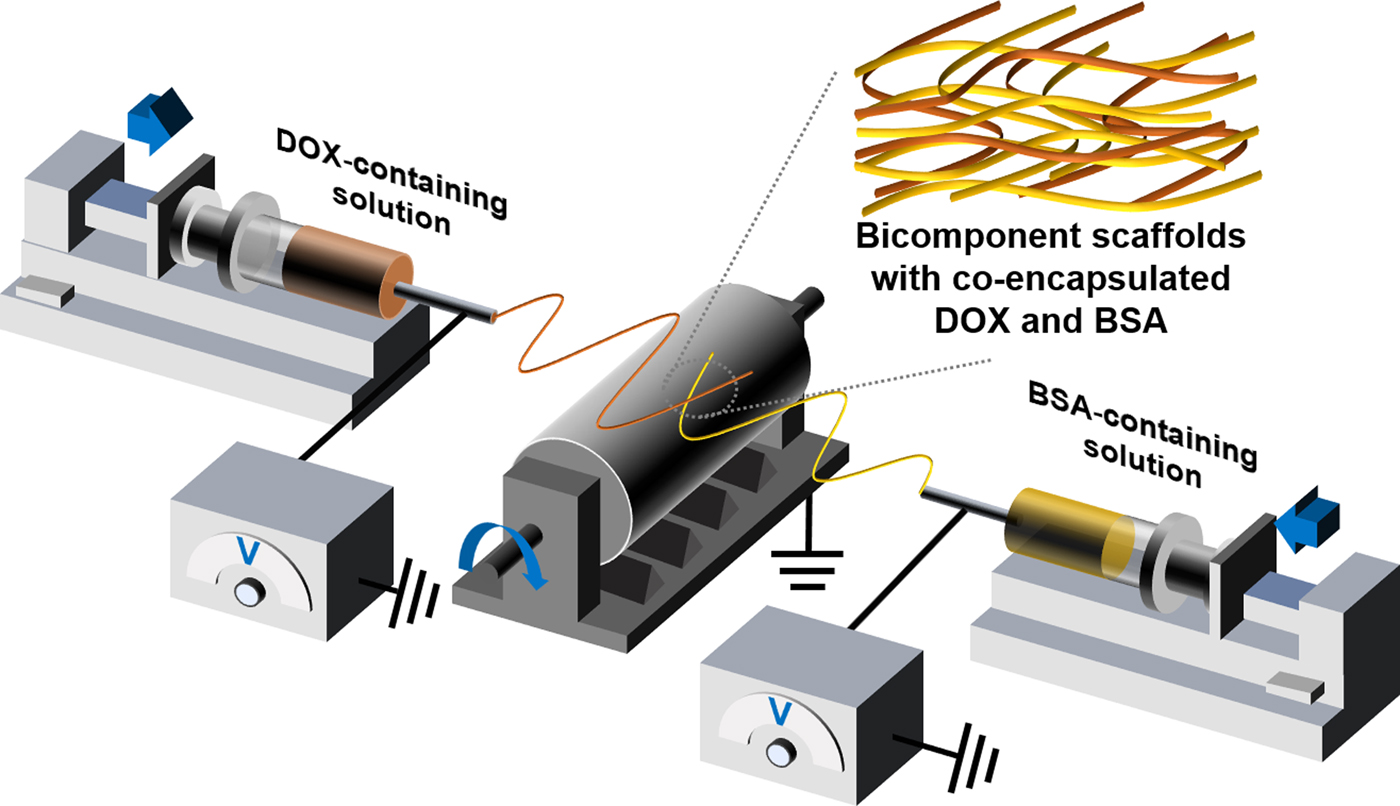
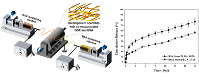
DSDP electrospinning was conducted for the fabrication of bicomponent nanofibrous scaffolds according to a previously established method,[Reference Wang and Wang15] where electrospun PLGA 75/25 fibers and PLGA 50/50 fibers were designed as delivery vehicles for DOX and BSA, respectively. The sources for the separate electrospinning of a BSA-containing solution and a DOX-containing solution were placed at the two sides of a grounded collecting drum. During electrospinning, DOX-encapsulated PLGA 75/25 fibers and BSA-encapsulated PLGA 50/50 fibers were concurrently and homogeneously deposited onto the collecting drum with constant rotation and reciprocated motion, therefore resulting in bicomponent scaffolds with intermeshed fibers formed from two distinct sources (Fig. 1). Both blend electrospinning and emulsion electrospinning were performed for preparing the DOX-encapsulated PLGA 75/25 fibers and BSA-encapsulated PLGA 50/50 fibers. For the blend electrospinning method, the cargo molecules (i.e., BSA or DOX) were directly suspended in a polymer solution (either a 15% w/v PLGA 75/25 solution in trifluoroethanol or a 15% w/v PLGA 50/50 solution in trifluoroethanol), resulting in either PLGA 75/25 scaffolds with incorporated 0.5 wt% DOX or the PLGA 50/50 scaffolds with incorporated 0.005 wt% BSA. As for the emulsion electrospinning method, a water-in-oil emulsion was first prepared according to an established method.[Reference Wang, Wang and Wang16] Briefly, stable emulsions were prepared by mixing a water-phased solution (aqueous solution of DOX or BSA) and an oil-phased solution (polymer solution of PLGA 75/25 or PLGA 50/50 at a polymer concentration of 15% w/v) at a volume ratio of 1:10 under vigorous mechanical stirring for 10 min with an addition of 20 µL of surfactant span 80. The PLGA 75/25 scaffolds with incorporated 0.5 wt% DOX and the PLGA 50/50 scaffolds with incorporated 0.005 wt% BSA were formed by the electrospinning of different emulsions. The same processing parameters (i.e., a solution feeding rate of 3.0 mL/h, an applied voltage of 15 kV and a working distance of 15 cm) were employed in both blend and emulsion electrospinning for scaffold manufacturing. The as-spun scaffolds were freeze-dried for 3 days for completely removing the residual solvents.
A schematic illustration of DSDP electrospinning for the fabrication of bicomponent nanofibrous scaffolds with co-encapsulated DOX and BSA.

Physiochemical characterization
The morphologies of DOX-incorporated PLGA 75/25 scaffolds and BSA-incorporated PLGA 50/50 scaffolds fabricated by blend electrospinning and emulsion electrospinning were separately visualized under a scanning electron microscope (SEM, Leo 1530, UK). Electrospun specimens for SEM observation were coated by a thin layer of gold by sputtering. Average fiber diameter of each scaffold was analyzed using an image analysis software, ImageJ at three separated experiments (N = 100). Different types of fibers formed by different electrospinning techniques were also collected onto a copper grid and then visualized under a transmission electron microscope (TEM, Philips CM200, the Netherlands) for comparing the difference in the metastructure between the blend electrospun fibers and emulsion electrospun fibers.
Static water contact angles on the DOX-incorporated PLGA 75/25 scaffolds and BSA-incorporated PLGA 50/50 scaffolds fabricated by blend electrospinning and emulsion electrospinning were measured on a contact angle measurement platform (SL200B, Shanghai Solon Tech Inc. Ltd, China) for assessing the wettability of each scaffold. In total, 5.0 µL water droplet was placed onto the flat specimen. The shape of water droplets stabilized on the surface of the specimen was recorded and the static water contact angles of each type of scaffold were analyzed (N = 6).
As for evaluating the in vitro degradation behavior of DOX-incorporated PLGA 75/25 scaffolds and BSA-incorporated PLGA 50/50 scaffolds formed by different electrospinning techniques, in vitro degradation tests were conducted. Pre-weighed specimens of different types of scaffolds were immersed in the immersion medium of 5 mL phosphate buffered saline (PBS, pH 7.4) with supplemented 0.02% w/v sodium azide for antibacterial purpose and stored in a shaking humid incubator at 37 °C. The immersion medium was refreshed every 2 days. The weight loss of specimens after immersion for 1, 2, and 3 weeks was statistically analyzed (N = 6). The morphology of the specimens upon the 3-week immersion treatment was examined by SEM.
In vitro release test
In order to study the release behavior of DOX from PLGA 75/25 scaffolds made by blend electrospinning and emulsion electrospinning, pre-weighed DOX-incorporated scaffolds were immersed in PBS (pH 7.4) as the immersion medium in a shaking humid incubator at 37 °C. The immersion medium was collected and refreshed at preset time points. The concentrations of the released DOX in the medium collected at specific time points were determined according to the optical absorbance of the medium at the wavelength of 483.5 nm using a UV-vis spectrophotometer (UV-2600, Shimadzu, Japan) and the calibration curve was obtained from the standard DOX solutions with gradient concentrations in PBS (pH 7.4). The curves demonstrating the cumulative release of DOX from different types of PLGA 75/25 scaffolds over an immersion period up to 21 days were plotted according to three separated experiments. The encapsulation efficiency of DOX was calculated through measuring the total content of DOX with respect to the theoretical value. To measure the total content of DOX encapsulated, pre-weighed DOX-incorporated specimens were completely dissolved in 0.05 mol/L Tri-HCl buffer solution (pH 8.6) containing 50 µg/mL of proteinase K at 37 °C. The absorbance of the extracted solution at the wavelength of 483.5 nm was monitored for calculating the total content of encapsulated DOX (N = 6).
In order to assess the release behavior of BSA from blend electrospun PLGA 50/50 scaffolds and emulsion electrospun PLGA 50/50 scaffolds, similar immersion treatments were conducted using a 0.02% w/v sodium azide supplemented PBS (pH 7.4) as the immersion medium. Within an immersion period of 21 days, the immersion medium was collected at preset time points and the contents of BSA in the immersion medium were determined using a BCA protein assay kit (Thermo Fisher Scientific Inc., USA) according to the manufacture's protocol, where BSA concentration was calculated according to the optical density (O.D.) at a wavelength of 562 nm measured by using a microplate reader (UVM 340, Asys HiTech GmbH, Australia) and the reference curve of the O.D. obtained from a standard BSA solution with gradient BSA concentration. The curves showing the cumulative release of BSA from different types of PLGA 50/50 scaffolds were subsequently plotted. Likewise, the total content of BSA encapsulated was quantified by BCA protein assay after completely dissolving pre-weighed BSA-incorporated scaffolds in dimethylsulfoxide, followed by the extraction of BSA into a 0.05 M NaOH aqueous solution. The encapsulation efficiency of BSA in different types of PLGA 50/50 scaffolds was hence calculated according to the total contents of BSA encapsulated with respect to the theoretical value (N = 6).
Statistical analysis
The statistical results are represented as mean ± standard deviation (SD). Statistical analyses are performed using analysis of variance with a Student's t-test. Results are regarded as having no significant difference (n.s.) with P > 0.05 or significantly different with *P < 0.05 and **P < 0.01.
Results and discussion
Morphology, structure, and wettability
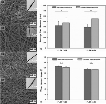
In this study, PLGA 75/25 scaffolds prepared by blend electrospinning and emulsion electrospinning were used for the delivery of DOX, while PLGA 50/50 scaffolds formed by blend electrospinning and emulsion electrospinning were employed for the delivery of BSA. To investigate the morphologies of these four types of scaffolds formulated using different polymers and different electrospinning techniques, SEM examinations were performed. All the four types of scaffolds show a ECM-like nanofibrous architecture with uniform and randomly oriented nanofibers [Figs. 2(a)–2(d)], which has been proven favorable to cell attachment and tissue regeneration.[Reference Stevens and George3] Since the identical processing parameters are applied in the different electrospinning processes, the overall nanofibrous architecture among different types of scaffolds exhibits limited differences regardless of different polymers (PLGA 75/25 or PLGA 50/50) and different electrospinning techniques (blend electrospinning or emulsion electrospinning). The average fiber diameters of each type of scaffold were analyzed, demonstrating slightly thinner fibers of the blend electrospun scaffolds (848.65 ± 156.63 nm for the DOX-incorporated PLGA 75/25 scaffolds and 790.10 ± 123.79 nm for the BSA-incorporated PLGA 50/50 scaffolds) than those of the scaffolds fabricated by emulsion electrospinning (958.18 ± 180.42 nm for the DOX-incorporated PLGA 75/25 scaffolds and 1109.83 ± 218.05 nm for the BSA-incorporated PLGA 50/50 scaffolds) [Fig. 2(e)], which is supposed to be governed by the increasing surface tension of the emulsion with the presence of water.[Reference Fridrikh, Yu, Brenner and Rutledge17] However, there is no significant statistical differences in the average fiber diameters of the scaffolds formed using the same electrospinning technique but different LA:GA ratios according to the similar inherent viscosities of PLGA 75/25 and PLGA 50/50. However, the metastructures of fibers fabricated by blend electrospinning and emulsion electrospinning display obvious difference (insets of Fig. 2). While the fibers formed by blend electrospinning exhibit monolithic metastructure [insets of Figs. 2a and 2(c)], clear discontinuous core–shell structures are formed in the emulsion electrospun fibers [insets of Figs. 2(b) and 2(d)] due to the Rayleigh instability of the emulsion under electrostatic stretch.[Reference Wang, Wang and Wang16] While the cargo molecules in the blend electrospun fibers with monolithic metastructure will be embedded in the polymer matrix, the core–shell structures of the emulsion electrospun fibers shall provide appropriate reservoirs for the encapsulation of cargo molecules. The wettability of scaffold determines the water permeability, subsequently affecting the hydrolysis-induced degradation behavior of scaffold and the diffusion-controlled release behavior of the cargo molecule.[Reference Cui, Li, Zhou and Weng18] To investigate the wettability of nanofibrous scaffolds formed by using different polymers and different electrospinning techniques, water contact angles on the four types of scaffolds were measured, demonstrating all hydrophobic property [Fig. 2(f)]. Slightly superior wettability is present for the scaffolds formed by PLGA 50/50 to those of PLGA 75/25 scaffolds owing to small variance in the chemical nature between LA and GA. The wettability of scaffolds formed by using the same polymer but different electrospinning methods shows no statistically significant difference.
Morphology, fiber metastructure (insets), and wettability of DOX-incorporated scaffolds and BSA-incorporated scaffolds prepared by different electrospinning techniques. (a) The morphology and fiber metastructure (insets) of DOX-incorporated PLGA 75/25 scaffolds formed by blend electrospinning. (b) The morphology and fiber metastructure (insets) of DOX-incorporated PLGA 75/25 scaffolds formed by emulsion electrospinning. (c) The morphology and fiber metastructure (insets) of BSA-incorporated PLGA 50/50 scaffolds formed by blend electrospinning. (d) The morphology and fiber metastructure (insets) of BSA-incorporated PLGA 50/50 scaffolds formed by emulsion electrospinning. (e) Average fiber diameters of different types of electrospun scaffolds. The error bars represent the SD of the average fiber diameters (N = 100) for different types of scaffolds. (f) Water contact angles of different types of electrospun scaffolds. The error bars represent the SD of the water contact angles (N = 6) for different types of scaffolds.

In vitro degradation behavior
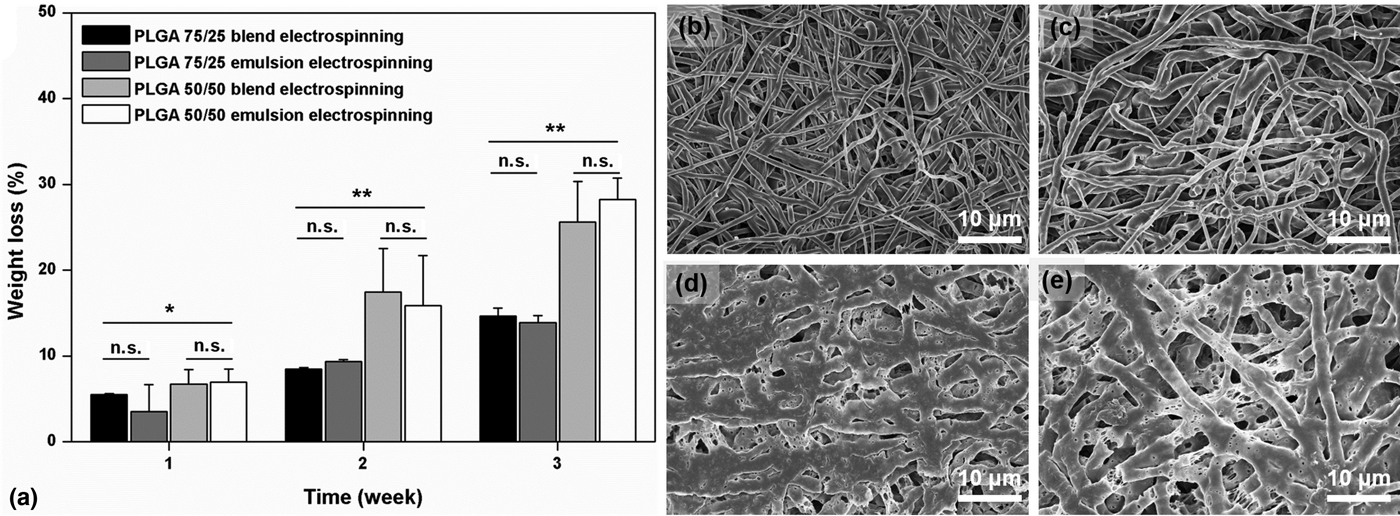
The degradation of the polymeric vehicles has been widely proven to significantly affect the release behavior of the cargo molecule.[Reference Szentivanyi, Chakradeo, Zernetsch and Glasmacher19] To evaluate the in vitro degradation behavior of DOX-incorporated PLGA 75/25 scaffolds and BSA-incorporated PLGA 50/50 scaffolds formed by different electrospinning approaches, their weight losses over time under the 37 °C PBS (pH 7.4) immersion treatments were analyzed [Fig. 3(a)]. While significantly different degradation rates exist between the PLGA 75/25 scaffolds and the PLGA 50/50 scaffolds, there is no statistically significant difference in the degradation behavior between the scaffolds formed using the same polymer yet different electrospinning techniques. PLGA degradation results from the hydrolysis of the ester bonds and follows a bulk erosion mechanism, generating fragments of oligomers and monomers with limited cytotoxicity, making it a desirable candidate biomaterial for tissue engineering.[Reference Nair and Laurencin14] The degradation rate of PLGA is affected by many factors such as molecular weight, LA:GA ratio, surface property, and the structure of PLGA scaffolds.[Reference Nair and Laurencin14] In this study, the scaffolds formed by using the same polymer (i.e., PLGA 75/25 or PLGA 50/50) yet different electrospinning techniques display similar nanofibrous architectures and surface wettability, therefore leading to similar degradation rates. However, PLGA 75/25 scaffolds and PLGA 50/50 scaffolds consisting of PLGA with different LA:GA ratios exhibit different wettabilities and different capabilities to resist hydrolysis, hence resulting in distinct degradation behaviors. The slightly better wettability of the PLGA 50/50 scaffolds favors the water permeability and subsequently the hydrolysis of the ester bonds. Also, it has been verified that the hydrolytic property of PLGA of amorphous nature is affected by the LA/GA arrangement during co-polymerization, where higher stability is present in the co-polymer with a higher ratio of LA compared with the intermediate co-polymers (i.e., PLGA 50/50).[Reference Nair and Laurencin14] Consequently, the PLGA 50/50 scaffolds exhibit faster degradation rates than those of the PLGA 75/25 scaffolds. The relatively rapid degradation of the PLGA 50/50 scaffolds can be also confirmed by the morphological changes among different types of scaffold upon the 3-week immersion treatments. The PLGA 75/25 scaffolds prepared by blend electrospinning [Fig. 3(b)] and emulsion electrospinning [Fig. 3(c)] upon immersion present slight fiber swelling indicated by the increasing fiber diameters while there is no significant difference between them. Obvious swelling and erosion of fibers as well as the formation of nanopores on the fiber surface are observed in the PLGA 50/50 scaffolds upon immersion [Figs. 3(d) and 3(e)], demonstrating their faster degradation rate. Importantly, for the emulsion electrospun PLGA 50/50 scaffolds, fiber erosion exposes the encapsulated cores once inside the fibers, offering possible routes for the release of the encapsulated cargo molecules.
In vitro degradation behavior of different types of scaffolds. (a) Weight loss percentages of different types of scaffolds for different PBS immersion time periods. The error bars represent the SD of the weight losses (N = 6) for different types of scaffolds. (b) The morphology of DOX-incorporated PLGA 75/25 scaffolds fabricated by blend electrospinning after the 3-week immersion treatment. (c) The morphology of DOX-incorporated PLGA 75/25 scaffolds fabricated by emulsion electrospinning after the 3-week immersion treatment. (d) The morphology of BSA-incorporated PLGA 50/50 scaffolds fabricated by blend electrospinning after the 3-week immersion treatment. (e) The morphology of BSA-incorporated PLGA 50/50 scaffolds fabricated by emulsion electrospinning after the 3-week immersion treatment.

Encapsulation efficiency and in vitro release behavior of DOX and BSA
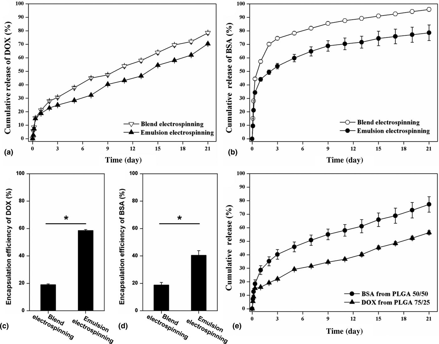
To investigate the influence of different polymers and different fiber structures on the encapsulation and delivery of different types of cargo molecules, the encapsulation efficiency and release behavior of DOX and BSA from different types of nanofibrous scaffolds were assessed. The release of DOX from blend electrospun scaffolds and emulsion electrospun scaffolds exhibits a two-stage characteristic, including an initially rapid release stage and a following sustained release stage [Fig. 4(a)], of which release kinetics follow a diffusion-controlled mechanism and a diffusion-and-erosion-controlled mechanism, respectively.[Reference Versypt, Pack and Braatz20] In the initial release stage (the beginning 24 h within the release period of 21 days), small amounts of DOX are rapidly released with the polymer fiber being hydrated and the subsequently rapid diffusion of DOX as some DOX molecules are dispersed near the surface of electrospun fibers due to the electrostatic repulsion effects during electrospinning.[Reference Chakraborty, Liao, Adler and Leong21] The sustained and steady DOX release is shown in the scaffolds prepared by blend electrospinning and emulsion electrospinning in the following release periods with the gradual degradation of polymer matrix, demonstrating desirable release profiles for potentially cancer treatment.[Reference Allen and Cullis22] As for the BSA-incorporated PLGA 50/50 scaffolds made by blend electrospinning and emulsion electrospinning, the release profiles of BSA exhibit the similar two-stage phenomena: initially rapid release at the beginning 24 h and subsequently sustained release within the following release periods [Fig. 4(b)]. The cumulative release percentages of BSA from the PLGA 50/50 scaffolds within the 1st day (over 40% for both blend electrospun scaffolds and emulsion electrospun scaffolds) are higher than those of the DOX from the PLGA 75/25 scaffolds (approximately 15%), which shall be attributed to the eases of water permeability and surface swelling for the PLGA 50/50 fibers compared with the PLGA 75/25 fibers revealed by the wettability assays and the degradation tests and hence the readily diffusion of the cargo molecules. Furthermore, compared with the severe burst release of BSA from the blend electrospun PLGA 50/50 scaffolds, the percentage of BSA released from the emulsion electrospun scaffolds in the initial stage (within the initial 1 day) is significantly reduced, which shall arise from the extending transport distance for the encapsulated BSA in the emulsion electrospun PLGA 50/50 fibers those possess larger average fiber diameters. Overall, the release of BSA from the PLGA 50/50 scaffolds made by emulsion electrospinning shows more sustained release behavior than that from the blend electrospun PLGA 50/50 scaffolds, therefore holding superior promise in stimulating cell functions in a high-efficiency and constant manner.[Reference Reed and Wu23] More importantly, it has been also found that emulsion electrospinning has a significant advantage in the cargo molecular encapsulation. While the scaffolds prepared by blend electrospinning show low encapsulation efficiency of DOX [Fig. 4(c)] and BSA [Fig. 4(d)], higher encapsulation efficiency of DOX [Fig. 4(c)] and BSA [Fig. 4(d)] has been achieved by emulsion electrospinning.
In vitro release behavior and encapsulation efficiency of DOX and BSA in different types of electrospun scaffolds. (a) In vitro release behavior of DOX from PLGA 75/25 scaffolds fabricated by blend electrospinning and emulsion electrospinning. The error bars represent the SD of the cumulative release percentages of DOX (N = 6) from PLGA 75/25 scaffolds formed by different electrospinning techniques. (b) In vitro release behavior of BSA from PLGA 50/50 scaffolds fabricated by blend electrospinning and emulsion electrospinning. The error bars represent the SD of the cumulative release percentages of BSA (N = 6) from PLGA 50/50 scaffolds formed by different electrospinning techniques. (c) Encapsulation efficiency of DOX in PLGA 75/25 scaffolds fabricated by blend electrospinning and emulsion electrospinning. The error bars represent the SD of the encapsulation efficiency of DOX (N = 6) in PLGA 75/25 scaffolds formed by different electrospinning techniques. (d) Encapsulation efficiency of BSA in PLGA 50/50 scaffolds fabricated by blend electrospinning and emulsion electrospinning. The error bars represent the SD of the encapsulation efficiency of DOX (N = 6) in PLGA 50/50 scaffolds formed by different electrospinning techniques. (e) In vitro dual release behavior of DOX and BSA from bicomponent scaffolds fabricated by DSDP emulsion electrospinning. The error bars represent the SD of the encapsulation efficiency of the cumulative release percentages of DOX (N = 6) and BSA (N = 6) from different nanofibers of the bicomponent scaffolds.

Owing to the superior performance of the emulsion electrospun scaffolds in the encapsulation and controlled release of cargo molecules, DSDP emulsion electrospinning was subsequently performed for preparing bicomponent nanofibrous scaffolds with dual release of DOX and BSA, where DOX and BSA were encapsulated in the emulsion electrospun PLGA 75/25 and PLGA 50/50 fibers, respectively. The in vitro dual release profiles of DOX and BSA were subsequently investigated, demonstrating both sustained release behavior within the whole 21-day release period [Fig. 4(e)]. The initially burst release behavior of BSA from the bicomponent scaffolds (cumulative release percentage of 28.66 ± 3.23% within the 1st day) is mitigated compared with that from the neat emulsion electrospun PLGA 50/50 scaffolds (cumulative release percentage of 44.41 ± 1.40% within the 1st day). By contrast, the release of DOX in the initial release periods from the bicomponent scaffolds (cumulative release percentage of 15.98 ± 0.51% within the 1st day) is slightly higher than that from the neat emulsion electrospun PLGA 75/25 scaffolds (cumulative release percentage of 13.16 ± 0.11% within the 1st day). The burst release of cargo molecules mainly results from the rapid diffusion of molecules located near the surface of electrospun fibers, where the swelling of polymeric fibers is one of key factors in determining the release kinetics of cargo molecules in the initial stage.[Reference Biondi, Ungaro, Quaglia and Netti24] Due to the more hydrophobic nature of PLGA 75/25 than that of PLGA 50/50, the bicomponent scaffolds are harder to swell compared with the PLGA 50/50 scaffolds yet easier to swell compared with the PLGA 75/25 scaffolds, therefore showing a slower BSA release and a slightly faster DOX release compared with those from the single-component scaffolds. For the overall release profiles of DOX and BSA from the bicomponent scaffolds, the release rate of DOX from the bicomponent scaffolds is obviously slower than that of BSA, which is mainly determined by the distinct degradation rates between the PLGA 75/25 and PLGA 50/50 fibers. The characteristics of the bicomponent scaffolds enabling sequential release of biomacromolecules and small molecular drugs owing to distinct degradation properties of different polymeric fibers hold promise in specific therapies, such as in postoperative cancer patients who need combinational care to assist in situ tissue regeneration within a relatively short-term period by the locally rapid release of bioactive biomacromolecules and to avoid cancer recurrence normally in a relatively long-term period by the locally sustained release of anticancer drugs.
Conclusions
In summary, nanofibrous scaffolds formed using different polymers (i.e., PLGA 75/25 and PLGA 50/50) and different electrospinning techniques (i.e., blend electrospinning and emulsion electrospinning) were investigated for the simultaneous delivery of anticancer drugs and biomacromolecules. It has been verified that the encapsulation efficiency and in vitro release behavior were affected by the fabrication methods and scaffold properties. Dual release of anticancer drugs and biomacromolecules with desirable encapsulation efficiency were achieved by the bicomponent nanofibrous scaffolds fabricated by DSDP emulsion electrospinning, presenting distinct release behaviors. The bicomponent nanofibrous scaffolds with dual release of small molecular drugs and biomacromolecules in controlled and sustained manners have potential in a variety of applications, such as combinational cancer postoperative care to inhibit cancer recurrence and simultaneously to assist in situ tissue regeneration by the appropriate administration of different therapeutic agents.
Acknowledgments
We would like to acknowledge the financial support from Hong Kong Research Grants Council General Research Funds (HKU717713E, 17201017), National Key R&D Program of China (2017YFA0701303), the China Postdoctoral Science Foundation (2017M612780), the Fundamental Research Program of Shenzhen (JCYJ20170818161757684, JCYJ20170307164610282), and a grant from HKU (201211159123).