The cow udder consists of four quarters, and most cows have two supramammary lymph nodes in each mammary half. Supramammary lymph nodes play an important role in lactation, especially in the fight against bacterial infections of the udder. In the B-mode ultrasonographic examination of the udder, supramammary lymph nodes appear to have an echogenic capsule, hypoechoic cortical region and hyperechoic inner medulla/hilus at the basal surface of the udder under the perineal region (Kofler et al., Reference Kofler, Schilcher and Buchner1998). However, the B-mode ultrasonographic appearance and measurements of the supramammary lymph nodes change according to the infection status of the udder. Khoramian et al. (Reference Khoramian, Vajhi, Ghasemzadeh-Nava, Ahrari-Khafi and Bahonar2015) reported that the length of the supramammary lymph nodes ranged between 5.77 and 12.90 cm, the width ranged between 2.07 and 7.41 cm, and the size of the nodes varied according to the infection status of the mammary quarters.
Mastitis causes lymphocyte proliferation and leads to morphological changes in the supramammary lymph nodes of cows. Khoramian et al. (Reference Khoramian, Vajhi, Ghasemzadeh-Nava, Ahrari-Khafi and Bahonar2015) reported that grey-scale ultrasonography was useful for detecting morphological changes in supramammary lymph nodes. However, there are few publications that report the use of Doppler ultrasonography in the examination of supramammary lymph nodes. Colour Doppler imaging is the most commonly used form of Doppler ultrasonography in radiological applications. Colour Doppler ultrasound provides qualitative information about the blood flow and perfusion of the tissues. In the colour Doppler technique, the direction of the blood flow in the vessels and tissues is shown as red and blue, which represent the blood moving towards and away from the probe, respectively. Accordingly, colour Doppler ultrasonography provides information on the vascular distribution category, vascularization intensity and morphological appearance of the vascularization of the target organ or tissue, such as lymph nodes. In this way, information about pathological changes in the target organ or tissue can be obtained (Mrdjen, Reference Mrdjen2013). The aim of this study was to investigate the relation between the B-Mode, Doppler ultrasonographic features of supramammary lymph nodes and mastitis in lactating cows.
Materials and methods
Animals
The animals were selected from 1000 animals brought to the Firat University Animal Hospital (Elazig, Turkey, 38°35′51.55″N, 39°16′53.28″E) between 01.05.2018 and 01.09.2018 for examination. A total of 102 cows (of various breeds) with body weights of 450–500 kg that were 3–5 years of age were included in this study. The body condition scores of the animals ranged between 3 and 3.5 points. The animals included in the study were divided into three groups:
Group 1 (n = 27): Cows whose mammary quarters were all CMT-negative.
Group 2 (n = 43): Cows with at least one CMT-positive quarter.
Group 3 (n = 32): Cows with clinical mastitis (acute, chronic or subacute mastitis) in at least one quarter.
Under the perineal region of the animals and on the dorsal surface of the udder, the B-Mode and colour Doppler modes were activated by a PW command via a 2.5-inch convex probe (Mindray DC-T6 Colour Doppler Ultrasound System; Shenzhen, China). After detecting the supramammary lymph node, the insonation angle was set to 60° for colour Doppler examination. Measurements were made on different days and post-milking.
B-mode ultrasonographic examination
This technique was performed as described by Khoramian et al. (Reference Khoramian, Vajhi, Ghasemzadeh-Nava, Ahrari-Khafi and Bahonar2015).
Colour Doppler ultrasonographic examination
This method was performed as described by Mrdjen (Reference Mrdjen2013). Accordingly, the vascular distribution category, vascularization intensity and morphological appearance of vascularization in the supramammary lymph nodes were evaluated. According to the localization of vascularization in the supramammary lymph node, the vascular distribution categories were determined as central, capsular, mixed, avascular, mosaic, peripheral or uncertain vascularization. According to the vascularization intensity, vascularization was classified from low to high intensity, as Type 1, Type 2, Type 3 or Type 4. Regarding the morphological appearance of vascularization, vascularization in the supramammary lymph node was divided into three groups: linear, branching and distortion.
Statistical analysis
The length, width, perimeter and cortex thickness measurements of the lymph nodes on B-mode ultrasonography were compared between the groups. In addition, the frequency (number and %) of echogenicity, calcification, border irregularity, cyst, honeycomb appearance, mass, vascular morphological appearance, vascular density and the vascular distribution category was determined. The effect of the study groups on the length, width, circumference and cortex thickness, was determined using the one-way analysis of variance (ANOVA) test. For the variables with significant variance analysis result, the Duncan post-hoc test was used for the multiple group comparisons. The χ 2 test in multiple orders (R × C) was used to determine the effect of groups on the frequency of echogenicity, calcification, border irregularity, cyst, honeycomb appearance, mass, vascular morphological appearance, vascular density and vascular distribution category. The Pearson correlation test was used to determine the correlation coefficients and the statistical significance level between some numerical variables. All statistical analyses were performed using the SPSS 11.5 version.
Results
When the B-mode ultrasonographic measurements of the supramammary lymph nodes were compared, there were statistically significant differences between the groups in terms of the length, width, circumference and cortex thickness of the lymph nodes. The width (4.33 ± 0.20 cm, P < 0.05), length (6.55 ± 0.23 cm, P < 0.01), circumference (17.59 ± 0.63 cm, P < 0.01) and cortex thickness (0.22 ± 0.03 cm, P < 0.01) of the lymph nodes were highest in the clinical mastitis group (data shown in Supplementary Table S1).
When the B-mode ultrasonographic appearance of the supramammary lymph nodes was compared between the groups, there were statistically significant differences between the groups in terms of the echogenicity, calcification and border irregularity of the lymph nodes. In the clinical mastitis group, hyperechogenicity (53.1%, P < 0.01), calcification (40.6%, P < 0.01) and irregular borders (12.5%, P < 0.05) in the lymph nodes were more frequent than in the other groups (data shown in Supplementary Table S2).
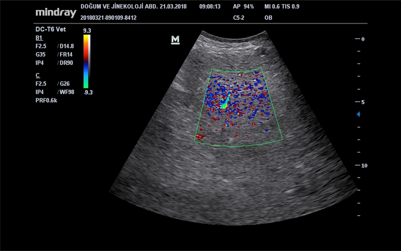
Colour Doppler ultrasonography measurements of the supramammary lymph nodes revealed that distortion-type vascular morphology (68%, P < 0.01), the rate of type 4 vascular densities (40.6%, P < 0.01) and the incidence of mixed-type vascular distributions were highest in the clinical mastitis group (37.5%, P < 0.01) (Fig. 1). Colour Doppler ultrasonography demonstrated that the frequency of avascularity in supramammary lymph nodes was highest in the CMT-negative group (55.6%, P < 0.01) (Table 1).
Colour Doppler ultrasonography image reveals mosaic type perfusion in supramammary lymph nodes with clinical mastitis.

Comparison of colour Doppler ultrasound measurements of the supramammary lymph nodes between the groups

**P < 0.01.
Discussion
B-Mode and colour Doppler ultrasonography are widely used as non-invasive methods to monitor lymph node pathologies in humans (Lee et al., Reference Lee, Choi, Baek and Lim2002). Additionally, colour Doppler and power Doppler ultrasonography are frequently used in the differential diagnosis of breast tumours in women (Lee et al., Reference Lee, Choi, Baek and Lim2002). In particular, power Doppler ultrasonography has been reported to be more sensitive to vascular signals in breast masses than colour Doppler ultrasonography. However, it is emphasized that colour Doppler ultrasonography is insufficient to determine the malignancy of breast masses. In animals, colour Doppler ultrasonography is generally used in cardiac and vascular assessments (King, Reference King2006). In addition, colour Doppler ultrasonography has been reported to be useful for examining blood flow to genital organs in the whole reproductive period (Dzięcioł et al., Reference Dzięcioł, Scholbach, Stańczyk, Ostrowska, Kinda, Woźniak, Atamaniuk, Skrzypczak, Niżański, Wieczorek, Scholbach and Kiełbowicz2014; Bollwein et al., Reference Bollwein, Heppelmann and Lüttgenau2016). To the best of our knowledge, there are no previously-published studies in the literature about the evaluation of supramammary lymph nodes in cows using the colour Doppler ultrasonography methods. Fragkou et al. (Reference Fragkou, Boscos and Fthenakis2014) reported that Doppler ultrasonography has not yet been used for evaluation of the udder in sheep.
On the other hand, ultrasonographic examination of the lymph nodes was reported to be highly important in terms of prognosis prediction and the treatment choice decision during the evaluation of some tumour metastases in dogs (King, Reference King2006). Perre et al. (Reference Perre, Koot, de Hooge, Weits and Leguit1996) could not diagnose 50% of malignant lymph nodes correctly when they considered the lymph nodes that showed peripheral blood flow (using colour Doppler ultrasonography) as malignant and the lymph nodes showing central blood flow as benign. Malignancy and infections cause neovascularization in tissues and organs. In Doppler ultrasonography, information about the indicators of neo-vascularization, such as luminal irregularities, scattered branching patterns, arterio-venous shunts and vascular structures that penetrate from the edge of the lesion to the centre, can be visualized (Milz et al., Reference Milz, Lienemann, Kessler and Reiser2001). In the present study, we found that supramammary lymph nodes had the highest length, height, circumference and cortex thickness in the lactating cows included in the clinical mastitis group. In addition, hyperechogenicity and calcification of the lymph nodes were more frequent in the clinical mastitis group and the borders of the lymph nodes were more irregular. The study groups had different results in the colour Doppler ultrasonography measurements. In the clinical mastitis group, the distortion-type vascular morphological appearance and the mixed-type vascular distribution category occurred more frequently and the type 4 vascular density rates were higher than in the other two groups. In addition, colour Doppler ultrasonography revealed avascularity of the supramammary lymph nodes most frequently in the CMT-negative group. There was also a significant negative correlation between the cortical thickness and calcification density of the supramammary lymph nodes and the vascular distribution category in the clinical mastitis group.
In conclusion, in this study, we found that the use of B-mode and colour Doppler ultrasonographic methods to determine the current mastitis condition in lactating cows is useful and also demonstrated that these non-invasive methods can provide essential information about the intranodal structure of the supramammary lymph nodes in healthy cows and those with clinical mastitis. The diagnostic potential of the technique remains to be determined.
Supplementary material
The supplementary material for this article can be found at https://doi.org/10.1017/S0022029919000530