Introduction
The order Acipenseriformes includes the members of two living families, Acipenseridae (sturgeons, ~25 extant species; Grande and Hilton, Reference Grande and Hilton2006; Hilton et al., Reference Hilton, Grande and Bemis2011) and Polyodontidae (paddlefishes, two extant species; see Grande and Bemis, Reference Grande and Bemis1991; Grande et al., Reference Grande, Jin, Yabumoto and Bemis2002), as well as two extinct families: †Chondrosteidae (two or three monotypic genera, Jurassic; see Hilton and Forey, Reference Hilton and Forey2009) and †Peipiaosteidae (five genera and six or seven species; see Grande and Bemis, Reference Grande, Bemis, Stiassny, Parenti and Johnson1996; Jin, Reference Jin1999). There is great interest in the evolutionary history of this group because it is the most species-rich group of basal Actinopterygii (i.e., non-neopterygian actinopterygians) among the extant diversity of ray-finned fishes. Further, Acipenseriformes has a relatively long fossil record, with known taxa dating back to the Early Jurassic. However, there is still relatively little information available for several fossil taxa, particularly within the family †Peipiaosteidae.
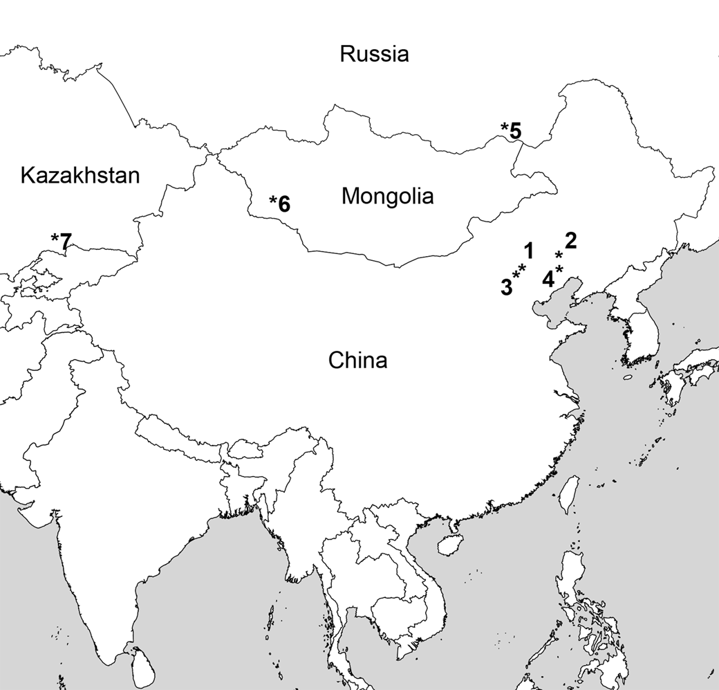
The family †Peipiaosteidae was first diagnosed by Liu and Zhou (Reference Liu and Zhou1965) with their description of †Peipiaosteus pani, at the time the only member of the family. The diagnosis offered for the family was generalized for Chondrostei (sensu Grande and Bemis, Reference Grande, Bemis, Stiassny, Parenti and Johnson1996) but did include several characters not seen in other chondrostean fishes, such as the absence of caudal fulcra and rhombic caudal scales. Grande and Bemis (Reference Grande, Bemis, Stiassny, Parenti and Johnson1996) provided a detailed osteological description of the species †P. pani on the basis of newly prepared and newly examined specimens. Jin (Reference Jin1999) provided further information on all species of fossil acipenseriforms found in the middle and late Mesozoic deposits of China (Haifanggou and Yixian formations), including three genera of †peipiaosteids. In the years between the studies of Liu and Zhou (Reference Liu and Zhou1965) and Grande and Bemis (Reference Grande, Bemis, Stiassny, Parenti and Johnson1996), several taxa were placed within the family (Jin, Reference Jin, Sun and Wang1995). These included two species in the genus †Stichopterus (†S. woodwardi Reis, Reference Reis1910 and †S. popovi Jakovlev, Reference Jakovlev1986; †S. popovi, from Boro-Nor, Gurvan-Eren-Nor, and Myangad, Lower Cretaceous, Mongolia, was assigned to a new genus †Boroichthys by Sytchevskaya [unpublished data, 2001], but this is a nomen nudem) and †Spherosteus scharovi Jakovlev, Reference Jakovlev and Menner1977 from the Late Jurassic Karatau locality in eastern Kazakhstan. Additional Chinese taxa were added to the family, including a second species of †Peipiaosteus (†P. fengningensis Bai, Reference Bai1983), †Yanosteus longidorsalis Jin et al., Reference Jin, Tian, Yang and Deng1995, and †Liaosteus hongi Lu, Reference Lu, Dong, Lu, Guo and Ji1995 (from Hebei and Liaoning, possibly Late Jurassic, Chang and Miao, Reference Chang, Miao, Arratia and Tintori2004). Although the family has taxonomically expanded, the amount of detailed comparative data for many of its taxa are lacking. In this paper, we describe the osteology of the monotypic genus †Yanosteus Jin et al., Reference Jin, Tian, Yang and Deng1995 from the Yixian Formation (Early Cretaceous) of China (Fig. 1) largely on the basis of a newly prepared, well-preserved specimen. We use the results of this study to discuss the characters of Grande and Bemis (Reference Grande, Bemis, Stiassny, Parenti and Johnson1996) and Jin (Reference Jin1999), who were the most recent to conduct phylogenetic analyses of the †Peipiaosteioidei. We also discuss the diversity of †peipiaosteioids, with suggestions for further study.
Type localities for †Peipiaosteidae. 1, †Yanosteus longidorsalis Jin et al., Reference Jin, Tian, Yang and Deng1995, Yixian Formation (Early Cretaceous). 2, †Liaosteus hongi Lu, Reference Lu, Dong, Lu, Guo and Ji1995, Haifanggou Formation (Late Jurassic). 3, †Peipiaosteus fengningensis Bai, Reference Bai1983, Yixian Formation (Early Cretaceous). 4,†Peipiaosteus pani Liu and Zhou, Reference Liu and Zhou1965, Yixian Formation (Early Cretaceous). 5, †Stichopterus woodwardi Reis, Reference Reis1910, Turgen Formation. 6, †Stichopterus popovi Jakovlev, Reference Jakovlev and Menner1977 (Early Cretaceous). Gurvaneren Formation (Early Cretaceous). 7, †Spherosteus scharovi Jakovlev, Reference Jakovlev and Menner1977, Karatau locality, ?Karabastausky Formation (Late Jurassic).

Materials and methods
Specimens examined
†Yanosteus longidorsalis: IVPP V10674, Yixian Formation at Tumen, Ningcheng County, Inner Mongolia; latex peel (FMNH PF 17555) of acid-etched specimen, whole body. Latex peels of specimens IVPP V10998.1 and IVPP V10998.7, both whole-body specimens, were also examined (Fig. 2).
†Yanosteus longidorsalis. (1) Latex peel of holotype (IVPP V10998.1), anterior facing right. (2) Latex peel of IVPP V10998.7, anterior facing right. (3) Specimen in the Museum of Toulouse (MHNT.PAL.2006.84.1) from Yixian Formation, Linyuan District, Liaoning, China (photo by Didier Descouens, via Wikimedia Commons, CC BY-SA 4.0), anterior facing left.

Comparative materials examined in this study include specimens listed in Grande and Bemis (Reference Grande, Bemis, Stiassny, Parenti and Johnson1996), Grande et al. (Reference Grande, Jin, Yabumoto and Bemis2002), Hilton and Forey (Reference Hilton and Forey2009), and Hilton et al. (Reference Hilton, Grande and Bemis2011). A dagger symbol (†) indicates taxa known only as fossils.
Preparation, study, photography, and illustration
The new specimen examined here was prepared by using a 25% HCl acid solution to clean away any remaining fossilized bone from the rock, resulting in a highly detailed negative impression of the skeleton of the fish. A black latex peel of the specimen was prepared from the impression, providing a highly detailed positive reproduction of the skeleton. Our osteological description that follows is based largely on this newly prepared specimen, although latex peels and photographs of two additional, newly prepared (or further prepared) specimens, including the holotype, were examined. We also refer to Jin's (Reference Jin1999) description for details of elements not preserved in these specimens.
The latex peels were coated with ammonium chloride sublimate before photography. Line drawings were prepared on the basis of photographs by EJH. Adobe Photoshop was used to crop the photos and adjust brightness and contrast. Figures were arranged and labeled using Adobe Illustrator.
Repositories and institutional abbreviations
The specimens examined and illustrated are deposited in the following institutions: The Field Museum (FMNH), Chicago, USA; Institute of Vertebrate Paleontology and Paleoanthropology (IVPP), Chinese Academy of Sciences, P.R. China.
Systematic paleontology
Class Osteichthyes Huxley, Reference Huxley1880
Subclass Actinopterygii Cope, Reference Cope1872
Infraclass Chondrostei Müller, Reference Müller1844
Order Acipenseriformes Berg, Reference Berg1940
Suborder †Peipiaosteoidei Grande and Bemis, Reference Grande, Bemis, Stiassny, Parenti and Johnson1996
Family †Peipiaosteidae Liu and Zhou, Reference Liu and Zhou1965
Genus †Yanosteus Jin et al., Reference Jin, Tian, Yang and Deng1995
Type and only known species
†Yanosteus longidorsalis Jin et al., Reference Jin, Tian, Yang and Deng1995
Etymology
Yan, from the ancient Chinese name of the present northern Hebei, China; osteus, from the Greek word osteon, meaning bone.
Diagnosis
As for species, monotypic.
†Yanosteus longidorsalis Jin et al., Reference Jin, Tian, Yang and Deng1995
Figures 2–9
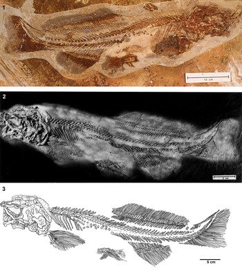
†Yanosteus longidorsalis, whole specimen (IVPP V10674). (1) Specimen prior to acid etching; anterior facing right. (2) Latex peel of specimen (cataloged as FMNH PF 17555); anterior facing left. (3) Line drawing of latex peel; anterior facing left.

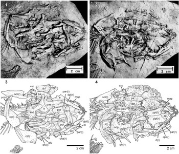
†Yanosteus longidorsalis, head. (1) Latex peel of IVPP V10998.1 (holotype). (2) Latex peel of IVPP V10998.7. (3, 4) Line drawings of (1) and (2), respectively. Anterior facing right in all. ap = autopalatine; br = branchiostegal; cb = ceratobranchial; cha = anterior ceratohyal; cl = cleithrum; clv = clavicle; d = dentary; dpl = dermopalatine; dpt = dermopterotic; dsp = dermosphenotic; exc = extrascapular; fr = frontal; h = hyomandibula; iclv = interclavicle; ihy = interhyal; l = left element (in parentheses); m = mentomeckelian; map = median anterior process of the parasphenoid; pa = parietal; par = prearticular; pas = parasphenoid; pcl = postcleithrum; ppt = palatopterygoid; pt = posttemporal; qj = quadratojugal; r = right element (in parentheses); scl = supracleithrum; sop = subopercle.

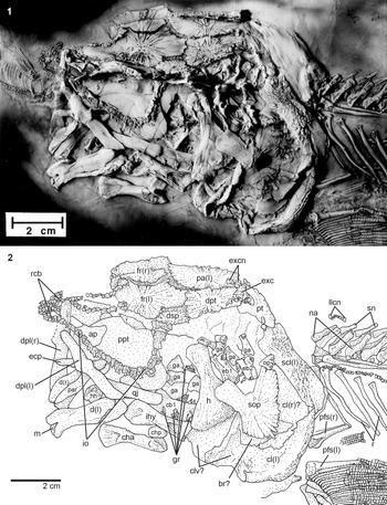
†Yanosteus longidorsalis, head and pectoral girdle from latex peel of IVPP V10674 (latex peel cataloged as FMNH PF 17555): (1) photograph; (2) line drawing. Anterior facing left in all. ap = autopalatine; br = branchiostegal; cb = ceratobranchial; cha = anterior ceratohyal; chp = posterior ceratohyal; cl = cleithrum; clv = clavicle; d = dentary; dpl = dermopalatine; dpt = dermopterotic; dsp = dermosphenotic; eb = epibranchial; ecp = ectopterygoid; exc = extrascapular; excn = canal bones lateral to the extrascapular; fr = frontal; ga = unidentified gill arch elements; gr = gill rakers; h = hyomandibula; hh = hypohyal; ihy = interhyal; io = infraorbitals; l = left element (in parentheses); llcn = lateral-line sensory canal; m = mentomeckelian; na = neural arch; op = opercle; pa = parietal; par = prearticular; pfs = pectoral-fin spine; ppt = palatopterygoid; pt = posttemporal; qj = quadratojugal; r = right element (in parentheses); r = rib; rcb = rostral canal bones; scl = supracleithrum; sn = supraneural; sop = subopercle.

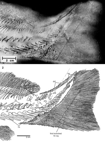
†Yanosteus longidorsalis, caudal-fin region of latex peel of IVPP V10674 (latex peel cataloged as FMNH PF 17555): (1) photograph; (2) line drawing. Anterior facing left in all. dcf = dorsal caudal fulcra; hs = haemal spine; hy = hypural; llcn = lateral line sensory canal; na = neural arch; ns = neural spine; phy = parhypural; rsc = rhomboidal caudal scales; sn = supraneural.

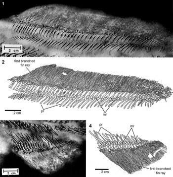
†Yanosteus longidorsalis, median fins. (1, 2) Dorsal fin from the latex peel of IVPP V10674 (latex peel cataloged as FMNH PF 17555): (1) photograph; (2) line drawing. (3, 4) Anal fin from the latex peel of IVPP V10674 (latex peel cataloged as FMNH PF 17555): (3) photograph; (4) line drawing. Anterior facing left in all. mr = middle radial of the dorsal or anal fin (the middle element of a pterygiophore); pr = proximal radial of the dorsal or anal fin (the middle element of a pterygiophore).

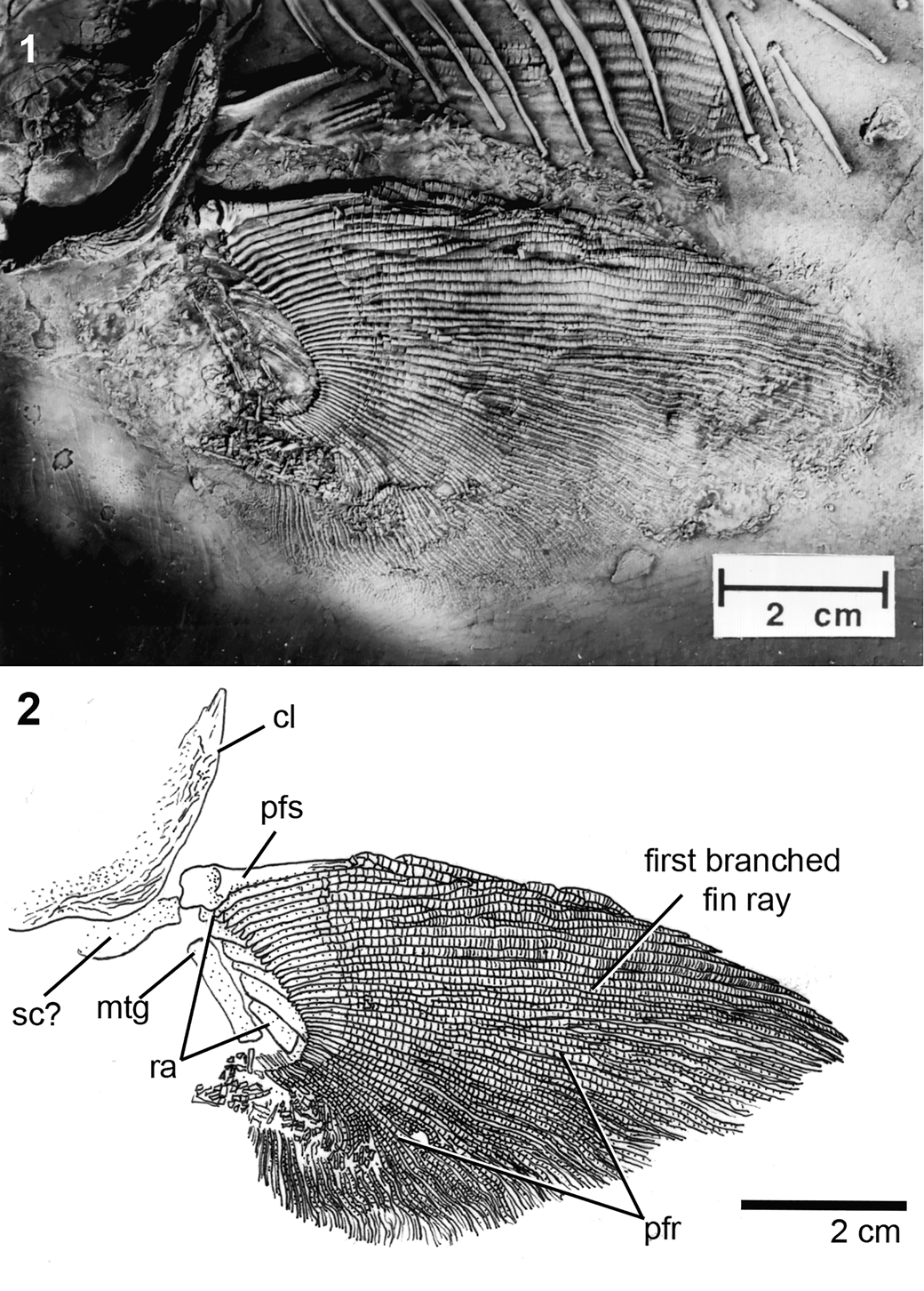
†Yanosteus longidorsalis, pectoral girdle and fin of IVPP V10674 (latex peel cataloged as FMNH PF 17555): (1) photograph; (2) line drawing. Anterior facing left in all. cl = cleithrum; mtg = metapterygoid; pfr = pectoral-fin ray; pfs = pectoral fin spine; ra = radials; sc = scapulocoracoid.

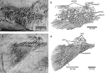
†Yanosteus longidorsalis, pelvic girdle and fin. (1, 2) IVPP V10998.1: (1) photograph; (2) line drawing. (3, 4) IVPP V10998.7: (3) photograph; (4) line drawing. Anterior facing right in all. be = basipterygial element; prm = middle radial of the pelvic fin.

Holotype
IVPP V10998.1, whole skeleton (Jin et al., Reference Jin, Tian, Yang and Deng1995, fig. 2A; Jin, Reference Jin1999, fig. 9, plate 8, fig. 1, plate 9, fig. 1, plate 12, fig. 3; latex peel Fig. 2.1).
Diagnosis
A †peipiaosteid (sensu Grande and Bemis, Reference Grande, Bemis, Stiassny, Parenti and Johnson1996) that differs from all other members of the family by possessing an extremely elongate dorsal fin. Other characters that, in combination, differentially diagnose †Yanosteus from other Acipenseriformes include short, stout pectoral fin spine, supraorbital sensory canal anterior to frontal surrounded by a series of small bony tubes, and the absence of epaxial caudal-fin rays.
Occurrence
Yixian Formation, Dongtuyao, Senjitu, Fengning County, Hebei Province, China; Early Cretaceous (late Hauterivian to early Aptian).
Etymology
Longi-, from the Latin word longus, meaning long; dorsalis, Latin, in reference to the dorsal fin; describing the elongate dorsal fin that is characteristic of this genus.
Remarks
†Yanosteus is represented by the most complete and well-preserved specimens of the three genera included within †Spherosteinae by Grande and Bemis (Reference Grande, Bemis, Stiassny, Parenti and Johnson1996) and Jin (Reference Jin1999), which include †Spherosteus, †Yanosteus, and †Liaosteus. The Yiaxian Formation, from which †Yanosteus was first described, is part of the Jeohl Group, which is late Hauterivian to early Aptian in age (Smith et al., Reference Smith, Evensen, York, Chang, Jin, Li, Cumbaa and Russell1995; Zhou et al., Reference Zhou, Barrett and Hilton2003). This geological group preserves a freshwater lacustrine habitat with exceptional preservation of plant, invertebrate, and vertebrate faunae, and is a mix of shale, sandstone, and conglomerate with breccia (Zhou et al., Reference Zhou, Barrett and Hilton2003). The ichthyofaunae of the Jianshangou and Dawangzhangzi beds of the Yiaxian Formation (ca. 125–111 Ma; see generally consistent but slightly older ages presented by Wang et al., Reference Wang, Olsen, Sha, Yao, Liao, Pan, Kinney, Zhang and Rao2016, which recover the Yixian Formation as ca. 131–124 Ma on the basis of 40Ar/39Ar dating), from which †Yanosteus is known, also have produced other Acipenseriformes (†Piapiaosteus pani and †Protopsephurus liui Lu, Reference Lu1994) and the osteoglossomorph teleost, †Lycoptera sinensis (Zhou et al., Reference Zhou, Barrett and Hilton2003).
Results
General body form and other observations
Measurements from the new specimen, as well as other specimens (from Jin, Reference Jin1999, table 7), are provided in Table 1. The new specimen (Fig. 3) is the largest for which measurement data are available, although proportionately it fits well with those data presented by Jin (Reference Jin1999). The head of †Yanosteus is 21.0%–27.0% of the standard length, which is comparable to other non-polyodontid Acipenseriformes. The eye is anteriorly positioned on the head, resulting in a short snout, although the precise margin of the orbit is unclear as the skull of most specimens is largely disarticulated (e.g., Fig. 4). The mouth is anteriorly directed (i.e., forward facing, as opposed to ventrally facing in acipenserids). It is noteworthy that the skeleton of a teleostean fish (†Lycoptera sp.) was found preserved in the mouth of our new specimen of †Yanosteus (unlabeled in Fig. 5.1). Although not conclusive evidence, this find is suggestive that this acipenseriform was piscivorous. The caudal peduncle is relatively short and stout. Although the specimens are compressed and the true form of the body is unknown, the profile of the body is reminiscent of that of modern Acipenseriformes, and †Yanosteus was likely rounded in cross section in life.
Morphometric data for a sample of †Yanosteus longidorsalis. SL = standard length, recorded as Jin's (Reference Jin1999) body length, which is from the tip of the snout to the ventral insertion of the caudal fin.

Bones of the skull roof and braincase
The bones of the skull roof in †Yanosteus are ornamented with ridges of bone radiating from the presumed center of ossification (Figs. 4, 5). The frontals (= parietals of some authors on the basis of its homology with the bone termed the ‘parietal’ in sarcopterygians; e.g., see Jollie, Reference Jollie1986; Arratia and Cloutier, Reference Arratia, Cloutier, Schultze and Cloutier1996; Schultze, Reference Schultze, Arratia, Schultze and Wilson2008; we follow the more commonly used term for this bone in actinopterygian studies) are the largest of the bones of the skull roof and are the most anteriorly positioned. They are slightly wider posteriorly than anteriorly and meet each other along the midline of the skull. Anteriorly the frontals are smooth, with a series of about seven bony elements on each side overlying this portion of the bones. These ossicles follow the supraorbital sensory canal anteriorly, after it exits the frontal (i.e., there is no true nasal bone, but rather a series of bony ossicles). Their relation to the rostral canal bones is unclear. Jin (Reference Jin1999, figs. 9, 10B) labeled small elements closely associated with the infraorbital ossicles anterior to the frontal bones as nasals. Posteriorly, the frontals meet the rectangular parietals (= postparietal of some authors on the basis of its homology with the bone termed the ‘postparietal’ in sarcopterygians; see also comments for frontal), which are also robust and ornamented over much of their exposed surfaces. A single large extrascapular is preserved on the left side of our specimen and is somewhat triangular in shape; it is unclear whether this is a lateral or median extrascapular. There is a small extrascapular exposed between the two limbs of the posttemporal of the new specimen, as well as in a similar position in other specimens (i.e., posterior to the skull roofing bones); only a single element is clearly present, although additional elements may be absent due to lack of preservation. Jin (Reference Jin1999, fig. 10) illustrated at most two (i.e., paired) extrascapular bones in his specimens. There is also a series of canal bones that lie lateral to the extrascapular (Figs. 4, 5); it is unclear whether these are lateral extrascapulars (i.e., incorporated into the skull roof) or just bony ossicles. The dermopterotic is badly damaged in our specimen, and little detail can be determined. However, there is a foramen on the lateral edge of the dermopterotic at about the level of the hyomandibula that likely represents the exit of the preoperculomandibular sensory canal. The dermosphenotic lies anterior to the dermopterotic and contacts both the frontal and parietal medially. It is somewhat drawn out posteriorly, so that it is almost teardrop shaped. The infraorbitals are small, relatively uniformly sized bones (the posteriormost two are the largest) that carry the infraorbital sensory canal from its exit from the dermosphenotic ventral and anterior to the orbit. The anterior infraorbital bones (i.e., rostral canal bones) appear to form a commissure at the tip of the snout, although their arrangement is disturbed in this region. About 30 infraorbital/rostral canal bones are preserved, but it is unclear where the distinction between the left and right sides should be made.
Neurocranium
No bones of the neurocranium, including the parasphenoid, are clearly preserved in our new specimen, but the shape of the parasphenoid can be seen in the other two specimens examined (Fig. 4). Jin (Reference Jin1999, figs. 9, 10, 11) also illustrated the parasphenoid of †Yanosteus. The parasphenoid of †Yanosteus extends posteriorly by two arms that form a deep aortic notch, as in other Acipenseriformes. There are large, anteriorly directed ascending rami of the parasphenoid at about the midpoint of the bone. The parasphenoid bears a distinct median anterior process (Fig. 4) that appears to be broader than that found in Acipenseridae (Hilton et al., Reference Hilton, Grande and Bemis2011, fig. 43).
Jaws and suspensorium
For consistency and ease of comparison with the skeletons of other Acipenseriformes, the terminology we use for the elements of the oral jaws and ventral hyoid arch of †Yanosteus follows that of our previous work on the anatomy of the order (e.g., Grande and Bemis, Reference Grande, Bemis, Stiassny, Parenti and Johnson1996; Grande et al., Reference Grande, Jin, Yabumoto and Bemis2002; Hilton and Forey, Reference Hilton and Forey2009; Hilton et al., Reference Hilton, Grande and Bemis2011). We acknowledge, however, the recent reevaluation of certain homology statements of Datovo and Rizzato (Reference Datovo and Rizzato2018), who suggested that the dermopalatine is homologous to the maxilla of non-acipenseriform actinopterygians. The elements of the upper jaw that are clearly preserved in our specimen include the dermopalatine, quadratojugal, palatopterygoid, and autopalatine (Fig. 5); we could not identify the entire outline of the ectopterygoid in our specimens (e.g., Fig. 5), but its anterior tip is a rounded point. The dermopalatine is elongate and forms the lateral margin of the anterior portion of the jaw. It is slightly broader anteriorly and bears a slight notch in its anterior margin. The quadratojugal is approximately the same width and length of the dermopalatine but is positioned more posteriorly and appears uniform in its width across its length. The precise shape and extent of the palatopterygoid (= endopterygoid of other authors; e.g., Arratia and Schultze, Reference Arratia and Schultze1991; Findeis, Reference Findeis1997; although this bone is likely homologous to the endopterygoid of other actinopterygians, we follow the terminology of Grande and Bemis, Reference Grande and Bemis1991, Reference Grande, Bemis, Stiassny, Parenti and Johnson1996 and Grande et al., Reference Grande, Jin, Yabumoto and Bemis2002 for consistency with our other publications on acipenseriform osteology) could not be determined, but it is the largest of the upper jaw bones, forming much of the roof of the mouth. Posteriorly, it has a rounded margin and appears to have a straight margin medially. Anteriorly, it is overlain by the autopalatine. Only the slightly rounded posterior margin of the autopalatine is exposed on our new specimen, but the entire outline of both the left and right autopalatine is seen in the specimens illustrated in Figure 4.
The dentary is the largest element of the lower jaw preserved in our specimen of †Yanosteus. It is a robust bone that is roughly rectangular in shape with a slight curve at its midpoint. The anterior end is forked, while the posterior end is slightly rounded. The medial surface of the dentary bears a groove, possibly housing Meckel's cartilage. The prearticular is broad and equal in length to that of the dentary (Fig. 5). The mentomeckelian is a small, rectangular bone (Fig. 4.3) that is visible in lateral view at the anterior end of the dentary (Fig. 5.2). Jin (Reference Jin1999, figs. 9, 10, 11) labeled an articular in †Yanosteus, but we could not confidently identify this bone in our specimens.
The largest bone of the hyoid arch that is preserved in our specimen of †Yanosteus is the hyomandibula, which is similar in its overall form to that of most other Acipenseriformes. There is a relatively narrow proximal head that would have articulated with the neurocranium. This head appears to be somewhat oval in cross section. Ventrally, the hyomandibula flares distinctly to become very broad and flat, with a gently curved posterior margin. The interhyal is large, and is roughly hourglass shaped, with deep concavities anteriorly and posteriorly. Anteriorly, the interhyal approaches the posterior corner of the jaw. More ventrally is an hourglass-shaped anterior ceratohyal and rounded posterior ceratohyal (the posterior ceratohyal is partially preserved and no significant details can be described). The anterior end of the anterior ceratohyal bears a deep, rounded concavity. Both ends of the anterior ceratohyal appear to be flattened in cross section. An element that corresponds to the hypohyal is preserved, but no details can be reported.
Opercular series
The opercle, the subopercle, and a single branchiostegal are clearly preserved (Figs. 4, 5). The opercle is a small, fan-shaped element that is much smaller than the subopercle and has a scalloped posterior margin; the posterior portion of the opercle is strongly ornamented. The subopercle is large and has a rounded posterior margin that is slightly scalloped, although this part of the element is not well preserved in our specimens. The posterior portion of the subopercle is slightly ornamented with fine ridges of bone. Anteriorly, the subopercle bears an unornamented process. This process is elongate, rounded, and distinctly set off from the main body of the subopercle.
There is a flat, broad fragment of dermal bone ventral to the subopercle in our specimens of †Yanosteus. This single branchiostegal is best seen in IVPP V10998.1 (holotype) and IVPP V10998.7 and has a broad horizontal body, positioned ventral to the subopercle, and a narrow anteroventrally directed extension that tapers to a point (Fig. 4).
Gill arches
Most elements of the gill arches are covered by overlying dermal bones of the skull, and therefore few aspects of their morphology can be described from our specimens of †Yanosteus (Fig. 5). In our new specimen, the first ceratobranchial from the left side, which bears short, triangular gill rakers on both its leading and trailing edges, is clearly preserved; the gill rakers are more slender and tightly packed on the trailing edge than on the leading edge. In the holotype, there are more posterior elements of the ceratobranchial series (unlabeled in Fig. 4.3). The preserved dorsal gill-arch elements of our new specimens include at least two epibranchials; other unidentified dorsal gill-arch elements likely include pharyngobranchials. Only one of the epibranchials preserves an association with four gill rakers. The first epibranchial bears a distinctly broad proximal head, which is likely the articulatory surface for the first pharyngobranchial, which has become displaced.
Vertebral column
The vertebral column is composed of ossified supraneurals, neural arches, ribs, and hemal arches (Fig. 3.3). There are 26 supraneurals preserved and 48 neural arches preserved, though there may be additional elements of both series preserved anteriorly that are covered by the dermal skull bones. The supraneurals extend from the anteriormost exposed vertebrae and continue posteriorly to under the anterior quarter of the dorsal-fin pterygiophores. Each supraneural flares proximally where it contacts the distal tip of the neural spines. Distally, the supraneurals taper to a blunt point. The neural arches are paired throughout the length of the series and are broad and rounded proximally but are drawn out to become spine-like elements, particularly more posteriorly in the series. There are approximately nine irregularly shaped ossified basiventral elements in the abdominal region, but this is likely an undercount because additional elements are probably hidden by the ribs anteriorly. The paired hemal arches, which are elongate spine-like elements, begin opposite the anterior insertion of the dorsal fin and continue into the caudal region as hypurals. There are 43 total hemal elements, including the 17 hypurals. The hemal elements mirror the neural elements, with a broadly rounded proximal end that tapers to a blunt or squared-off end (i.e., the hemal spine portion of the element). There are no ossified centra.
There are 11 ribs on both sides of our new specimen of †Yanosteus, although those of the right side are displaced and the count is approximate. As for the neural arches and supraneurals, there may be additional ribs that are covered anteriorly in our specimen.
Caudal fin and supports
The caudal fin of †Yanosteus is broad and only weakly forked (Fig. 6). There are 17 ossified hypurals that, together with the posterior hemal arches, support the caudal-fin rays. There are eight unsegmented, unbranched fin rays at the anteroventral end of the caudal fin and 20 segmented unbranched caudal-fin rays, which extend almost the entire ventral margin of the caudal fin; the longest fin ray of the ventral caudal lobe is the second branched caudal-fin ray. There are 61 branched caudal-fin rays on our specimen, all of which are ventral to the tip of the notochord (i.e., there is no trace of epaxial caudal-fin rays). In total, there are 89 caudal-fin rays, which fits within the range provided by Jin (Reference Jin1999, p. 86–98). There are six ossified epurals dorsal to the space the notochord would occupy in life. Farther anterior there are two bones, partially overlying one another, that are of unclear homology but possibly are caudal supraneural elements. There are also two unidentified bones posterior to the caudalmost neural arches; these are oval and flat. Along the dorsal margin of the caudal region are 29 dorsal caudal fulcra (30 reported by Jin, Reference Jin1999) that smoothly grade from short, leaf-shaped elements to more elongate fulcra typical of Acipenseriformes (Hilton et al., Reference Hilton, Grande and Bemis2011). At least anteriorly in the series (i.e., the anterior ~15 dorsal caudal fulcra), these are paired. It is unclear more posteriorly whether they are paired or median structures (as found in other Acipenseriformes; e.g., Hilton et al., Reference Hilton, Grande and Bemis2011). There are a few (~10) rhombic caudal scales located near the posterior tip of the caudal region of our large specimen of †Yanosteus.
Dorsal and anal fins and supports
One of the most remarkable aspects of the morphology of †Yanosteus is its elongate dorsal fin (Fig. 7.1, 7.2), which is unusual among Acipenseriformes and otherwise is found only in †Spherosteus and †Liaosteus (†Peipiaosteidae; Jin et al., Reference Jin, Tian, Yang and Deng1995) and the acipenserid †Priscosturion longipinnis (Grande and Hilton, Reference Grande and Hilton2006). The dorsal fin of †Yanosteus inserts posterior of the midpoint of the body (54.7%–59.3% standard length [SL]) but extends for a large portion of the body (37.0%–46.7% SL). The dorsal fin of our large specimen has 144 segmented branched rays as well as four unsegmented and 30 segmented unbranched rays (178 rays total). This is slightly more dorsal-fin rays than reported by Jin (Reference Jin1999), who found between about 160 and 170 dorsal-fin rays. The dorsal-fin rays are supported by 48 proximal radials and 49 middle radials; no trace of distal radials was found on our specimen. This number of radials agrees with that reported by Jin (Reference Jin1999), who reported 42–51 proximal and 45–52 middle dorsal-fin radials. As seen in other acipenseriforms, some of the middle and proximal dorsal-fin radials are forked (e.g., the proximal end of the twelfth middle radial in Fig. 7.1, 7.2).
The anal fin of †Yanosteus is relatively short and appears to show ontogenetic variation in its proportional size (11.9% SL in the smallest specimen and 19.5% in the largest; Table 1). The anal fin is positioned far posteriorly on the body (78.0%–73.3% SL) and is roughly opposite the midpoint of the dorsal fin. In our large specimen, there are one unsegmented and 18 segmented unbranched anal-fin rays and 55 branched segmented rays (74 anal-fin rays total; Fig. 7.3, 7.4). Jin (Reference Jin1999) found there to be 62 fin rays total in the anal fin for the two specimens that could be confidently counted. These fin rays are supported by 21 proximal and 23 middle radials; Jin (Reference Jin1999) reported the same counts for his specimens, with the exception of one specimen, which had 21 middle radials in the anal fin. As found in the dorsal fin, there is no trace of the distal radial series preserved on our specimens.
Pectoral and pelvic girdles and fins
The exposed surface of the posttemporal of †Yanosteus is strongly ornamented with ridges of bone (Fig. 5). The posttemporal articulates with the posterior margin of the skull roof but is not as tightly interlocked with the skull roofing bones as that of other acipenseriforms (e.g., Acipenseridae). The anterior end of the posttemporal is forked, with an elongate dorsal branch and a shorter, more lobate ventral limb. Ventrally, the posttemporal contacts the robust supracleithrum, which is about two times the length of the posttemporal. Much of the surface of the supracleithrum is roughly ornamented; the only smooth external surface of the supracleithrum is the posteroventral region. The cleithrum is large and, as in other acipenseriforms, is curved along its posterior margin where it defines the posterior extent of the pectoral girdle. This thickened curved region is ornamented with ridges. Medially, the so-called cleithral wall is smooth and gently bowl shaped and defines the posterior limits of the branchial chamber. The clavicle is not well preserved, but there is broken dermal bone present between the left and right cleithra that likely corresponds to the clavicles. No detail of shape of these elements can be distinguished. Jin (Reference Jin1999, figs. 9, 10) illustrated the clavicle as bearing a rounded anterior margin and a narrow, dorsally projected process. The lack of an interclavicle was interpreted as a general character of †Peipiaosteidae by Jin (Reference Jin1999). However, we identify a poorly preserved dermal element, found in both the holotype and another specimen (Fig. 4), as likely an interclavicle. There is no trace of a postcleithrum in our new specimen, but this is likely due to preservation as a slender postcleithrum straddling the articulation between the supracleithrum and cleithrum was found in our other specimens (Fig. 4; see also Jin, Reference Jin1999, figs. 9, 10B).
The pectoral fin of our specimen of †Yanosteus is supported by a metapterygium and six ossified radials (Fig. 8). This is a greater number of pectoral radials than reported by Jin (Reference Jin1999; 2–3 radials), but this may due to the specimen described here being larger and possibly more ossified. Our specimen bears 13 unbranched and 29 branched fin rays posterior to the short but well-formed pectoral-fin spine (Fig. 8). The base of the pectoral-fin spine is robust and bears a concavity that would have articulated with the elements of the pectoral girdle. Among Acipenseriformes, a fin spine is otherwise seen in †Chondrosteus acipenseroides Egerton, Reference Egerton1858 (Chondrosteidae; Hilton and Forey, Reference Hilton and Forey2009), †Protopsephurus liui (Polyodontidae; Grande et al., Reference Grande, Jin, Yabumoto and Bemis2002), and all members of Acipenseridae (Bemis et al., Reference Bemis, Findeis and Grande1997; Hilton et al., Reference Hilton, Grande and Bemis2011). The spine of †Chondrosteus appears different from the other acipenseriform taxa in being much shorter and not fused to the fin rays of the leading edge of the pectoral fin. By contrast, in †Protosephurus, †Yanosteus, and Acipenseridae, the pectoral-fin spine forms at least in part by the fusion of adjacent anterior pectoral-fin rays (Findeis, Reference Findeis1994; Dillman and Hilton, Reference Dillman and Hilton2015). The pectoral-fin spine of †Yanosteus appears to include two or three fin rays in its formation.
The pelvic fins of †Yanosteus are positioned just posterior to the midpoint of the body (53.%–58.0% SL) and bear around 17 unbranched and 18 branched fin rays (35 fin rays total; countable only on a single specimen examined here; Fig. 9), which agrees well with the counts reported by Jin (Reference Jin1999; 34 in the one reliably reported individual). We found nine radials that are partially embraced by the proximal ends of the fin rays. In addition, there is a series of ossified basipterygial elements that are moderately narrow bars of bones with flared anterior and posterior ends. The precise orientation and relation to other elements of the pelvic girdle are unclear. There are six of these basipterygial elements preserved in our large specimen, and we suspect that they were arranged as three per side.
Scales
The body of †Yanosteus is largely devoid of scales, with the exception of tubular elements that are associated with the lateral line sensory canal. In addition, there are a few (approximately nine) irregularly shaped rhomboid scales at the very posterior tip of the caudal region. We did not find the round-based scales associated with the pectoral girdle in our new specimen, although this is likely due to lack of preservation as they have been reported in †Yanosteus by Jin (Reference Jin1999, fig. 10A) and can be seen in the holotype (observed, but not illustrated in Fig. 4.1).
Discussion
Characters of †Peipiaosteidae
†Peipiaosteidae is broadly recognized as a member of the Acipenseriformes on the basis of the presence of palatoquadrates with an anterior symphysis and a broad autopalatine portion, palatoquadrate bridge, and quadrate flange and a subopercle with a well-developed anterior process and for having the infraorbital sensory canal in a series of tube-like ossicles, all of which are synapomorphies for the order (Grande and Bemis, Reference Grande, Bemis, Stiassny, Parenti and Johnson1996; Bemis et al., Reference Bemis, Findeis and Grande1997). However, its precise position has been disputed either as a sister group of all remaining Acipenserformes, including †Chondrosteidae, or as sister to Acipenseridae + Polyodontidae (see discussion in Hilton and Forey, Reference Hilton and Forey2009). Grande and Bemis (Reference Grande, Bemis, Stiassny, Parenti and Johnson1996) were the first to differentially diagnose the family on the basis of the results of a cladistic analysis and found several characters to define the family, including extreme reduction or loss of the rhombic caudal scales, absence of interclavicle, and a unique shape of the opercular bones in having a ‘ctenoid’ posterior margin. To this list, Jin (Reference Jin1999) added two characters of the caudal fin to the diagnosis: having the dorsal caudal fulcra present as slender and paired elements and having the ventral caudal fulcrum either reduced or lost altogether. The results of our osteological study of †Yanosteus are consistent with these five characters as defining the family †Peipiaosteidae, with some caveats. Although we agree that the absence of an interclavicle appears to be shared by most members of the family, we found an element that may correspond to an interclavicle. In our specimens of †Yanosteus, this region of the pectoral girdle is damaged or covered by overlying elements. Among all of the material that we have examined, only specimens of †Pepiaosteus pani have the pectoral region preserved in a manner that this statement can be made unambiguously. The other character that warrants comment is the form of the posterior margin of the opercular bones (inclusive of the opercle, subopercle, and branchiostegals). While we agree that the posterior margin of the opercular bones is similar among †peipiaosteids, and different than the condition found in other groups of Acipenseriformes (e.g., the finger-like process of bone extending from the posterior margin of polyodontids; Grande and Bemis, Reference Grande and Bemis1991), we note that some acipenserids have a posterior margin that is gently scalloped and reminiscent of the condition found in †Peipiaosteidae. One character that relates to the subopercle that should be explored across the diversity of Acipenseriformes is the shape of its anterior process. In most members of the order, there is an anterior process that is of varying degrees of distinctiveness from the main (i.e., ornamented) body of the subopercle. This process typically has a smooth surface and is often rounded, although there is a wide variation in shape, ranging from small, indistinct, and angular (e.g., in Huso huso (Linnaeus, Reference Linnaeus1758); personal observation, EJH) to long, narrow, and somewhat pointed (e.g., in †Priscosturion, Grande and Hilton, Reference Grande and Hilton2006, †Protopsephurus, Scaphirhynchus, and Pseudoscaphirhynchus). However, in both †Peipiaosteus and †Yanosteus, the anterior process of the subopercle is very distinctly set off from the main portion of the element so that there is a notch along the anterodorsal margin of the subopercle. Further, the anterior end of the process is broadly rounded. The condition of the subopercle should be examined in other members of the family before reaching conclusions on the diagnostic value of this character.
†Peipiaosteidae is divided into two subfamilies (Grande and Bemis, Reference Grande, Bemis, Stiassny, Parenti and Johnson1996): †Stichopterinae (including †Peipiaosteus and †Stichopterus) and the †Spherosteinae (including †Spherosteus, †Yanosteus, and †Liaosteus). Grande and Bemis (Reference Grande, Bemis, Stiassny, Parenti and Johnson1996) diagnosed the subfamily †Stichopterinae as †peipiaosteids with the series of dorsal caudal fulcra reduced to a single element and the ventral caudal fulcrum either lost or greatly reduced in size. Jin (Reference Jin1999) found the †stichopterines to also share similarly shaped plank-like palatopterygoids with slightly concave anterior margins and having elongate, densely packed triangular gill rakers. The gill rakers of Acipenseriformes vary widely in shape, and the variation in this last character should be broadly evaluated. Grande and Bemis (Reference Grande, Bemis, Stiassny, Parenti and Johnson1996, p. 90) suggested that many of the differences between †Peipiaosteus and †Stichopterus may reflect ontogenetic variation (i.e., all specimens of †Stichopterus are relatively large whereas those of †Peipiaosteus are relatively small) and that the two genera may prove to be synonymous.
The subfamily †Spherosteinae, comprising †Spherosteus, †Yanosteus, and †Liaosteus, is diagnosed by the presence of an elongate dorsal fin (Grande and Bemis, Reference Grande, Bemis, Stiassny, Parenti and Johnson1996; Jin, Reference Jin1999). Among Acipenseriformes, a dorsal fin that is more than 20% the total length and has more than 100 fin rays is otherwise seen only in the acipenserid †Priscosturion. In that taxon, the dorsal fin approaches 50% of the total length and is surely a homoplastic occurrence (see Grande and Hilton, Reference Grande and Hilton2006). Jin (Reference Jin1999) also found the loss of epaxial caudal-fin rays, having the postcranial skeleton mostly ossified, and the presence of a unique bone he interpreted to represent a fusion of the prearticular and coronoids in the lower jaw to be additional synapomorphies of the subfamily. We agree with the first two of these characters, although we note that having a mostly ossified postcranial skeleton could be considered to be plesiomorphic for actinopterygians generally, and this character should be more clearly defined in future analyses (e.g., scoring the presence of ossification of individual series of elements, such as the neural arches, rather than a global condition for the entire postcranial skeleton). The third character described by Jin (Reference Jin1999) pertaining to the presence of a fused prearticular and coronoid needs to be reevaluated. In comparing our specimen of †Yanosteus with the illustrations provided by Jin (Reference Jin1999), we believe that the element he identified as a prearticular + coronoid is rather an elongate prearticular. This enlarged prearticular (relative to that of other Acipenseriformes) would be consistent with other aspects of the skeleton of †Yanosteus, which are more heavily ossified and larger than in most other members of the order. For example, the interhyal of †Yanosteus is also much longer and apparently much more heavily ossified than those found in other Acipenseriformes.
Diversity of †Peipiaosteidae and future studies of fossil Acipenseriformes
With the description of the skeleton presented here for †Yanosteus, we have continued to build the comparative data available for †Peipiaosteidae. However, several members of the family still lack the detailed data to be broadly included in systematic analyses. For example, specimens of †Liaosteus, †Spherosteus, and †Stichopterus require new preparation and new study to develop complementary data to those that are now available for †Yanosteus and †Peipiaosteus. The presence of ontogenetic versus taxonomic variation within the family must be reevaluated on the basis of direct study of additional specimens. This fundamental source of morphological variation may have direct impact on the taxonomy of †Peipiaosteidae at both the genus level (evaluating the potential synonymy of †Stichopterus and †Peipiaosteus) and the species level (the validity of †P. fengningensis).
Acknowledgments
We thank J. Weinstein for photographic assistance. Financial support of the National Science Foundation (DEB-1754483, to EJH and LG) and a Field Museum Bass Fellowship (to EJH) are gratefully acknowledged. We thank W.E. Bemis and M. Friedman for helpful reviews of the manuscript. This is contribution number 3923 of the Virginia Institute of Marine Science, William & Mary.