Introduction
Antarctica is geographically isolated and experiences extreme climatic conditions compared to lower latitudes (Laws Reference Laws1984). In Antarctic terrestrial and freshwater ecosystems, microorganisms (i.e. bacteria, algae, fungi and viruses) may comprise the majority of the biomass and biodiversity, and this may be the case particularly in habitats subject to more climatically extreme conditions, where higher organisms do not survive (Cowan & Ah Tow Reference Cowan and Ah Tow2004, Yergeau et al. Reference Yergeau, Bokhorst, Huiskes, Boschker, Aerts and Kowalchuk2007). Of the c. 1000 fungal species reported from the Antarctic and sub-Antarctic region, only 2–3% are considered to be endemic (Bridge & Hughes Reference Bridge and Hughes2010, https://legacy.bas.ac.uk/bas_research/data/access/fungi/Speciespublic2.html accessed 26 December 2017), but research suggests endemism in microorganisms may be greater and more widespread than thought previously (Lawley et al. Reference Lawley, Ripley, Bridge and Convey2004, De Wever et al. Reference De Wever, Leliaert, Verleyen, Vanormelingen, Van Der Gucht, Hodgson, Sabbe and Vyverman2009, Vyverman et al. Reference Vyverman, Verleyen and Wilmotte2010). Antarctic communities may be at risk if fungal strains are introduced from outside Antarctica or moved between different Antarctic regions (Cowan et al. Reference Cowan, Chown, Convey, Tuffin, Hughes, Pointings and Vincent2011, Hughes et al. Reference Hughes, Cowan and Wilmotte2015a). Introduced fungi may compete with native Antarctic species, alter existing nutrient cycling or cause disease in native flora and fauna (Bridge & Hughes Reference Bridge and Hughes2010, Augustyniuk-Kram Reference Augustyniuk-Kram, Chwedorzewska, Korczak-Abshire, Olech and Lityńska-Zając2013). For example, on sub-Antarctic Marion Island, the non-native Botrytis cinerea Pers. 1797, which was probably transferred to the island on fresh vegetables, now infects Kerguelen cabbage (Kloppers & Smith Reference Kloppers and Smith1998). Fungi and other microbes are likely to be transferred to Antarctica by natural means, for example on wind currents or on ocean debris and feathers of birds (Marshall Reference Marshall1998, Hughes & Convey Reference Hughes and Convey2010, Reference Hughes and Convey2012), with strains that are able to grow in the Antarctic natural environment potentially becoming established. Alternatively, fungi may be introduced to Antarctica by humans (e.g. Wicklow Reference Wicklow1968, Bridge & Hughes Reference Bridge and Hughes2010, Osyczka et al. Reference Osyczka, Mleczko, Karasinski and Chlebicki2012), with, as yet, little known consequences for endemic microbial communities (Vyverman et al. Reference Vyverman, Verleyen and Wilmotte2010).
The Antarctic continent has been subject to human visitation for less than 200 years, but the extent of national Antarctic operator and tourism industry activity is increasing, resulting in greater risk of non-native species introductions (Frenot et al. Reference Frenot, Chown, Whinam, Selkirk, Convey, Skotnicki and Bergstrom2005, Tin et al. Reference Tin, Fleming, Hughes, Ainley, Convey, Moreno, Pfeiffer, Scott and Snape2009, Hughes et al. Reference Hughes, Fretwell, Rae, Holmes and Fleming2011a, Reference Hughes, Pertierra, Molina-Montenegro and Convey2015b). While research attention has focused on the importation of plant propagules and invertebrates (Whinam et al. Reference Whinam, Chilcott and Bergstrom2005, Lee & Chown Reference Lee and Chown2009, Hughes et al. Reference Hughes, Convey, Maslen and Smith2010a, Reference Hughes, Lee, Ware, Kiefer and Bergstrom2010b, Chown et al. Reference Chown, Huiskes and Gremmen2012, Lityńska-Zając et al. Reference Litynska-Zając, Chwedorzewska, Olech, Korczak-Abshire and Augustyniuk-Kram2012, Tsujimoto & Imura Reference Tsujimoto and Imura2012, Chwedorzewska et al. Reference Chwedorzewska, Korczak-Abshire, Olech, Lityńska-Zając and Augustyniuk−Kram2013, Huiskes et al. Reference Huiskes, Gremmen and Bergstrom2014), less attention has been paid to the threat of importing non-native microorganisms to Antarctica (Broady & Smith Reference Broady and Smith1994, Cowan et al. Reference Cowan, Chown, Convey, Tuffin, Hughes, Pointings and Vincent2011, Augustyniuk-Kram Reference Augustyniuk-Kram, Chwedorzewska, Korczak-Abshire, Olech and Lityńska-Zając2013) and in particular the risk associated with contaminated timber packing materials (Osyczka Reference Osyczka2010, Osyczka et al. Reference Osyczka, Mleczko, Karasinski and Chlebicki2012).
This paper reports the unintentional importation of fungi to Rothera Research Station, Antarctica, in association with softwood cargo packaging. The hyphal extension rates of the imported fungi at different temperatures are examined, and the potential for their establishment in Antarctica is discussed.
Methods
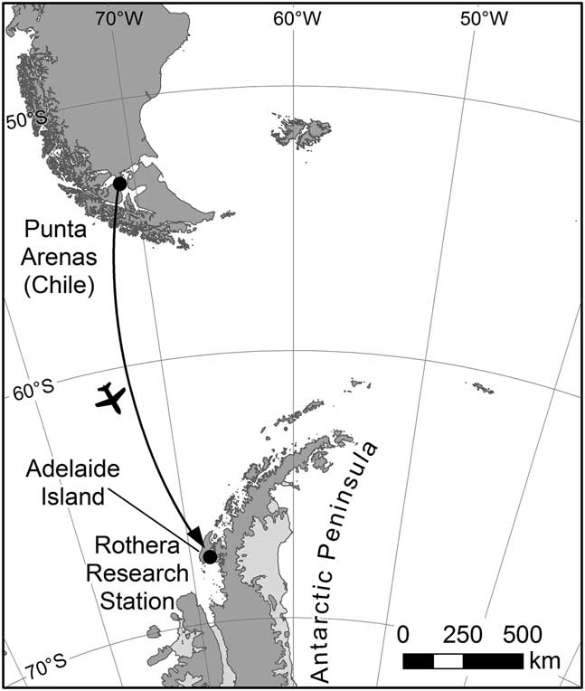
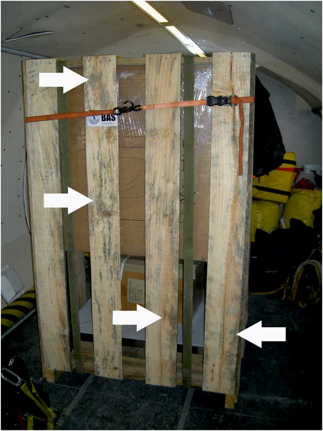
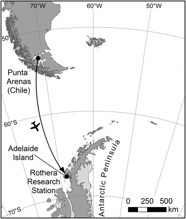
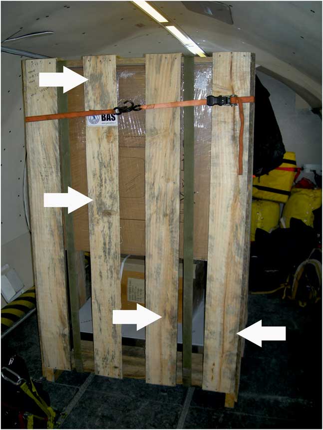
In January 2015, an item of cargo contained in a softwood crate was flown from Punta Arenas, Chile, to the British Antarctic Survey’s Rothera Research Station, Adelaide Island, Antarctic Peninsula (Fig. 1). In general, the British Antarctic Survey specifies the use of wood for packaging that conforms to the International Standards on Phytosanitary Measures 15 (ISPM 15), and this is routinely used for cargo originating from the UK. However, in this case the cargo was sourced and packaged in Chile, and normal packing practices were not observed. The bark-free wood that was used to produce the bespoke crate was heavily contaminated with fungal hyphae prior to construction of the crate, possibly as a result of inappropriate storage practices. During the six-hour flight it was observed that white and darkly pigmented mycelia were present on the outer surfaces of the packaging crate, except where it had been freshly sawn during construction (Fig. 2). Darkly pigmented fungal structures were also visible on the plank surfaces. Upon arrival at Rothera Research Station, the station management was informed of the importation of the contaminated crate. The crate was off-loaded from the aircraft, the catering equipment unpacked and the packing crate quarantined. Following sampling, the wooden packing crate was incinerated.
Map of the Antarctic Peninsula and southern South America, showing the transfer route of the contaminated crate from Punta Arenas to Rothera Research Station.
Fungal-contaminated packing crate aboard the British Antarctic Survey’s De Havilland DASH 7 en route to Rothera Research Station, Antarctica, from Punta Arenas, Chile. Areas of the wood colonized by dark pigmented mycelia are visible on the plank surfaces (indicated by white arrows).
Fungal isolation
Wood fragments were sampled aseptically from below the surface of the wooden crate and placed in sterile polyethylene bags. In the laboratory, the wood fragments were cut into small fragments (c. 1×2 mm) aseptically and washed for 5 min in 10 changes of sterile distilled water. Individual wood fragments were placed onto potato dextrose agar medium (PDA; Oxoid; Unipath Ltd, Basingstoke, UK) and incubated at 15°C. Single fungal colonies were isolated after five days. Following isolation, three strains were grown routinely on PDA in 90 mm diameter Petri dishes.
Molecular identification
Fungal cultures were maintained on PDA medium at 4°C for 10 weeks while in transit back to the UK from Antarctica. Fresh cultures were prepared for DNA extractions on half-strength PDA and incubated at 15°C. A DNeasy Power Soil Kit (Qiagen, Manchester, UK) was used to extract genomic DNA from the fungal cultures, following the manufacturer’s protocol. DNA was extracted from c. 0.3–0.5 g of fungal colony. The DNA extracts were stored at -20°C prior to polymerase chain reaction (PCR) amplification. Internal transcribed spacer (ITS) regions of ribosomal RNA genes were amplified using ITS1F and ITS4 primers (White et al. Reference White, Bruns, Lee and Taylor1990, Gardes & Bruns Reference Gardes and Bruns1993). Polymerase chain reactions consisted of 4 µl of 5×PCRBIO Reaction Buffer (PCR Biosystems, London, UK), 0.2 µl of each primer (20 pmol), 1µl of template DNA and 0.5 U of PCRBIO Taq DNA polymerase (PCR Biosystems, London, UK), diluted with PCR grade water to a final volume of 20 µl. The PCR reactions were run in a PCRmax thermocycler (AlphaCycler 4, PCRmax, Stone, UK) for 35 cycles, with each cycle consisting of 95°C for 30 s, 56°C for 30 s and 72°C for 30 s, with an initial denaturation step of 95°C for 5 min and a final extension step at 72°C for 5 min. The PCR products were separated by agarose gel electrophoresis and visualized after staining with GelRed.
The PCR products were bidirectionally Sanger sequenced (Source Bioscience, Cambridge, UK). Forward and reverse chromatograms were edited and assembled, and primer sequences trimmed using Geneious v. 6.1.6. The resulting consensus ITS sequences were deposited in GenBank under accession numbers MH279746 and MH279747. The sequences were subjected to blastn searches against the National Center for Biotechnology Information (NCBI) nucleotide collection, with closest named matches being noted.
Growth rate analysis at different temperatures
Agar medium plugs taken from the colony margins of all three fungi were inoculated onto PDA and incubated in the dark at 0, 4, 15 and 25°C. Hyphal extension rate analyses were carried out on three replicate Petri dishes of each fungus per treatment, with a mean value per dish being derived from three measurements made with sliding calipers to an accuracy of 0.1 mm. Hyphal extension was measured at regular intervals for up to 38 days and extension rates calculated by linear regression.
Air temperature data
Mean monthly air temperature data were obtained for Punta Arenas using World Meteorological Organization CLIMAT data (https://www.wmo.int/cpdb/chile), and data for Rothera Research Station were obtained from the SCAR READER database (https://www.scar.org/data-products/ref-data-environmental-research/). Temperatures in moss beds on Anchorage Island, Ryder Bay (67°36'18"S, 68°12'20"W), 5 km from Rothera Research Station, were recorded with a Campbell CR10X Logger and IMKO temperature probes.
Results
Strain identification
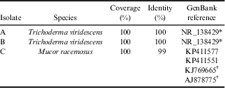
Three fungal strains were isolated from the wooden crate. Molecular analyses showed that the ITS region sequences of two isolates both matched at 100% identity and 100% coverage with that of Trichoderma viridescens (A.S. Horne & H.S. Will.) Jaklitsch & Samuels reference material (CBS 433.34), and the third matched at 99% identity and 100% coverage with isolates of Mucor racemosus Bull. (Table I).
Identification of the three fungal strains isolated from the imported softwood crate.
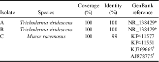
*T. viridescens reference material (CBS 433.34); †f. sphaerosporus.
Hyphal extension rate at different temperatures
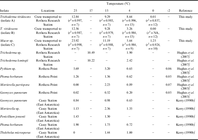
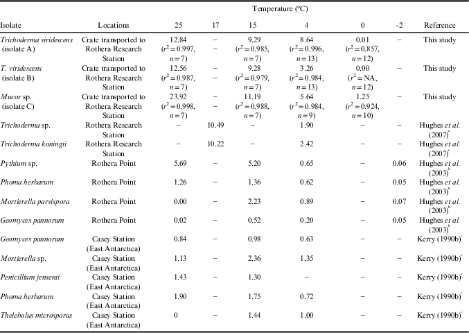
Hyphal extension rate experiments showed differences in the abilities of the three fungal isolates to grow at the temperatures tested (Table II). All three strains grew at 4, 15 and 25°C. The two isolates of Trichoderma viridescens displayed similar hyphal extension rates at 25°C and 15°C, but isolate A grew at over twice the rate of isolate B at 4°C and was able to grow at a rate of 0.01 mm d-1 at 0°C, whereas isolate B did not grow at this temperature (Table II). Mucor racemosus grew faster than either of the T. viridescens isolates at all temperatures and was able to grow at a rate of 1.25 mm d-1 at 0°C (Table II).
Hyphal extension rates (mm d-1) at different temperatures of the three fungal strains imported in this study and fungi isolated from Antarctica in other studies.
a Fungi grown on minimal medium supplemented with glucose.
b Fungi grown on potato dextrose agar.
c Fungi grown on tomato agar and Lilly Barnett’s synthetic medium.
Air temperature data
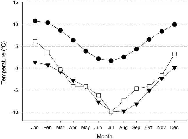
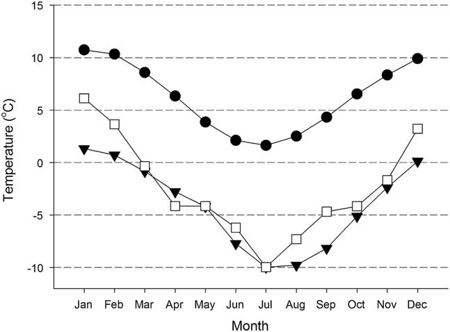
Figure 3 shows the mean monthly air temperatures recorded at Punta Arenas and Rothera Research Station and the temperatures at 5 cm depth in moss on Anchorage Island (5 km from Rothera Research Station). In general, there was a difference in air temperature of c. 10°C between Punta Arenas and Rothera Research Station throughout the year, with winter mean monthly air temperatures in Punta Arenas being roughly equivalent to summer mean monthly air temperatures at Rothera Research Station. However, mean monthly temperatures of moss beds on Anchorage Island were up to 5°C higher than mean air temperatures at nearby Rothera Research Station.
Mean monthly air temperature data for Punta Arenas, Chile (1888–2017) (●) and Rothera Research Station, Antarctica (1977–2017) (▼), and temperature recorded at 5 cm depth in moss at Anchorage Island, Ryder Bay, located 5 km from Rothera Research Station (□).
Discussion
Three fungal stains were isolated from the cargo packaging, although it is highly likely that other fungi were also imported on the wood. Sequencing of ITS regions of ribosomal RNA genes indicated that one of the isolates was Mucor racemosus, the others being identified as two strains of Trichoderma viridescens exhibiting different hyphal extension rates at low (≤4°C) temperatures. Trichoderma and Mucor are both globally ubiquitous in soils and are thought to contribute to the degradation of plant debris through the production of extracellular enzymes but are generally not able to degrade lignocelluloses (Eida et al. Reference Eida, Nagaoka, Wasaki and Kouno2011, Błaszczyk et al. Reference Błaszczyk, Strakowska, Chełkowski, Gąbka-Buszek and Kaczmarek2016). Both genera have been isolated previously from sub-Antarctic islands and maritime and continental Antarctica (Corte & Daglio Reference Corte and Daglio1964, Sugiyama Reference Sugiyama1970, Hurst et al. Reference Hurst, Pugh and Walton1983, Kerry Reference Kerry1984, Fletcher et al. Reference Fletcher, Kerry and Weste1985, McRae & Seppelt Reference McRae and Seppelt1999, Bradner et al. Reference Bradner, Sidhu, Yee, Skotnicki, Selkirk and Nevalainen2000, Kochkina et al. Reference Kochkina, Ivanushkina, Ozerskaya, Chigineva, Vasilenko, Firsov, Spirina and Gilichinsky2012, Saili et al. Reference Saili, Siddiquee, Vui Ling, González and Vijay Kumar2014). Trichoderma spp. were transported previously to Antarctica in association with national operator activities and detected in Antarctic soils subjected to human impacts (Kerry Reference Kerry1990a, Hughes et al. Reference Hughes, Bridge and Clark2007, Osyczka et al. Reference Osyczka, Mleczko, Karasinski and Chlebicki2012), and, along with other fungi, were also isolated from soils subjected to human activity near Mawson, Davis and Rothera research stations, but were not found locally in more pristine soils (Line Reference Line1988, Hughes et al. Reference Hughes, Bridge and Clark2007). Furthermore, both Trichoderma and Mucor spp. have been reported in studies examining non-native fungi introduced to research stations on clothing and equipment (Augustyniuk-Kram Reference Augustyniuk-Kram, Chwedorzewska, Korczak-Abshire, Olech and Lityńska-Zając2013) and fresh foods (Hughes et al. Reference Hughes, Lee and Tsujimoto2011b), with a Trichoderma strain also having been isolated previously from wood packaging (Kerry Reference Kerry1990a). Moreover, the high isolation frequency of Mucor spp. and other fungal genera near Mawson Station (East Antarctica) could have been due to introductions from temperate regions in the time since the station was established in 1954 (Kerry Reference Kerry1990a).
Previous studies have also suggested that fungi other than Trichoderma and Mucor spp. may have been introduced to the Antarctic. For example, introduced wood decay fungi have been isolated from timber on Deception Island, South Shetland Islands (Held & Blanchette Reference Held and Blanchette2017), and Phialophora fastigiata Lagerb. & Melin, potentially introduced on softwood packing crates, has been found in contaminated soils around Australian Antarctic research stations (Kerry Reference Kerry1990a, Bölter et al. Reference Bölter, Kandeler, Pietr and Seppelt2002). It has also been suggested that Aspergillus fumigatus Fresen., 1863, which causes aspergillosis in birds, was introduced to near an Adélie penguin Pygoscelis adeliae (Hombron & Jacquinot) colony at Cape Hallett in Victoria Land during the period in which Hallett Station was present (Wicklow Reference Wicklow1968, Bridge & Hughes Reference Bridge and Hughes2010). Osyczka et al. (Reference Osyczka, Mleczko, Karasinski and Chlebicki2012) identified 13 ascomycetes and basidiomycetes imported to Arctowski Station (King George Island, South Shetland Islands) associated with timber, but suggested, without confirmatory checks, that the probability of the imported fungi becoming established and spreading was rather negligible. However, research by Farrell et al. (Reference Farrell, Arenz, Duncan, Held, Jurgens and Blanchette2011), who examined introduced and indigenous fungi of the Ross Island historic huts and pristine areas of Antarctica, found that although non-Antarctic fungal species were almost certainly introduced to the continent in association with wood and other organic materials, it was indigenous soil fungi that exploited these substrates for nutrients. In contrast, Held & Blanchette (Reference Held and Blanchette2017) studied wood rot fungi in timber buildings on Deception Island and suggested that brown and white rot Basidiomycota were introduced with the wood and building materials and were thriving under maritime Antarctic conditions.
What is the likelihood of introduced fungi becoming established in the Antarctic natural environment? In this study of hyphal extension rates, all of the isolated fungi exhibited psychrotolerance, with all three strains growing at 4°C and Mucor racemosus showing relatively rapid growth at 0°C. Mean summer soil temperature measured at Anchorage Island (5 km from Rothera Point) was reported as 2.8°C at 5 cm soil depth (Yergeau et al. Reference Yergeau, Bokhorst, Kang, Zhou, Greer, Aerts and Kowalchuk2012), which is close to the January mean temperature reported here of 4.3°C in moss at the same depth (Fig. 3). Furthermore, it has been reported that coastal Antarctic soils can attain summer temperatures of >15°C when subjected to high levels of solar radiation (Smith Reference Smith1988, Davey et al. Reference Davey, Pickup and Block1992). Therefore, soil temperature may not be a limiting factor for the establishment of some of the species imported to Rothera Research Station, although other factors, such as salinity, water availability and UV radiation, may have an influence (Kerry Reference Kerry1990b, Arnold et al. Reference Arnold, Convey, Hughes and Wynn-Williams2003, Hughes et al. Reference Hughes, Lawley and Newsham2003, Ruisi et al. Reference Ruisi, Barreca, Selbmann, Zucconi and Onofri2007). The two Trichoderma viridescens strains isolated in this study had faster hyphal extension rates at 4°C than two Trichoderma strains isolated previously from contaminated soils at Rothera Research Station (Hughes et al. Reference Hughes, Bridge and Clark2007), as well as other strains isolated from pristine vegetated sites near Rothera Research Station (Hughes et al. Reference Hughes, Lawley and Newsham2003) and Casey Station (Wilkes Land, coastal East Antarctica; 64°17'S, 100o32'E) (Kerry Reference Kerry1990b) (Table II). Given that the three strains studied here originated from Punta Arenas (53°10'S, 70°56-W), which experiences mean winter (June–August) temperatures of 2.1°C (Fig. 3), it is unsurprising that the fungi showed some pre-adaptation to growth at low temperatures. As the physiologies of the three introduced fungal strains studied here are apparently compatible with the Antarctic summer climate, it is possible that they might have become established in soil and other substrates close to Rothera Research Station had steps not been taken to destroy the packaging on which they were introduced to the Antarctic. However, detecting the presence of introduced microbes in the Antarctic natural environment represents a considerable challenge. It is possible that sequencing of ITS regions of precursor ribosomal RNA (Rajala et al. Reference Rajala, Peltoniemi, Hantula, Mäkipää and Pennanen2011), a much shorter-lived molecule than DNA that is predominantly present in metabolically active tissue, could be used to identify whether introduced, non-endemic fungi are present in Antarctic microbial communities.
Cargo packing material management
In 1991, the Antarctic Treaty Consultative Parties (ATCPs) agreed Annex II to the Protocol on Environmental Protection to the Antarctic Treaty (entered into force in 1998), which stipulates that non-native species are not to be intentionally introduced, unless in accordance with a permit issued by an appropriate national authority. More specifically, the Protocol states that ‘each Party shall require that precautions are taken to prevent the accidental introduction of microorganisms (e.g. viruses, bacteria, yeasts, fungi) not present naturally in the Antarctic Treaty area’ and prohibits the deliberate introduction of non-sterile soil.
All 29 Antarctic Treaty Consultative Parties participate in the International Standards for Phytosanitary Measures No. 15 (ISPM 15) developed by the International Plant Protection Convention. The Measure stipulates that wood materials of a thickness greater than 6 mm, used to ship products between countries, should be heat treated or fumigated to prevent transfer of insects and disease-causing microorganisms. In agreement with the 'Checklists for supply chain managers of national Antarctic programmes for the reduction in risk of transfer of non-native species' (COMNAP & SCAR 2010), it is recommended that equivalent wood heat-treatment standards are employed by national operators importing cargo into Antarctica. Furthermore, it is suggested that particular attention be given to the adequate storage of wood to prevent fungal contamination prior to transport into the Treaty area. Antarctic microbial diversity may be impacted irreversibly unless fuller consideration is given to the routes and risks of microbial introductions and the agreement of internationally applicable biosecurity measures (Cowan et al. Reference Cowan, Chown, Convey, Tuffin, Hughes, Pointings and Vincent2011, Convey et al. Reference Convey, Hughes and Tin2012, Hughes et al. Reference Hughes, Cowan and Wilmotte2015a).
Acknowledgements
This paper is a contribution to the SCAR State of the Antarctic Ecosystem (AntEco) research programme. The authors are supported by NERC core funding to the British Antarctic Survey’s Polar Science for Planet Earth programmes 'Biodiversity, Evolution and Adaptation' and 'Environment Office - Long Term Monitoring and Survey' (EO-LTMS). The authors are grateful to Laura Gerrish for map production, Alison Massey for technical assistance, Will Goodall-Copestake for help with DNA sequence analyses, and to three anonymous reviewers, who provided helpful comments on the manuscript.
Author contributions
KAH led the experimental work and the conceptual development and writing of the paper. MM led the molecular biological work. YU contributed to the environmental sampling. KKN contributed expert advice and helped with the drafting and revision of the manuscript.