Implications
The potential beneficial effect of administering non-pathogenic strains of Escherichia coli and Clostridium perfringens type A (CPA) strains to piglets in a herd with a high prevalence of neonatal diarrhoea was investigated. The aim was to stabilize gut health by competitive bacterial colonization before contact with pathogenic bacteria and to achieve protection initiated by competition for nutrients. The effect of the cocktail of non-pathogenic microbes was not clearly visible. The mortality and presence of pathological lesions was decreased in the treated litters. By contrast, daily weight gain (DWG) from birth to weaning was higher in the control group. However, this could also at least partly have been dependent on the reduced litter size and thus better access to the udder.
Introduction
Piglets are born free from any microbiota but colonization of their gut starts immediately after birth by ingestion of bacteria from the vaginal fluids and the environment (Smith, Reference Smith1965). The amount, composition and metabolism of these early colonizing microbial species in the gastrointestinal tract have a strong impact on the piglets’ postnatal development (Mazmanian et al., Reference Mazmanian, Liu, Tzianabos and Kasper2005). The outcome of gut colonization in piglets is highly dependent on the microbial load of the environment and, therefore, influenced by hygiene measures (Thompson et al., Reference Thompson, Wang and Holmes2008; Schokker et al., Reference Schokker, Zhang, Zhang, Vastenhouw, Heilig, Smidt, Rebel and Smits2014). It was shown that the load with C. perfringens and E. coli in the early gut flora of newborn piglets was higher and of lower homogeneity compared with the gut flora at the end of the suckling period. Until weaning, number of coliforms and clostridia decrease and flora becomes more homogeneous (Melin et al., Reference Melin, Jensen-Waern, Johannisson, Ederoth, Katouli and Wallgren1997). Aside from harmless commensals and ubiquitous microorganisms, pathogenic and facultative pathogenic microorganisms are also taken up by suckling piglets. This often results in neonatal colibacillosis or enteritis, if lactogenic immunity via the sows’ entero-mammary immunological link is disturbed or piglets do not ingest enough colostrum (Salmon et al., Reference Salmon, Berri, Gerdts and Meurens2009).
Neonatal diarrhoea is commonly present in pig farms and leads to high economic losses due to increased morbidity and mortality of piglets (Fairbrother and Gyles, Reference Fairbrother and Gyles2012). The high incidence of suckling piglet diarrhoea and the public and scientific demand for a reduction in the use of antimicrobials in veterinary medicine reveal the urgent need for alternatives in disease prevention and treatment. Since the ban on antibiotic growth promoters, intensive research has focussed on the development of alternative gut-health-stabilizing strategies, mainly around weaning time. Positive effects of probiotics, prebiotics, organic acids, enzymes and phytobiotics administered around weaning have been shown, but were, in most cases, not convincing with regard to cost–benefit analyses under field conditions (Thacker, Reference Thacker2013).
The health-stabilizing effects of probiotics are assessed by an improvement in average DWG, feed conversion rate and a reduction of secondary disease problems. Probiotics promote the establishment of beneficial gut flora and inhibit the growth of pathogenic bacteria in the intestine (Fuller, Reference Fuller1989). Most in vivo studies were performed after weaning, when the incidence of diarrhoea was reduced significantly due to treatment (Melin and Wallgren, Reference Melin and Wallgren2002).
A limited number of studies have shown the positive effects of the administration of known probiotic strains during first days of life (Abe et al., Reference Abe, Ishibashi and Shimamura1995; Genovese et al., Reference Genovese, Harvey, Anderson and Nisbet2001). In the present study, E. coli and C. perfringens strains originating from healthy pigs were tested for their potential beneficial effects in suckling piglets under field conditions. The hypothesis was that gut microflora would be stabilized due to the effect of exclusion of pathogenic isolates of C. perfringens and E. coli by competition. The strains used were previously collected from faeces of healthy piglets in their first weeks of life on pig farms with a high health status in newborn piglets.
Material and methods
The experimental protocol has been approved by the Lower Saxony State Office for Consumer Protection and Food Safety (certificate: 41.3-63003 – 01/2013).
Bacterial cocktail composition
A bacterial cocktail hypothesized to influence gut microflora as a potential competitive exclusion culture was produced by microbiological experts (Innovative Veterinary Diagnostic GmbH, Hannover, Germany). Faeces from healthy suckling piglets aged less than 2 weeks on farms with a high health status was used to isolate bacterial strains.
Four E. coli strains (without genes for toxins or fimbriae) were isolated and characterized by molecular typing for fimbriae F4, F5, F6, F17, F41, F18ab, F18ac, heat-stable toxins I and II (STII), heat-labile toxin (LT), shiga toxin 2e, as well as other factors such as enteroaggregative heat-stable enterotoxin, porcine attaching and effacing-associated factor, adhesin involved in diffuse adherence, intimin, bundle-forming pili and enzyme lysine N-6-monooxygenase involved in biosynthesis of dihydroxamate siderophore aerobactin.
In addition, two different CPA strains, originally isolated from faeces of healthy pigs, were included in the bacterial cocktail. Both CPA strains were positive for α-toxin and negative for β2-toxin genes and showed no toxin production in vitro (data not shown). Toxin expression of CPA strains in vitro was analyzed, following a routine diagnostic protocol of the Innovative Veterinary Diagnostic GmbH. In brief, proteins in bacterial liquid culture supernatant were separated by SDS-PAGE and transferred to a nitrocellulose membrane for western blot assay (Sambrook et al., Reference Sambrook, Fritsch and Maniatis1989). After overnight blocking at 4°C, protein blots were incubated for 60 min at room temperature with primary polyclonal rabbit antibodies, which had been raised against recombinant α and β2-toxins and were used in dilutions of 1 : 2500 (anti-α-toxin) and 1 : 5000 (anti-β2-toxin). After washing, protein blots were incubated with the secondary antibody (alkaline phosphatase-coupled anti-rabbit conjugate) for 60 min, washed and developed for 25 min using the substrates nitrotetrazolium blue chloride and 5-bromo-4-chloro-3-indolyl-phosphate p-toluidine. In all, 10 reference CPA strains with different toxin gene expression profiles in vitro as well as two concentrations of purified α-toxin (0.5 and 1.5 µg) and β2-toxin (1 and 3 µg) standards had been used for validation of toxin production of CPA strains from diagnostic samples. CPA strains lacking clearly visible toxin bands were assessed as negative, visible toxin bands weaker than reference protein bands of 0.5 µg (α-toxin) and 1 µg (β2-toxin) were assessed as weakly positive, between 0.5 to 1.5 µg (α-toxin) and 1 to 3 µg (β2-toxin) as positive and >1.5 µg (α-toxin) and >3 µg (β2-toxin) as strongly positive.
Liquid cultures of isolates were stored as single-dose freeze-dried aliquots in light protected glass bottles. In total, the bacterial cocktail dose for an individual piglet consisted of six different bacterial strains containing 106 colony-forming units of each isolate. At least 10 min before oral application, each dose was dispersed in 1.5 ml 0.9% sterile sodium chloride, mixed well and stored at +5°C to +7°C for no more than 20 min until application to guarantee stability of the bacterial cocktail.
Animals and management
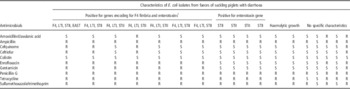
The study was performed in a 1500 sow (Landrace×Large White) farrowing farm in Mecklenburg, Western Pomerania, Germany. Production was with all-in all-out farrowing units followed by cleaning, disinfection and vacancy for 24 h following a group farrowing scheme of ~60 sows at a weekly interval. Replacement gilts were produced in-house, so that no animals of foreign origin entered the herd. Dominant herd health problems consisted of a high incidence of postpartum dysgalactia syndrome (PPDS) in sows and neonatal diarrhoea in piglets. Both CPA positive for β2-toxin gene and enterotoxigenic E. coli (positive for F4 fimbriae and enterotoxins LTI and STII) had frequently been isolated from diarrhoeic piglets during the months before study start. Most of the isolated pathogenic E. coli strains were found to be resistant to several antimicrobial substances (Table 1).
Antimicrobial susceptibility of various Escherichia coli isolates from faeces or intestinal content of suckling piglets suffering from diarrhoea

1 F4=fimbriae 4; LTI=heat-labile toxin I; STII=heat-stable toxin II; EAST=enteroaggregative heat-stable enterotoxin; S=susceptible; R=resistant.
Assessment of bacterial resistance was performed according to the Clinical and Laboratory Standards Institute guidelines by determination of minimal inhibitory concentrations and comparison with published clinical breakpoints.
Listed antimicrobial substances are representatives of different classes of antimicrobials. Respective licensed products for use in swine exist and clinical breakpoints are available.
Sows and gilts were routinely vaccinated against porcine parvovirus and swine erysipelas using commercially inactivated vaccines (Parvoruvac®; Merial, Saint-Priest, France). Because commercial vaccines against E. coli and C. perfringens type C, which had been administered in the past, failed to improve clinical problems, herd-specific vaccination of sows and gilts against isolated E. coli and CPA strains had been introduced before this study was initiated.
Before 5 days of the expected farrowing date, sows were moved to well cleaned and disinfected individual farrowing crates (MS Megades®; Shippers, Kerken, Germany) on slatted floor and were orally dewormed with fenbendazole in feed (Panacur® 4% powder for swine; Intervet GmbH, Unterschleissheim, Germany). Sows had free access to water and were fed twice daily.
On day 114 of gestation, parturition was induced by i.m. injection of 0.7 ml of a synthetic prostaglandin analogue (Estrumate® 250 µg/ml; Intervet GmbH), unless parturition had already started on this day or was imminent. After 24 h of completion of farrowing, sows received an i.m. injection of 2 ml of prostaglandin F2α (10 mg Dinoprost, Dinolytic®; Pfizer, Karlsruhe, Germany) for mechanical emptying of the uterus.
Standard management procedures for piglets on the farm were as follows: grinding of piglets’ canine teeth on the 1st day of life, tail docking and oral administration of 0.7 ml (35 mg) toltrazuril (Baycox®; Bayer Animal Health, Leverkusen, Germany) on the 3rd day of life, i.m. injection of 200 mg iron dextrane (Ursoferran 200 mg/ml; Medistar GmbH, Bernburg, Germany) and castration on day 4 or 5 of life. Before the study was initiated, individual piglets suffering from diarrhoea in the first days of life had been treated either with enrofloxacin, colistin, trimethoprim–sulphonamide or amoxicillin. Piglets were only treated once with one antimicrobial substance and if diarrhoea was still present on the next day, another antimicrobial substance was administered once. This inadequate treatment procedure had been changed before study start.
Experimental design
The study was performed in a subgroup of 29 sows and their 418 newborn piglets out of one farrowing group. Before parturition, sows were randomly assigned to two groups, with 15 sows in group A (treatment) and 14 sows in group B (control). Each group contained two first parity sows.
Farrowings were supervised day and night. Immediately after birth and before first colostrum intake, individual piglets of both groups were marked with an ear tag, weighed, clinically checked for overall health and orally treated with either 1.5 ml of the bacterial cocktail (A, n=228) or 1.5 ml sterile 154 mM sodium chloride (B, n=190). Treatment was repeated on days 2 and 3 of life.
The original litter was kept with the sow during the first 24 h postpartum. If the number of piglets was higher than the functional teats of the sow, heaviest piglets were cross-fostered. Sows with fewer piglets than functional teats were receivers. During the study, piglet numbers did not exceed the capacity of farrowing sows. All piglets had free access to sows’ milk at all times. In case of diarrhoea, individual piglets were orally treated with 1.7 mg of enrofloxacin (Baytril® 0.5% oral solution; Bayer Animal Health)/kg BW.
BW of all piglets were recorded on days 1, 2, 3, 7 and 27 of life. DWG was calculated as gram gained per day. A clinical examination of individual piglets was performed daily and a faecal score was recorded (0=normal, 1=soft faeces, 2=mild diarrhoea, 3=severe diarrhoea) throughout the first 7 days of life. The number of total piglets born, piglets born alive, stillborn and nursed piglets as well as piglet losses were recorded for each litter. Within the 1st week of life, all dead piglets were necropsied following a standardized examination scheme. On day 7, pooled faecal samples of suckling piglets of eight different litters not involved in the study and without toltrazuril treatment on the 3rd day of life were collected for Cystoisospora suis detection.
A clinical examination of sows was performed daily starting 1 day before farrowing until day 7 after farrowing. The examination included rectal temperatures in the morning, adspection, palpation of the mammary glands and evaluation of vaginal discharge. PPDS was defined as body temperature >39.5°C in combination with vaginal discharge, mastitis or lack of milk.
Statistical analyses
Statistical analyses were performed using the statistical software SAS® (SAS Institute Inc., Cary, NC, USA). Because data were not distributed normally, significant differences between the litters of different groups were calculated using the Wilcoxon’s test. Differences in frequencies between both groups such as piglet losses and dead piglets with visual gut alterations were compared using the χ 2 test.
Results
Reproductive performance and growth performance of piglets
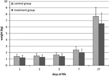
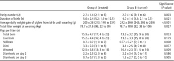
The number of live born, dead and weaned piglets are shown in Table 2. The number of live born piglets per litter did not differ significantly between both groups (15.2±4.4 (A) v. 13.6±3.3 (B)), whereas more stillborn piglets and a longer duration of birth occurred in group A. Significantly, more piglets per litter were weaned in group A (12.5±3.6 (A) v. 10.4±2.3 (B), P=0.009). Overall, piglet losses of 18.0% in group A and 24.7% in group B were not significantly different. The average DWG from birth until weaning was lower in group A piglets (200±26 g (A) v. 242±20 g (B), P<0.001) resulting also in different BW at weaning (6.50±0.99 kg (A) v. 7.86±1.42 kg (B), P=0.003, Figure 1).
Weight of suckling piglets of group A and B on days 1, 2, 3 and 7 after birth and at weaning. Piglets of group A showed lower average daily weight gains from birth until weaning than group B piglets (P<0.001) resulting in different BW at weaning (group A: 6.5±0.99 kg; group B: 7.86±1.42 kg). Bars represent the mean values and the top lines denote the respective SD.

Details of litters, including production data, daily weight gain and incidence of diarrhoea during suckling time

Mean±SD, and median and range within brackets.
Piglet health status
None of the 228 piglets treated showed symptoms of vomiting, diarrhoea or shock, nor did any of them die following oral administration of the cocktail.
On day 2, diarrhoea was observed in 17% of group A piglets compared with 20% of group B piglets. In both groups an average of 2.2 piglets per litter displayed diarrhoea (2.2±2.3 (A) v. 2.2±3.4 (B)). On day 3, 5.3% (group A) and 12.7% (group B) of piglets exhibited diarrhoea. Differences between the two groups were not significant. C. suis was not detectable in any of the pooled samples.
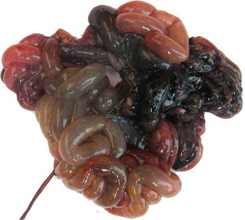
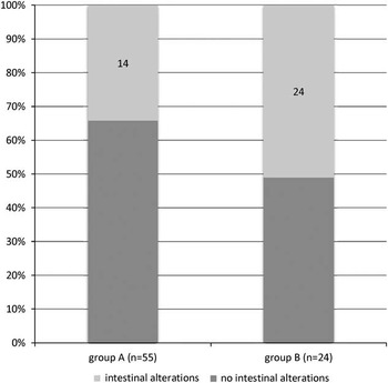
Small intestinal mucosal alterations characterized by a thickened, congested intestinal wall, were found in dead, euthanized and crushed piglets. These findings were less frequent in group A piglets (P=0.004). Necrotizing enteritis was diagnosed (Figures 2 and 3) in three group B piglets.
Pathological gut alterations observed in dead piglets with a higher frequency in group B (P=0.004). Necrotizing enteritis occurred only in group B piglets.

Piglets which died during the 1st week of life due to different reasons (lack of energy, being crushed, being euthanized) were necropsied. The intestinal mucosa was examined for pathomorphological alterations. Significantly fewer gut alterations were found in group A compared with group B (34% v. 51%, P=0.004).

Sows’ health status
The number of functional teats, parity number, occurrence of sows suffering from PPDS and being treated did not differ between the two groups. Overall, 70% of all sows showed symptoms of PPDS. Sows from group A had on average significantly longer durations of birth (5.6±2.4 h (A) v. 4.0±1.4 h (B), P=0.02) and more stillborn piglets (group A: range 0 to 2, group B: range 0 to 1, P=0.008).
Discussion
The present study was inspired by previous experiments in swine, in which either specific bacterial strains (Davidson and Hirsh, Reference Davidson and Hirsh1976) or porcine-derived diverse bacterial cultures (Anderson et al., Reference Anderson, Stanker, Young, Buckley, Genovese, Harvey, DeLoach, Keith and Nisbet1999) had successfully been administered to reduce colonization with pathogenic bacteria (Nurmi et al., Reference Nurmi, Nuotio and Schneitz1992).
In this study, no effect on the incidence of diarrhoea as the main target parameter was observed with the prophylactic administration of potentially beneficial bacteria to suckling piglets. However, it is noteworthy that more piglets were weaned and significantly fewer pathological tissue alterations in the small intestine were observed in treated pigs. DWG from birth to weaning was higher in the control group, so that a negative treatment effect cannot be excluded. However, reduced litter sizes in the control group might have favoured access of individual piglets to the udder and the uptake of milk. Finally, the weaning weights of the whole litters did not differ between the two groups.
Four significant differences were observed in production parameters between the groups: duration of birth and stillborn piglets per litter were interdependent, not influenced by treatment, but may have influenced the outcome of the study, because long duration of birth negatively impacts upon the vitality of piglets.
The other two parameters such as number of weaned piglets per litter and the average DWG until weaning may have been influenced by the treatment, but the causality remains hypothetical.
Several external factors influenced the outcome of the study: litter sizes were large and piglets suffered from colostrum and milk deficiency as main predisposing factors to suckling piglet diarrhoea. A negative effect of farrowing induction on day 114 of gestation due to shorter gestation length and lower birth weight of piglets was reported (Gunvaldsen et al., Reference Gunvaldsen, Waldner and Harding2007). Because piglets from large litters have a higher risk of low birth weight and growth retardation, farrowing induction in this herd can be assessed as an additional risk factor (Baxter et al., Reference Baxter, Rutherford, d’Eath, Arnott, Turner, Sandoe, Mousten, Thorup, Edwards and Lawrence2013). It cannot be excluded that long-term reproductive performance of breeding stock was negatively impacted, because sows were not selected appropriately for physiological reproductive behaviour.
On this farm, piglet diarrhoea had been treated inconsistently with various antimicrobial substances for several months before study start. Selective pressure caused by the implementation of antibiotics results in an increase in resistant bacterial strains on swine farms (Varga et al., Reference Varga, Rajic, McFall, Reid-Smith, Deckert, Checkley and McEwen2009; Gibbons et al., Reference Gibbons, Boland, Egan, Fanning, Markey and Leonard2016) and was also reflected in the resistance spectrum of pathogenic E. coli strains isolated from diarrhoeic animals in this study. This failure of antibiotic therapy of suckling piglet diarrhoea was the first impulsion of the used approach.
During the study, individual treatment of diseased piglets with enrofloxacin very likely interfered with the probiotic cocktail, but had to be accepted for reasons of welfare. Antibacterial treatment of diseased pigs, which interfered with the administered bacteria was not relevant for the assessment of the prophylactic effect of the cocktail.
The second factor which might have interfered with the effect of the bacterial cocktail was the routinely performed herd-specific vaccination of sows. Whether vaccine-induced colostral antibodies had a neutralizing effect on the administered bacterial strains cannot be excluded, but is not probable. The vaccine contained classic enterotoxic E. coli (ETEC) with fimbria as the major antigenic component. In general, colonization factors known from different E. coli strains show high antigenic heterogeneity lacking any cross-protection, which hampers its efficacy under field conditions, where pigs are exposed to various strains (Luo et al., Reference Luo, Qadri, Kansal, Rasko, Sheikh and Fleckenstein2015). For CPA, no immunogenic components aside from the toxins are known so far and attachment does not seem to be a precondition for CPA-associated disease (Songer and Uzal, Reference Songer and Uzal2005). Anti-toxin antibodies have been found to protect against the disease (Springer et al., Reference Springer, Finzel, Florian, Schoepe, Woitow and Selbitz2012), but these might not interfere with the CPA strains administered, which produced no toxins.
The main reason herd-specific sow vaccination failed might have been the high proportion of sows suffering from PPDS leading to deficient uptake of protective colostral and lactogenic antibodies by piglets. Other predisposing factors limiting the effect of vaccines might have been the high daily work load due to weekly group farrowings in separate units resulting in farrowings nearly every day. Hygiene measures might not have been effective enough to reduce the bacterial burden adequately.
Aside from herd factors, the study design might also not have been suitable to detect effects of the bacterial cocktail. The early application of bacteria before first colostrum intake differs from other studies administering probiotics 12 h (Genovese et al., Reference Genovese, Harvey, Anderson and Nisbet2001) or 1 h after birth (Davidson and Hirsh, Reference Davidson and Hirsh1976). Although an optimal interaction of the introduced strains with host cells before contact with pathogens can be assumed with this approach, immune modulation by colostral components, which physiologically balance bacterial gut colonization, was lacking and negative effects on piglet development cannot be excluded (Nguyen et al., Reference Nguyen, Yuan, Azevedo, Jeong, Gonzalez and Saif2007; Levast et al., Reference Levast, Berri, Wilson, Meurens and Salmon2014).
Different mechanisms to exclude pathogens from the gut using probiotics have been proposed: (1) stimulation of the immune system, (2) production of antibacterial substances, (3) modification of the environment which favours the development of a beneficial bacterial population and (4) competition for attachment sites and for nutrients (Anderson et al., Reference Anderson, Stanker, Young, Buckley, Genovese, Harvey, DeLoach, Keith and Nisbet1999; Wassenaar et al., Reference Wassenaar, Beimfohr, Geske and Zimmermann2014). Adverse clinical effects might occasionally happen even with established probiotics with a long history of safe use (Gronbach et al., Reference Gronbach, Eberle, Muller, Olschlager, Dobrindt, Leithauser, Niess, Doring, Reimann, Autenrieth and Frick2010; Guenther et al., Reference Guenther, Straube, Pfister, Guenther and Huebler2010). Genome sequencing can facilitate the decision as to whether a bacterial strain can be classified as harmless, but revealed virulence-associated genes might in part contribute to the positive effects seen in vivo and have also been found in established probiotic strains (Vejborg et al., Reference Vejborg, Friis, Hancock, Schembri and Klemm2010; Wassenaar and Gunzer, Reference Wassenaar and Gunzer2015; Wassenaar et al., Reference Wassenaar, Zschüttig, Beimfohr, Geske, Auerbach, Cook, Zimmermann and Gunzer2015). The strains used in this clinical field trial were only characterized by routine diagnostic methods, because a further pre-selection of promising strains using functional assays in vitro is not yet available. No diagnostic system to differentiate between CPA strains belonging to the physiological gut flora and disease-causing strains is available as yet (Amtsberg et al., Reference Amtsberg, Bisping, El Sukhon, Matthiesen and Krabisch1976; Springer et al., Reference Springer, Finzel, Florian, Schoepe, Woitow and Selbitz2012). The observations of Melin et al. (Reference Melin, Jensen-Waern, Johannisson, Ederoth, Katouli and Wallgren1997), that piglets already early in life were colonized with CPA, led to the decision to include two CPA strains with no toxin production in vitro in the bacterial cocktail.
Compared with all other studies where piglets were euthanized at 36 h of life (Davidson and Hirsh, Reference Davidson and Hirsh1976), 40 to 48 h of life (Siggers et al., Reference Siggers, Siggers, Boye, Thymann, Molbak, Leser, Jensen and Sangild2008) or 5 days after challenge (Genovese et al., Reference Genovese, Anderson, Harvey and Nisbet2000 and Reference Genovese, Harvey, Anderson and Nisbet2001), this trial can be considered a long-time study under field conditions.
Conclusion
The gut flora stabilizing effect of the bacterial cocktail administered to newborn piglets was not clearly visible in this study, although the treatment possibly led to a reduction in intestinal pathoanatomical alterations. A higher number of weaned piglets and a lower average DWG were found in group A, but finally litter weights at weaning did not differ. Further studies should be performed to optimize bacterial doses, cocktail composition and treatment intervals in herds with a good sow health status.
Acknowledgements
The authors thank Dr. Katrin Strutzberg-Minder and Dr. Karen Dohmann, Innovative Veterinary Diagnostic GmbH, for their excellent support in characterization of bacterial isolates. We thank Dr. Hartmut Rohrmann for the fruitful collaboration on farm.