Implications
Lipomatous muscle myopathy is a degenerative muscle pathology sporadically reported in Piedmontese breed and characterized by a different grade of infiltration of adipose tissue, involving multiple or single muscles or rarely the whole carcass. The present data are the first detailed contribution to understand the aetiology of this disease that has a profound economic effect on the cattle industry.
Introduction
The muscular hypertrophy, or double-muscle phenotype, is a heritable condition in cattle that primarily results from an increase in number of muscle fibres (hyperplasia) rather than the enlargement of individual muscle fibres (hypertrophy; Hanset et al., Reference Hanset, Michaux, Dessy-Doize and Burtonboy1982). The relative increase in fibre number is observed early in pregnancy (Swatland and Kieffer, Reference Swatland and Kieffer1974) and results in a calf possessing nearly twice the number of muscle fibres at birth. The Piedmontese breed is characterized by a high frequency of ‘double-muscled’ character and is mainly known for its superior yields of lean and tender meat (Kambadur et al., Reference Kambadur, Sharma, Smith and Bass1997). The meat from the double-muscled Piedmontese compared with normal animals and to other breeds has higher water and protein content. The intramuscular fat content is very low, ∼1%, and consequently the triacylglycerol content is greatly reduced, as a result of lower fat deposition, with a positive increase of the polyunsaturated/saturated fatty acid ratio (Barge et al., Reference Barge, Brugiapaglia, Destefanis and Mazzocco1993).
The meat of the hypertrophied Piedmontese animals is also very tender because of a large reduction in muscle collagen and a lower proportion of stable non-reducible cross-links (Boccard, Reference Boccard1981; Destefanis et al., Reference Destefanis, Barge and Brugiapaglia1993).
Italian literature shows the occasional presence of lipomatous muscle dystrophy, also called ‘steatosis’ by Fontana (Reference Fontana1963), in this breed. This disease, characterized by the substitution of muscle tissue with adipose tissue, which can appear as large white veins or as a normal marbling, is sporadically reported in cattle, pigs, and rarely in sheep, horses and dogs (Marcato et al., Reference Marcato, Rosmini and Bazzo2002).
In cattle, this disease is usually considered an incidental finding during necropsy or slaughter, but sometimes it can be so extensive as to damage the whole carcass. It can affect individual muscles of one limb or more frequently several muscles, also may present as bilateral, symmetrical or asymmetrical.
The muscles have a macroscopic reticulated appearance in the cross sections, whereas in the longitudinal sections a white streak is quite abundant. The muscle volume remains unchanged, and the border between healthy and pathological parts of the same muscle results to be nuanced (Van Vleet and Valentine, Reference Van Vleet and Valentine2007).
To date, no specific research has been aimed to understand the aetiology of the disease. Various authors (Fontana, Reference Fontana1963; Link et al., Reference Link, Cassens, Bray and Kowalczyk1967; Guarda and Castagnaro, Reference Guarda and Castagnaro2002; Marcato et al., Reference Marcato, Rosmini and Bazzo2002) have proposed several hypotheses, but did not provide a definitive answer.
The authors stated that the pathology might be due to abnormal development and distribution of blood vessels or alteration of the spinal marrow or resulting from nutritional disturbance or a congenital defect. None of these theories have evidence or proof to back it up, but probably the disease is multifactorial, with genetic, vascular and nutritional factors involved.
Currently, there is no specific classification of this muscular disorder, nor a detailed assessment of the chemical and physical characteristics of the meat affected.
The aim of the present study was to report the macroscopical and histopathological features of the muscle affected by this pathology and to describe the changes of the chemical and physical characteristics of the meat.
Material and methods
The muscle samples of 40 Piedmontese cattle from different herds of Piedmont region were examined. The animals included 37 beef cattle (19 females and 18 males) aged from 14 to 32 months, and 3 cows aged 8, 10 and 11 years. The control animals were five Piedmontese subjects, aged from 15 months to 11 years, reared in the Piedmont region, with no evident neuromuscular disorders.
Muscle tissues were examined macroscopically and then collected for histopathological investigations, chemical and physical analysis.
To investigate the possible genetic background of the disorder, a preliminary analysis of the genetic relationship between 32 affected animals was carried out.
Histopathology
Muscle samples were collected at the slaughterhouse and immediately sent to the laboratories of the Section of Pathologic Anatomy of the Department of Animal Pathology of Torino University. They were cut into 1 cm3 pieces (parallel to the muscle fibres), frozen in isopentane and cooled using liquid nitrogen and stored at −80°C until subsequent analyses. Slide sections (10 μm thick) of muscle tissue were obtained using a cryostat and stained by the following histological and histochemical techniques: haematoxylin and eosin (H&E); modified Gomori trichrome; periodic acid-Schiff (PAS); red oil stains, Sudan black, Sudan III, adenosine triphosphatase (ATPase), cytochrome C oxidase (COX); succinate dehydrogenase (SDH) and reduced nicotinamide adenine dinucleotide–tetrazolium reductase (NADH-TR).
Immunohistochemical stainings were also made on selected 8 μm sectioned frozen muscle specimens. Primary antibodies employed in IHC included reagents specific to characterize the inflammatory cells (B lymphocytes, CD79 – mouse anti-CD79, 1 in 25; Dako, Glostrup, Denmark; code no. M7051 – (Mason et al., Reference Mason, Cordell, Tse, van Dongen, van Noesel, Micklem, Pulford, Valensi, Comans-Bitter, Borst and Gatter1991); T lymphocytes, CD4 – mouse anti-CD4, 1 in 100; VMRD Inc., Pullman, WA, USA, cell line CACT138A – (Davis et al., Reference Davis, Hamilton, Park, Larsen, Wyatt and Okada1990); T lymphocytes, CD8 – mouse anti-CD8, 1 in 100; VMRD Inc., Pullman WA, USA, cell line CACT80C – (Gutierrez et al., Reference Gutierrez, Forster, McConnell, Cassidy, Pollock and Bryson1999) and major histocompatibility complex (MHC) type I, MHCI – mouse anti-MHCI,1 in 200; VMRD Inc., Pullman, WA, USA, cell line H58A (Davis et al., Reference Davis, Marusic, Lewin, Splitter, Perryman, McGuire and Gorham1987) and to identify the different types of muscular fibres (F18-mouse anti-myosin heavy chain, 1 : 1; Developmental studies Hybridoma Bank, Iowa, IA, USA, Mab1628 – mouse anti-slow muscle myosin (Millipore Corporation, Billerica, MA, USA), 1 : 30; Millipore, UK and A4.74 – mouse anti-myosin (human fast fibres), 1 : 1; Developmental studies Hybridoma Bank, Iowa, IA, USA). The sections were incubated with 1% hydrogen peroxide in methanol for 20 min at room temperature (RT), followed by incubation with 5% normal goat serum for 1 h at RT, then 2 h at RT with primary antibodies. Secondary detection was made by use of an avidin–biotin complex (ABC) kit (Pierce, 32020, Thermo Fisher Scientific Inc., Rockford, IL, USA) or a MACH Universal HRP-Polymer Detection kit (Biocare Medical, Concord, CA, USA, code no. M4U534). Immunolabelling was ‘visualized’ with diaminobenzidine–tetrahydrochloride (Sigma-Aldrich, St Louis, MO, USA).
All the slide sections were examined under a light microscope by three pathologists. The severity of the most important microscopical lesions, evaluated on H&E stained sections (fat infiltration, neoangiogenesis and inflammatory cells), was classified using a following semiquantitative scoring system: no lesions (0), a low number of focal to multifocal lesions (1), a moderate number of lesions, disseminated through one or more areas (2) or diffuse and severe lesions (3).
Chemical and physical characteristics
Chemical and physical characteristics were performed on nine beef samples.
Ultimate pH measurements were taken in duplicate using a Crison portable pH meter (Crison Instruments, S.A., Alella, Spain) fitted with a spear type electrode and an automatic temperature compensation probe.
Proximate analyses were carried out according to the Association of Official Analytical Chemists (AOAC, 1970) methods. Tissue samples were weighed, dried at 125°C for 5 h and reweighed to determine the water content. The meat was further lyophilized and ground in a blender for analyses of protein and intramuscular fat. Nitrogen was determined by the Kjeldahl method using a Büchi System apparatus (Büchi Labortechnik, Flawil, Switzerland). The CP was then calculated by multiplying N × 6.25. Lipid extraction of intramuscular fat was determined by the Soxhlet method using a Büchi Extraction System.
Surface Lightness (L*), redness (a*) and yellowness (b*) colour indices were obtained in duplicate after 1 h of blooming at 4°C with a Minolta CR331C Colorimeter (Minolta Camera Co., Japan) calibrated on D65 illuminant. Hue angle, which describes the fundamental colour of a substance, and Chroma, which describes the vividness of a colour, were calculated as tan−1 (b*/a*) and (a*2 + b*2)0.5, respectively (International Commission on Illumination CIE, 1978).
Two methods for assessing texture characteristics of meat, Warner–Bratzler Shear Force and Texture Profile Analysis, were employed. For each sample, 8 to 10 blocks 1 cm2 cross-section area cut parallel to the muscle fibre direction were obtained. An Instron 5543 universal testing machine (Instron Corporation, USA) was used in both instrumental tests. The shear force evaluation was assessed using a Warner–Bratzler device, which measures the peak force required to cut the parallelepiped in half perpendicular to its length. The samples were sheared perpendicular to the fibre at 200 mm/min crosshead speed. The peak force (N) generated was recorded for each sample, and when necessary, the mean of all the samples for a given muscle was used for statistical analysis. Texture Profile Analysis was evaluated using a modified compression device that avoids transversal elongation of the samples (Lepetit and Culioli, Reference Lepetit and Culioli1994). Each sample underwent two cycles of 80% compression. The parameters determined were: hardness (N), the maximum peak force required to compress the sample during the first compression cycle; cohesiveness, the area of work during the second compression cycle divided by the area of work during the first compression cycle (dimensionless); springiness (dimensionless), the ratio of the distance of contact with the sample during the second compression (L2) to the same distance for the first compression (L1); gumminess (N), the product of hardness x cohesiveness; chewiness (N), the product of hardness x cohesiveness x springiness.
Results
All the animals showed an excellent state of nutrition and none of them had clinical signs related to muscular or nervous disease.
Gross pathology revealed a different grade of infiltration of adipose tissue, involving multiple or single muscles. Only in four animals (9 months, 8, 10 and 11 years) the whole carcass was involved.
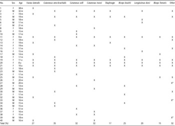
The most affected regions were the ventral abdomen and the shoulder, especially the cutaneous colli and trunci, the muscles of the thoracic group, the diaphragm and vastus lateralis. Table 1 shows the infiltrated muscles in different animals.
Affected muscles in different animals

*not identified muscles.
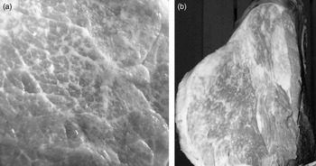
Macroscopically the lesions consisted in a variable deposition of fat tissue in muscle stroma, in order to enhance the longitudinal streak in sagittal section and giving a cross-linked appearance in cross section (Figure 1 a and b). The limits of the infiltrative process were not clear. However, the thickness of the muscle remained intact and no changes in the contours of the muscles were present.
(a) cross section of vastus lateralis muscle with a severe fatty infiltration giving a cross-linked appearance. (b) posterior limb showing a variable deposition of fat tissue in muscle stroma.

The preliminary analysis of the genetic relationships, considering the average of 3.125 complete generations, revealed no ancestors potentially responsible for the transmission of the disorder. The mean inbreeding coefficient observed was very low (0.0196).
Histopathology
The following features were recorded: fat tissue deposition, morphology and metabolic changes of the muscle fibres, characteristic of the interstitial tissue, blood vessels and inflammatory cells.
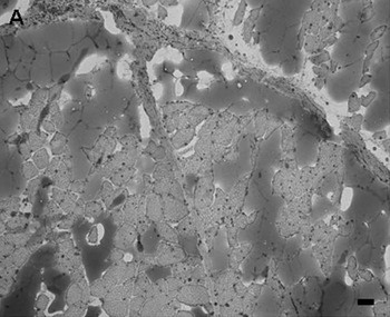
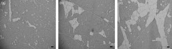
The most important feature was a variable infiltration of adipose tissue changing in distribution and severity between the samples. Histochemical stains (Sudan black, Sudan III and oil red stainings) confirmed this fat deposition (Figure 2). Only 13 cases (32.5%) showed a severe infiltration (score 3). The majority of the muscles (45%) showed a slight infiltration (score 1), whereas nine cases (22.5%) showed a moderate fat deposition (score 2; Figure 3a, b and c).
Fat deposition. Sudan III (100x) Bar = 50 μm.

Score system for fat infiltration. (a) slight infiltration; (b) moderate fat deposition; (c) severe infiltration. Haematoxylin and Eosin (100x) Bar = 50 μm.

The fibres, especially near fatty infiltration, frequently showed an increase in cross-sectional diameter accompanied by compression of neighbouring fibres. Size variation of fibres (small and hypertrophic fibre) was observed in 30 cases (75%). Increased numbers of fibres with centrally located nuclei were present in 25 cases (62.5%), pyknotic nuclear clumps in 1 case (2.5%) and necrotic fibres in 9 cases (25.5%).
Histochemical stainings did not reveal metabolic changes in affected muscles compared with the control samples.
No differences in size were observed between the muscle fibres.
Increase of connective tissue was detected in 27 cases (67.5%).
Twenty samples (50%) showed different grade of neoangiogenesis. The semiquantitative score classifies the vascular neoformation as grade 1 (16 cases, 40%), grade 2 (2 cases, 5%) and grade 3 (2 cases, 5%). This feature was especially reported at the periphery of the areas infiltrated by adipose tissue and was frequently associated with the presence of inflammatory cells or necrotic fibres.
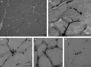
Non-suppurative inflammatory cells infiltrations were also seen in 26 cases (65%), often organized in small clusters (Figure 4a). According to the distribution and numbers of non-suppurative cells, the inflammatory infiltrates were classified in score 1 (52.5% of cases), score 2 (7.5% of cases) and score 3 (5% of cases). Sporadic focal immunopositivity for MCHI, CD8, CD4 and CD79 were detected (Figure 4b, c, d and e).
Muscle tissue. (a) non-suppurative inflammatory cells infiltrations organized in small clusters. Haematoxylin and Eosin (400x) Bar = 100 μm. (b) sarcolemmal immunopositivity for MHC1 (←). Immunohistochemistry mouse anti-MHC1, counterstained with haematoxylin (600x) Bar = 100 μm. (c) focal T lymphocytes positive for CD8 (←). Immunohistochemistry mouse anti-CD8, counterstained with haematoxylin (600x) Bar = 100 μm. (d) focal T lymphocytes positive for CD4 (←). Immunohistochemistry mouse anti-CD4, counterstained with haematoxylin (600x) Bar = 100 μm. (e) focal B lymphocytes positive for CD79 (←). Immunohistochemistry mouse anti-CD79, counterstained with haematoxylin (600x) Bar = 100 μm. MHC = major histocompatibility complex.

Chemical and physical characteristics
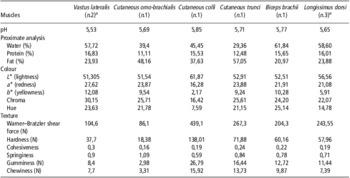
The results of chemical and physical analyses are shown in Table 2.
Chemical and physical characteristics of the affected muscles

amean value.
Muscle pH has a large effect on muscle colour and plays an important role in the stability and acceptability of meat. Muscle pH affected by the myopathy ranged from 5.53 (vastus lateralis) to 5.85 (cutaneous colli). The pH of longissimus dorsi muscle was slightly higher than that observed by Destefanis et al. (Reference Destefanis, Barge and Brugiapaglia1994) in the same muscle of normal animals of Piedmontese breed (5.65 v. 5.48). In any case, the pH value fell into the normal range.
As regards proximate composition, it is important to stress the extremely high percentage of fat and the low water and protein content.
The biceps brachii muscle had the lowest fat content (20.97%), whereas the cutaneous trunci muscle showed the highest percentage (57.05%). The intramuscular fat exhibited also a large variation.
Of the six muscle types, the vastus lateralis and the cutaneous omo-brachialis had the highest (16.83%) and the lowest (11.11%) protein content, respectively.
In comparison with these data, Destefanis et al. (Reference Destefanis, Barge and Brugiapaglia1993) observed in the longissimus dorsi muscle of Piedmontese animals a very low fat content (0.71%) and a water and protein content of 74.97% and 22.25%, respectively.
As regards the colour, it must be underlined the high lightness values. Lightness ranged from 51.31 of vastus lateralis to 61.87 of cutaneous colli. Compared with longissimus dorsi muscle of normal animals (Destefanis et al., Reference Destefanis, Barge and Brugiapaglia1994) lightness was very high (35.05 v. 56.56).
Tenderness was estimated indirectly as Warner–Bratzler shear force. The cutaneous omo-brachialis muscle had the lowest shear force value, whereas the cutaneous colli muscle had the highest value. Mean shear force value of the longissimus dorsi muscle affected by the myopathy was extremely higher than that value reported by Destefanis et al. (Reference Destefanis, Barge and Brugiapaglia1993) in the same muscle of normal animals of Piedmontese breed (244 N v. 87 N).
Compared with the meat not showing the syndrome, also hardness (at 80% compression), which represents a measure of connective tissue strength, showed higher values (57.96 N v. 16.02 N, unpublished data).
The gumminess and chewiness values had also high values. The values were ∼4 and 3 times respectively higher in comparison to normal tissue.
Discussion
The present study examined the pathological features and the chemical and physical characteristics of several muscles of Piedmontese cattle affected by lipomatous muscular myopathy.
Currently, no specific classification of this muscular disorder exists. Most of the muscular pathology veterinary books call this condition as muscle steatosis or ‘lipomatosis’ (Valentine, Reference Valentine2007), whereas the Italian literature classifies this pathology as a muscular dystrophy (Marcato et al., Reference Marcato, Rosmini and Bazzo2002) or considers it as a pseudohypertrophy of the muscle (Guarda and Castagnaro, Reference Guarda and Castagnaro2002). Few data are reported on histological and histochemical features (Fontana, Reference Fontana1963); generally only an increase of fat deposition within muscles replacing the muscle fibres is described, sometimes accompagnied by variable degree of fibrosis and peripheral fibre atrophy (Guarda and Castagnaro, Reference Guarda and Castagnaro2002; Marcato et al., Reference Marcato, Rosmini and Bazzo2002; Valentine, Reference Valentine2007).
No data are reported about the chemical and physical characteristics of muscles affected by this systemic disorder.
Only a few number of case reports are present in the literature about fatty degeneration of single portion of muscles or groups of muscles in ruminants, most of which are assumed to be caused by denervation (Hartman and Shorland, Reference Hartman and Shorland1957; Cook, Reference Cook1963; Link et al., Reference Link, Cassens, Bray and Kowalczyk1967; Beck et al., Reference Beck, Marchello and Ray1971; Marchello et al., Reference Marchello, Brown and Ray1971; George et al., Reference George, Morgan, Glock, Tatum, Schmidt, Sofos, Cowman and Smith1995).
In literature, a muscle steatosis indicates a condition characterized by abnormal retention of lipids within the cell generally associated to acquired condition (Marcato et al., Reference Marcato, Rosmini and Bazzo2002). Common usage of the word myopathy is restricted to describe a disorder of muscle generally characterized by different grade of degeneration of muscle fibres and high serum level of muscle enzymes. In humans, myopathy is divided into several entities, such as diseases of the lower motor neuron, dystrophies, congenital myopathies, nutritional myopathies, metabolic and endocrine myopathies, inflammatory myopathies and a miscellaneous group (Goedegebuure et al., Reference Goedegebuure, Hartman and Hoebe1983). Similarly to humans the classification of myopathies in animals is subject to modification based on findings of on-going genetic and pathogenetic studies as well as on changes in disease definitions (Van Vleet and Valentine, Reference Van Vleet and Valentine2007). Therefore, a muscular dystrophy represents a degenerative myopathy due to faulty nutrition of the muscles frequently genetically determined, although the way it is inherited is not the same for all types of the disease. The one unifying feature of the dystrophies is regarded as their muscle histological findings, with variations in fibre size, fibre necrosis, invasion by macrophages, and ultimately, replacement by fat and connective tissue; they are not diagnostic for any particular type (Jeffrey et al., Reference Jeffrey, Hopper and Bradley1985; Marden et al., Reference Marden, Connolly, Siegel and Rubin2005; Kobayashi et al., Reference Kobayashi, Nakamura, Hasegawa, Fujita, Orima and Takeda2009). The replacement of muscle tissue with fat is also observed in human congenital muscular myopathies and innervation defects (Dubowitz and Sewry, Reference Dubowitz and Sewry2007), but the distribution of large and small fibres in clusters or large groups could be a useful criteria for differentiating neurogenic changes secondary to denervating process (McGavin and Baynes, Reference McGavin and Baynes1969; Jeffrey et al., Reference Jeffrey, Hopper and Bradley1985).
Exogenous stimuli, such as pharmacological agents injected, may be applied to the bovine muscles with resulting increased connective and fat deposition (Beck et al., Reference Beck, Marchello and Ray1971; George et al., Reference George, Morgan, Glock, Tatum, Schmidt, Sofos, Cowman and Smith1995). This condition is considered a ‘pathologically induced lipid deposition’ that according to Gutmann and Zelena (Reference Gutmann and Zelena1962) expressed the concept that fat cells replace atrophied muscle tissue during the late stages of denervation atrophy.
In the present study, the affected muscles showed atrophic and hypertrophic fibres randomly distributed. ATPase and immunochemistry confirmed that variation in size affected both fibre types. The multifocal distribution of muscles affected, different from case to case, represents another feature that make unlikely a neurogenic or vascular aetiopathogenesis.
The changes occurring in sarcolemmal nuclei, such as internal nuclei and nuclear clumps, are considered dystrophic changes. Numerous fibres with centrally located nuclei were reported in the present study, sometimes associated with pyknotic nuclear clumps and necrotic fibres.
Internal nuclei were reported in cattle dystrophy of the diaphragmatic muscles (Goedegebuure et al., Reference Goedegebuure, Hartman and Hoebe1983) and in some human dystrophy as myotonic dystrophy (Casanova and Jerusalem, Reference Casanova and Jerusalem1979; Nadaj-Pakleza et al., Reference Nadaj-Pakleza, Lusakowska, Sułek-Piątkowska, Krysa, Rajkiewicz, Kwieciński and Kamińska2011). The presence of nuclear clumps represents a marker of fibres atrophy and it also occurs in limb girdle and chronic dystrophies (Dubowitz and Sewry, Reference Dubowitz and Sewry2007).
Focal mononuclear cells, classified by immunohistochemistry as macrophages or lymphocytes, were detected in the present samples too. The presence of these cells, the increase of connective tissue and the neoangiogenesis reported in the 46.6% of cases, confirm the existence of an inflammatory reaction and of a process of repair in response to the dystrophic process. Activated macrophages infact can phagocytize the necrotic tissue and neovascularization accelerates this action by providing a vascular supply to the regenerating and granulation tissue.
In the present research, morphological lesions in muscle tissue are associated with severe changes of chemical and physical characteristics of the meat affected compared with standard parameters of the Piedmontese cattle. Marbling, normally, appears in fresh meat as white flecks or a streak of fat, as subjectively assessed by meat graders, and is related to muscle fat content. Among breeds, marbling flecks are different in the quantity, structure and distribution in different muscles (Albrecht et al., Reference Albrecht, Teuscher, Ender and Wegner2006). The Piedmontese breed is infact characterized by a very low fat content, whereas the Japanese Black breed contains enormous amounts of fat within the muscle compared with Holstein cattle that, even if kept in the same production system, store about 14% more fat in the longissimus dorsi (Albrecht et al., Reference Albrecht, Gotoh, Ebara, Xu, Viergutz, Nürnberg, Maak and Wegner2011).
Intramuscular fat content of cattle muscle is an important component of traits that influence eating quality, such as meat tenderness, juiciness and taste (Hovenier et al., Reference Hovenier, Brascamp, Kanis, Van der Werf and Wassenberg1993).
In the present research a variable deposition of fat tissue in muscle stroma could sometimes only increase the marbling flecks, but in cases of severe infiltration the muscle masses may appear largely replaced by fatty tissue to render them unfit for sale.
Considering the very low fatness of the hyperthrophied animals, like the Piedmontese breed, the intramuscular fat content of muscles affected by this pathology is remarkably high.
As regards colour, all the muscles appeared pale because of the great amount of marbling, which reflects back most of the light.
Also, the rheological properties were markedly influenced by this pathology. The high values related to texture indicated that the meat was extremely tough, probably owing to the high concentration of the connective tissue.
In general, considering the meat characteristics of hypertrophied animals, even if we have reference data only for longissimus dorsi muscle (Tatum et al., Reference Tatum, Gronewald, Seideman and Lamm1990), nevertheless we can reasonably suppose that the lipomatous myopathy induced similar alteration also in the other muscles.
Several findings observed in the present cases lead one to classify this disease as a muscular dystrophy characterized by variations in fibre size, necrosis, mononuclear cells infiltration, increase of connective tissue and, especially, replacement by fat, even if some authors report the absence of degenerative aspects of fibres (Marcato et al., Reference Marcato, Rosmini and Bazzo2002). Muscular dystrophies are an heterogeneous group of degenerative muscle disorders confirmed or suspected to have a hereditary predisposition (Campbell, Reference Campbell1995; Sewry, Reference Sewry2010). Considerable progresses have been made in recent years in the identification of causative genes, but in veterinary medicine the number of inherited muscle disorders in which the gene defect is known remain very limited (Shelton and Engvall, Reference Shelton and Engvall2002; Van Vleet and Valentine, Reference Van Vleet and Valentine2007).
The role of genetic factors in the aetiopathogenesis of this muscular disorder remains to be evaluated even if a preliminary analysis of the genetic relationships performed on 32 cattle seems to exclude a simple Mendelian inheritance. A large-scale study to investigate the existence of loci associated with this disorder on Piedmontese cattle of both sexes would be appropriate.
Acknowledgements
The authors thank Dr O. Paciello from University of Naples, Italy and Dr R. Piercy from Royal Veterinary College of London, UK, for some immunohistochemical investigations. They extend their special thanks to the ANABORAPI for providing the data need for the genetic analysis and to Dr S. Capaldo and Dr V. Panero for providing the majority of the samples.