Tetralogy of Fallot is a complex form of congenital heart disease first described by Neils Stensen, but identified as a clinical entity by Arthur Louis Etienne Fallot.Reference Fallot1, Reference Marquis2 He described the coexistence of four discrete cardiac malformations, namely a ventricular septal defect, subpulmonary stenosis, overriding of the aorta, and right ventricular hypertrophy. Tetralogy of Fallot is the most common form of cyanotic congenital cardiac disease, with a prevalence of 4.7 for each 10,000 births per year in some regions of the United States of America.Reference Reller, Strickland, Riehle-Colarusso, Mahle and Correa3 As described in the previous review, there is a wide range of anatomic phenotypes within tetralogy of Fallot. An accurate anatomical assessment of the intracardiac abnormalities, therefore, is crucial prior to consideration of any surgical intervention. Echocardiography with the use of colour and spectral Doppler modalities provides the most clinically useful non-invasive test for complete anatomical and functional assessment of the heart in patients with tetralogy of Fallot. Echocardiograms can define intracardiac anatomy, anatomical variations, associated congenital cardiac defects, and biventricular function, allowing physicians to provide surgical and medical care for the patients diagnosed with the entity.
Anatomy
As emphasized in the previous review, tetralogy of Fallot is distinguished from the normal heart on the basis of antero-cephalad malalignment of the muscular outlet, or conal, septum, or its remnant, which together with an abnormal relationship with the septoparietal trabeculations produces subpulmonary infundibular stenosis.Reference Anderson and Weinberg4 Variations in the phenotypic morphology depend on the margins of the ventricular septal defect, the extent of aortic override, and the degree and level of pulmonary obstruction.Reference Anderson and Weinberg4 The ventricular septal defect is typically perimembranous, but may have exclusively muscular margins when viewed from the right ventricle, or can extend to become doubly committed and juxta-arterial. The aortic valve typically overrides the crest of the ventricular septum. If the degree of override is such that more than half of the circumference is supported within the right ventricle, then tetralogy co-exists with the ventriculo-arterial connection of double outlet right ventricle.Reference Anderson and Weinberg4 Others support the notion that tetralogy of Fallot cannot coexist with double outlet right ventricle and that by definition, the aorta is assigned to the left ventricle in tetralogy since there is continuity between the leaflets of the aortic and mitral valves, in other words absence of a completely muscular subpulmonary infundibulum. There are multiple substrates for obstruction to outflow from the right ventricle. In addition to the infundibular stenosis, pulmonary valvar stenosis is common, with many patients having bifoliate pulmonary valves (also called bicuspid pulmonary valves or bileaflet pulmonary valves). Additional stenosis can be found at the pulmonary sinutubular junction, or within the branches of the pulmonary arteries, which can be additionally hypoplastic. Peripheral pulmonary stenosis can be seen, most commonly within the left pulmonary artery at the level of the insertion of the arterial duct.Reference Anderson and Weinberg4, Reference Suzuki, Ho, Anderson and Deanfield5 Tetralogy of Fallot can be associated with many other common congenital cardiac malformations. Recognition of the various associated anomalies is important prior to pre-operative planning of the surgical repair. Anomalies include a right aortic arch, which occurs in about one-third of patients with tetralogy of Fallot.Reference Suzuki, Ho, Anderson and Deanfield5, Reference Ramaswamy, Lytrivi and Thanjan6 An aberrant subclavian artery may also coexist.Reference Ramaswamy, Lytrivi and Thanjan6 These arch anomalies are particularly common in children with 22q11 deletion syndrome. It is important to establish the side and pattern of branching of the aortic arch prior to contemplating any palliative procedures. Coronary arterial abnormalities are frequent, with origin of the anterior interventricular artery from the right coronary artery being particularly significant. Indeed, the presence of any major coronary artery crossing the right ventricular outflow tract may preclude transjunctional incisions, complicating the surgical reconstruction. (A transjunctional incision is often called a “transannular incision”.) Atrioventricular septal defect can also occur, especially in children with Down’s syndrome. This association can affect the timing of the repair, as most surgeons would prefer to repair an atrioventricular septal defect after three months of age, which can lead to the need for surgical palliation with a shunt if significant cyanosis develops at less than three months of age. Tetralogy can also exist in association with absent pulmonary valve leaflets whereby the pulmonary leaflets are vestigial. As a result of this anomaly, there is both pulmonary stenosis and significant pulmonary regurgitation which produces right ventricular outflow tract dilation. The central pulmonary arteries are frequently markely dilated and the arterial duct is frequently absent. Mortality for this lesion is higher than for the usual form of tetralogy of Fallot. Additional sources of flow of blood to the lungs, such as aorto-pulmonary collateral arteries or bilateral arterial ducts, may also exist, especially in patients with pulmonary atresia. The precise delineation of all sources of pulmonary flow helps planning of surgical pulmonary arterial reconstruction. Many times echocardiography alone is insufficient for this purpose, and the use of cardiac magnetic resonance imaging, computerized tomographic angiography, or cardiac catheterization with angiography, may be necessary to evaluate fully the pulmonary arterial pathways.
Other associated defects to be excluded prior to surgical palliation or repair include systemic venous anomalies, such as a persistence of the left superior caval vein. This occurs in approximately one-tenth of patients with tetralogy of Fallot, and will influence the choice of cannulation for cardio-pulmonary bypass.Reference Suzuki, Ho, Anderson and Deanfield5 Defects within the oval fossa, and additional ventricular septal defects, should also be indentified if present, since these may need to be addressed during the surgical repair. Rarely, obstruction within the left ventricular outflow tract, such as bifoliate aortic valves, aortic valvar stenosis, or coarctation, can co-exist with tetralogy.Reference Aru, Juraszek, Moskowitz and Van Praagh7, Reference Tateishi, Koide and Mizukami8
Surgical intervention
The surgical approach to the patient with tetralogy of Fallot can be as varied as the spectrum of co-existing disease. The individual preference of the surgeon, the age and size of the patient, the experience of the institution, and the support provided by the regional cardiovascular team are all important factors in pre-surgical planning. The goals of surgical intervention are to establish adequate flow of blood to the lungs, and to limit the degree of pulmonary regurgitation, along with the morbidity and mortality associated with surgical procedures. Ideally, the number of future surgical interventions required by the patient over their life-time should also be limited. Tetralogy can exist with varying degrees of pulmonary stenosis, with a common atrioventricular junction, with so-called absence of the leaflets of the pulmonary valve, or with pulmonary atresia rather than stenosis.Reference Jacobs, Jacobs, Mavroudis, Lacour-Gayet and Tchervenkov9 For this discussion, we will review only the surgical approach to tetralogy of Fallot with varying degrees of pulmonary stenosis.
The initial surgical intervention may be a complete repair, with closure of the ventricular septal defect and creation of laminar flow across the right ventricular outflow tract. Alternatively, initial palliation may be achieved with creation of a systemic-to-pulmonary arterial shunt, delaying subsequent repair until later in life. Shunts are typically performed in patients less than three months of age. Recent trends have been to push towards earlier complete repairs. Many centres, nonetheless, continue to opt for palliative procedures in young infants.Reference Jacobs, Jacobs, Mavroudis, Lacour-Gayet and Tchervenkov9–Reference Kantorova, Zbieranek, Sauer, Lilje, Haun and Hraska11 Recently, many teams have pursued so-called valve-sparing repairs, as opposed to insertion of transjunctional patches.Reference Stewart, Backer, Young and Mavroudis12 (A transjunctional patch is often called a “transannular patch”.) Such patches, usually considered transannular, are placed across the ventriculo-pulmonary junction to relieve the pulmonary stenosis, but this usually creates significant pulmonary regurgitation. A valve-sparing approach is achieved by resecting the infundibular muscle bundles, repairing the native pulmonary valve, and using infundibular and/or supravalvar patches. The aim of this approach is to maintain a functional pulmonary valve, and reduce the degree of pulmonary regurgitation.
No patient with tetralogy of Fallot is cured after undergoing cardiac surgical intervention, and all patients require life-long follow-up. The primary long-term issues include progressive pulmonary regurgitation, residual infundibular or peripheral pulmonary stenosis, tricuspid regurgitation, right ventricular dilation or dysfunction, residual septal defects, aortic regurgitation, and arrhythmias. Many patients eventually require replacement of the pulmonary valve due to chronic pulmonary insufficiency. The use of Gore-Tex bifoliate pulmonary valves may have advantages over standard pulmonary homografts or porcine valves when used as replacements.Reference Quintessenza, Jacobs, Morell, Giroud and Boucek13, Reference Quintessenza, Jacobs, Chai, Morrell, Giroud and Boucek14 In addition, the transcatheter delivery of stented bovine pulmonary valves is now possible, although their long-term results are still being evaluated.
Echocardiography
Echocardiography is the most comprehensive and readily available modality for interrogating infants, children, and adults with congenitally malformed hearts. The use of a reproducible and methodical segmental approach allows for the complete evaluation of the complex intracardiac anatomy in patients with tetralogy of Fallot. Echocardiography is a non-invasive study that can be performed in most countries throughout the world. It allows for the accurate diagnosis and serial assessment of the cardiac anatomy and function of patients with tetralogy of Fallot from fetal life through adulthood. The typical echocardiographic examination utilizes a collection of sweeps from the subcostal, parasternal, apical, and suprasternal approaches.
Fetal echocardiography
Transabdominal fetal echocardiograms are routinely performed between 18 and 22 weeks gestation. Using this approach, it is now possible to detect more than half of major forms of congenital cardiac disease. The indications for fetal echocardiography include, but are not limited to, an abnormal obstetric ultrasonic examination, family history of congenital cardiac disease, or fetal arrhythmias. Tetralogy can be distinguished on fetal echocardiograms by the finding of a large ventricular septal defect associated with overriding of the aorta as seen in both the short axis and four-chambered views. Antero-cephalad malalignment of the muscular outlet septum can typically be seen from the short axis view, albeit that the degree of pulmonary stenosis can vary depending upon the timing of the fetal echocardiogram. Infundibular and valvar pulmonary stenosis may progress throughout gestation, and can be followed with serial echocardiograms. The identification of severe pulmonary stenosis also allows for the potential for fetal intervention with balloon valvoplasty, though this has not been routinely performed for this defect.Reference Matsui and Gardiner15 Fetal echocardiography has its limitations, and it may not be possible to exclude additional small septal defects, minor valvar abnormalities, partially anomalous pulmonary venous return, coronary arterial abnormalities and varying degrees of aortic coarctation. One marker of severe pulmonary outflow obstruction during fetal life is the recognition of reversal of flow in the arterial duct. If tetralogy of Fallot is identified during fetal life, genetic counseling with possible amniocentesis using fluorescent in situ hybridization (FISH) analysis should be performed to identify chromosomal anomalies that may be associated including 22q11 deletion and Down’s syndrome. The advantages of fetal echocardiography include prenatal counseling for the family, and the ability to deliver the child at a tertiary centre of care capable of providing immediate treatment with prostaglandin and cardiac surgical intervention after birth.
Transthoracic echocardiography
Transthoracic echocardiograms are routinely used throughout the world for the pre-operative assessment of infants and young children with tetralogy of Fallot. The ventricular septal defect is typically located in the perimembranous region, and can be seen in multiple echocardiographic planes. The sweeps performed in the subcostal transverse, parasternal short axis, and apical four or five chamber views are useful in delineating the margins of the defect. A non-traditional view, the right anterior oblique plane, which is 45 degrees rightward of the frontal view, highlights the infundibular septum and readily identifies anterior deviation into the right ventricular outflow tract. The ventricular septal defects typically are present under the septal leaflet of the tricuspid valve and extend anterior to the aortic valve. The perimembranous ventricular septal defect may rarely be partially occluded by aneurysmal tricuspid valve tissue, and may have inlet or outlet extension. The prognosis is worse in those with restrictive ventricular septal defect. An apical four-chamber view is useful for excluding an additional inlet ventricular septal defect, or overriding and straddling of the tricuspid valve, while a parasternal short axis view can exclude extension of the defect so that it is doubly committed. In these instances, the muscular outlet septum may be hypoplastic or absent and may be seen up to the margin of the pulmonary valve annulus. The use of colour Doppler is beneficial in assessing the defect, and screening for additional muscular ventricular septal defects. The latter defects can be found anywhere in the muscular septum and so all views should be used for their detection. The parasternal long, short, and apical four or five-chambered views should be used to identify and define the location of any additional muscular defects. Those performing echocardiography should remember that muscular ventricular septal defects may not be seen until the pulmonary vascular resistance starts to drop. Special care should be taken to ensure that the ventricular septum is well visualized in both the apical four or five-chamber view, and in the parasternal long axis view.
The relationship of the ventricular septal defect to the left ventricular outflow tract, in other words the degree of aortic override, is well seen in both the parasternal long and apical four or five-chambered sweeps. The differentiation between concordant and double outlet ventriculo-arterial connections can readily be achieved with these images. Mitra-to-aortic continuity is well seen in the parasternal long-axis view, and this feature is used by some to assign the aorta to the left ventricle. In the tetralogy type of double outlet right ventricle, a subaortic infundibulum can be identified in this view. If present, this feature can promote the development of subaortic obstruction after repair.
The right ventricular outflow tract and the remainder of the right ventricular morphology are best evaluated in the parasternal short axis and subcostal sagittal sweeps. A systematic approach should always be taken to ensure that the right ventricle, its infundibular region, the pulmonary valve, the sinotubular junction, and the pulmonary trunk and its branches are fully evaluated in every patient. The right ventricular inlet and the tricuspid valve are best seen in the apical four or five chamber views. Pulse Doppler assessment of the jet of tricuspid regurgitation can be used to estimate the right ventricular pressure, though pre-operatively in those with an unrestrictive ventricular septal defect, the right ventricular pressure is systemic. The degree of right ventricular hypertrophy should be assessed using the subcostal sagittal, parasternal short axis, and apical views. The muscular outlet septum and the infundibular regions are best seen in the subcostal sagittal and parasternal short axis planes. The use of colour and spectral Doppler can aid in the evaluation of the severity and location of right ventricular outflow tract obstruction. The presence of acceleration prior to the pulmonary valve is diagnostic for infundibular obstruction. Pulsed Doppler interrogation can also demonstrate and quantitate dynamic acceleration of flow in the right ventricular outflow tract, especially from the subcostal sagittal plane. The relationship of the ventricular septal defect to the right ventricular outflow tract obstruction can also be identified in these views.
The size of the pulmonary valve, the pulmonary trunk and the peripheral pulmonary arteries is probably best assessed in the parasternal or suprasternal short axis views. Measurements of the diameter of these structures should be obtained with cross-sectional imaging, and are crucial in the pre-operative surgical planning. The presence of a patent arterial duct can also be detected with these imaging planes. Further assessment of the pulmonary valvar leaflets can be obtained by clockwise rotation leading to an “en face” view. This view will help exclude a bifoliate pulmonary valve (in other words, a pulmonary valve with 2 leaflets).
The identification of associated congenital cardiac defects is critical in the pre-operative echocardiographic assessment. Interatrial communications, if present, are best seen in the subcostal sagittal and coronal views. The distinction of a defect within the oval fossa from a sinus venosus defect is best made in the subcostal sagittal view or in the high right parasternal view in those with poor subcostal windows. The sidedness of the aortic arch is best determined using a sweep from the suprasternal notch. The sidedness of the arch can then be determined based on the pattern of branching of the brachiocephalic arteries, with the brachiocephalic artery being right-sided when the arch is to the left, or left-sided with a right aortic arch. Care must be taken if using this approach to ensure that the brachiocephalic artery branches into subclavian and carotid arteries. Absence of such branching is suggestive of presence of aberrant origin of the right subclavian artery from the descending aorta and possibly a vascular ring that may need to be addressed during the surgery.
The coronary arterial anatomy is well visualized in the parasternal short axis view. The origins of the right and left coronary arteries, along with the origin of the anterior interventricular coronary artery, should be imaged prior to surgical intervention. Origin of the anterior interventricular artery from the right coronary artery, or presence of dual interventricular coronary arteries, or major infundibular branches crossing the right ventricular outflow tract, can significantly complicate reconstruction of the right ventricular outflow tract.
Presence of a persistent left superior caval vein draining to the coronary sinus should be suggested by dilation of the coronary sinus, this being readily seen from the subcostal frontal view apical four chamber view with posterior angulation or from the parasternal long axis view. Suprasternal or high parasternal views can then be used to demonstrate the left superior caval vein, and either a smaller brachiocephlaic vein or, in some patients, no brachiocephalic vein at all.
Transoesophageal echocardiography
Transoesophageal echocardiograms are important adjunctive tests in patients with tetralogy of Fallot, with the images being collected using sweeps similar to those providing transthoracic echocardiograms. The technique has its limitations, and cannot reliably be used to image the aortic arch, the right and left pulmonary arteries, patency of the arterial duct, or aorto-pulmonary collateral arteries.Reference Ayres, Miller-Hance and Fyfe16 Transoesophageal echocardiograms are typically indicated when transthoracic images are not obtainable, or else are deemed inadequate. The most common indications are during open heart surgery, during cardiac catheterizations, or when there are unsatisfactory transthoracic images. Some of the contraindications for transoesophageal echocardiography include oesophageal abnormalities or prior oesophageal surgery, active gastrointestinal bleeding or coagulopathy, respiratory decompensation or inadequate control of the airway, or abnormalities of the cervical spine or pharynx. Prophylaxis against endocarditis is not routinely recommended in patients undergoing transoesophageal echocardiography.Reference Ayres, Miller-Hance and Fyfe16
Intra-operative transoesophageal echocardiograms are routinely performed in patients undergoing cardiac surgical repair of tetralogy of Fallot. Pre-operatively, these images aid in the confirmation of the intracardiac anatomy. They should not be considered the initial diagnostic test, as delineation of the intracardiac anatomy should be completed, usually by transthoracic echocardiography, prior to entering the operating suite. The post-operative echocardiograms are used to confirm successful surgical repair. They should exclude residual atrial or ventricular septal defects, residual obstruction in the right ventricular outflow tract, assess the degree of pulmonary or tricuspid regurgitation, and evaluate the right and left ventricular function following the repair. Of note, some surgeons leave the foramen open to allow for right-to-left shunting immediately after surgical intervention. The right ventricle is non-compliant from the ventricular incision and the residual atrial level shunt augments cardiac output during the first few days to weeks after surgery. Over time these small defects either start to shunt left to right or close on their own. Rarely, these defects require device closure at a later date.
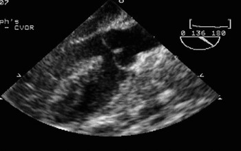
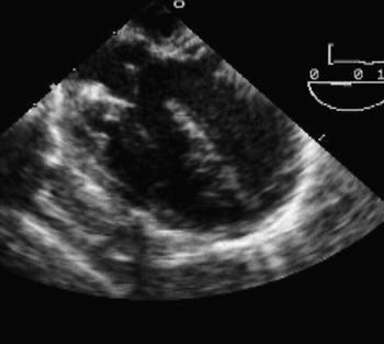
A routine, systematic, approach to transoesophageal echocardiography will allow the clinician to evaluate effectively all the intracardiac anatomy. The long axis and four chamber sweeps are useful for evaluating the relationship of the ventricular septal defect to the tricuspid and aortic valves (Figs 1 and 2). They can also be used to exclude additional ventricular septal defects, and to show the extent of inlet or outlet extension of the ventricular septal defect.Reference Suzuki, Ho, Anderson and Deanfield17 Restrictive defects, if present, can be identified.Reference Flanagan, Foran, Van Praagh, Jonas and Sanders18
The transesophageal echocardiogram demonstrates a perimembranous ventricular septal defect with overriding of the aorta as seen from a long-axis view with angulation.

The transoesophageal echocardiogram demonstrates the relationship of the ventricular septal defect to both the tricuspid and aortic valves in a four chambered view at 0°.

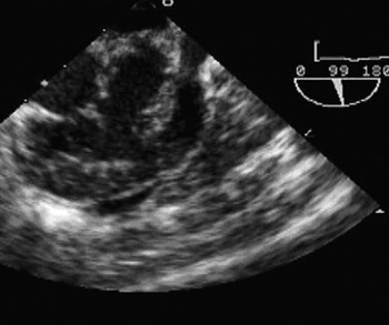
The aortic short-axis view allows for evaluation of the aortic valvar leaflets and the coronary arterial anatomy. The right ventricular inflow and outflow tracts are usually obtained with a multiplane angle of about 60 degrees (Fig. 3). This view allows for assessment of the tricuspid valve, the ventricular septal defect, the body and infundibulum of the right ventricle, and the pulmonary valve. This view is useful in both pre- and post-operative studies. Colour and pulse Doppler interrogation aid in the assessment of the degree of obstruction across the right ventricular outflow tract and pulmonary regurgitation. Though this information is useful, the degree of cyanosis is more important in the determination of timing of surgery for these patients. The aortic long-axis view, at around 120 degrees, is especially useful for assessing the relationship of the ventricular septal defect to the aortic valve and the degree of aortic override. Post-operatively, the integrity of closure of the ventricular septal defect is well visualized in this view. The bicaval view allows for assessment of the systemic veins and atrial septum. Transgastric views are excellent for assessment of the right and left ventricular function; and also provide the best angles for Doppler assessment of the right and left ventricular outflow tracts.
This transesophageal echocardiogram demonstrates the right ventricular outflow tract and ventricular septal defect in a short axis view with 60° angulation.

New methods of echocardiography
The advent of 3-dimensional echocardiography has provided for improved spatial delineation of the relationships of the intracardiac abnormalities in patients with tetralogy of Fallot. This modality is especially useful in the assessment of the ventricular septal defect and the right ventricular outflow tract. It also has the potential for volume-based assessment of ventricular function and size.Reference Niemann, Pinho and Balbach19 Quantitation of the right ventricular size and function is important for determining the timing of replacement of the pulmonary valve in patients with previously repaired lesions. Many consider that 3-dimensional echocardiography will change the fashion of future acquisition of images. A single 3-dimensional sweep contains all the information provided by serial cross-sectional sweeps. The time spent on acquisition of images may be drastically reduced as centres become more accustomed to 3-dimensional imaging.
Other new modalities offer further insight into both systolic and diastolic ventricular function. These include tissue Doppler imaging, and analysis of strain rate.Reference Mulder, Vliegen and van der Wall20, Reference Knirsch, Dodge-Khatami and Kadner21 These modalities have improved the assessment of ventricular function, providing the ability to quantitate systolic and diastolic functional parameters for both the right and left ventricles. Further clinical research is now needed to define the best non-invasive echocardiographic marker for future replacement of the pulmonary valve.
Long-term follow-up
All patients with tetralogy, regardless of the type of surgical repair, need to have ongoing follow-up by well-trained cardiologists throughout their lifetimes. As already discussed, these patients are at risk for progressive pulmonary regurgitation, recurrent pulmonary infundibular or peripheral pulmonary stenosis, tricuspid regurgitation, residual septal defects, aortic regurgitation, right ventricular dilation or dysfunction, and late arrhythmias. The use of annual or semi-annual transthoracic echocardiograms is helpful in evaluating such patients on an outpatient basis.
As children enter adolescence and adulthood, their acoustic windows continue to decline, and adjunctive studies will be required for ongoing assessment of their cardiac anatomy and function. Magnetic resonance imaging and angiography have had increasing use in patients with tetralogy of Fallot.Reference Alpendurada and Kilner22 These modalities are especially useful for quantification of the right ventricular size and function, as well as delineation of the anatomy of the pulmonary trunk and its branches. Other modalities used in the long-term assessment include ultrafast computerized tomographic scans, with angiography to assess coronary arterial abnormalities, abnormalities of the pulmonary trunk and its branches, and ventricular function. Lung perfusion scans are also useful, as they allow quantification of the amount of blood flowing to each lung, revealing pulmonary arterial stenosis that may not have been apparent on transthoracic or transoesophageal echocardiograms.
Conclusions
Echocardiography is essential in the care of patients with tetralogy of Fallot. The use of its different modalities allows for a continuum of care from the womb to adult life. The use of a methodical and systematic approach to acquisition of images, whether fetal, transthoracic or transoesophageal, allows for complete and reliable assessment of the intracardiac anatomy and any associated congenital cardiac defects. The newer modalities of echocardiography, such as 3-dimensional echo, tissue Doppler, and strain rate imaging, hold further promise as noninvasive tools for better anatomical and functional assessment.