Behçet’s disease is a multi-systemic, chronic, inflammatory disease that involves small and large arteries and its aetiology is unknown. It was first described as a clinical picture consisting of recurrent oral aphthous, genital ulcer, and hypopyon iridocyclitic complex in 1937. Behçet’s disease can involve the skin, joints, central nervous system, gastrointestinal system, urogenital system, eyes, lungs, and cardiovascular system and is considered a vasculitis that can affect any site within the arterial and venous systems. Diagnosis is based on clinical criteria, primarily because there is no specific diagnostic laboratory test and histopathological findings. Vascular involvement occurs in one-third of patients and often consists of recurrent superficial or deep vein thrombosis. Cardiac findings have been found in 7–46% of patients with Behçet’s disease in previous adult series, and these include endocarditis, myocarditis, pericarditis, endomyocardial fibrosis, coronary arteritis with or without myocardial infarction, aneurysm of coronary arteries, valve dysfunction, conduction system disorders, and intracardiac thrombosis. Reference Gürgün, Ercan and Ceyhan1 Intracardiac thrombosis is a rare finding mostly seen in adult young men. 2 In the literature today, there are less than 10 paediatric cases diagnosed with Behçet’s disease and intracardiac thrombosis.
In this case report, we described a child who was first presented with chest pain and then was diagnosed with an intracardiac thrombosis related to Behçet’s disease.
Case
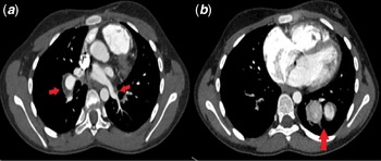
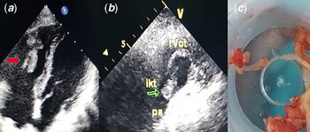
A 12-year-old girl presented with the complaint of fatigue, weakness, and chest pain for 1 year. She had a history of cough and light-coloured blood coming from the mouth during the past 15 days. Physical examination was normal, there are no typical clinical features of Behçet’s disease. Echocardiography showed a large mobile mass originating from the right ventricular apex, filling half of the right ventricular cavity and extending to the pulmonary valve. Mild degree tricuspid regurgitation was also observed. Cardiac MRI was also compatible with the presence of a 32 x 20 mm mobile thrombus in the right ventricle. This was considered to be chronic intracardiac thrombosis, and subcutaneous enoxaparin treatment was started. Chest CT imaging showed multiple filling defects compatible with peripheral pulmonary arterial embolism and pulmonary infarct areas (Fig 1). Ventilation-perfusion scintigraphy showed defects compatible with multiple embolism in both lungs. Biochemical and thrombophilia tests were normal except for low serum protein C value (51.93 IU/dL, normal:70–140 IU/dL). There was no vasculitic lesion on ocular examination. Pathergy test (which is one of the diagnostic criteria of Behçet’s disease and is a non-specific hypersensitivity skin reaction test induced by pinprick) and HLA-B27 and HLA-B51 screening tests were negative. Autoimmune screening tests were normal, except serum amyloid A value (930 mg/L, upper limit of normal: 0–6.4 mg/L), and the erythrocyte sedimentation rate was elevated (78 mm/h). Familial Mediterranean fever mutation tests were negative. No pathological clinical or laboratory findings were detected for malignancy. We considered Behçet’s disease as the most likely diagnosis since the patient had a history of recurrent oral ulcers, intracardiac thrombosis, and pulmonary arterial embolism. Pulsed steroid, colchicine, and cyclophosphamide treatment were started. Due to the absence of reduction in the size of intracardiac thrombosis with enoxaparin and immunosuppressive treatment, and after considering the risks and benefits of invasive versus continued medical management, the intracardiac mass was removed surgically. The pathology result was compatible with the organised thrombosis (Fig 2). Steroid medication, colchicine, cyclophosphamide (once a month for 6 months), and low-dose enoxaparin treatments were continued after surgery. In the early post-operative period, estimated systolic pulmonary artery pressure calculated according to tricuspid regurgitation was 50 mmHg by echocardiographic examination, falling to 40 mmHg at the review 6 weeks after surgery. Pulmonary hypertension was not considered significant enough to start specific treatment, but enalapril treatment was started for moderate-degree tricuspid valve regurgitation. One month after surgery, chest CT imaging showed no filling defects compatible with embolic events in either lung. During hospitalisation and follow-up at 6 months after surgery, haemorrhagic complication or thrombosis were not observed. Recurrent thrombosis was not observed by echocardiographic examinations performed at monthly intervals for 6 months but moderate tricuspid regurgitation that developed secondary to surgery persisted. Although she had no clinical or laboratory evidence of recurrences when reviewed (6 months after surgery), she died at home due to massive haemoptysis 10 days after the last control examination. Post-mortem examination could not be performed because when we called her for a check-up we learned from her family that she could not be transferred to the hospital and died at home. We thought that she died due to pulmonary arterial haemorrhage, which is the most mortal complication of pulmonary involvement in Behçet’s disease.
Chest CT shows ( A ) partially thrombosed aneurym (red arrows) in right lower lobar pulmonary artery and pulmonary thromboembolism in left lower lobe segmental arteries, ( B ) two segmental pulmonary artery aneuryms (red arrow) in left lower lobe.

An echo density in the right ventricle and attached to the moderator band (red arrow) at apical four chamber view (A), the magnified echo density (green arrow) in the pulmonary artery at modified short axis view (B) shown by echocardiography and the surgically removed fibrous tissue was compatible with the organised thrombosis (C). rvot: right ventricle outflow tract, ikt: intracardiac thrombus, pa: main pulmonary artery.

Discussion
Behçet’s disease is a multi-systemic disease that affects young adults, especially in Mediterranean, Middle East, and Far East countries. Since there is no specific laboratory test, it is very difficult to diagnose. Clinical features such as orogenital aphthae, ocular and skin lesions, arthritis and neurological, gastrointestinal, vascular, and pulmonary symptoms are helpful for diagnosis. Cardiovascular involvement in Behçet’s disease is estimated to range from 7 to 46% in adult Behçet’s disease patients. Reference Gürgün, Ercan and Ceyhan1 In Behçet’s disease, vasculitis lesions may affect veins and arteries of all sizes. It has been reported that venous involvement is 29%, and arterial involvement varies from 8 to 18%. Reference Gürgün, Ercan and Ceyhan1 Intracardiac thrombosis is an extremely rare complication of Behçet’s disease. It is more common in male gender and advanced ages, but it is very rare in the children. 2 The diagnosis of Behçet’s disease in childhood is difficult due to its rarity and lack of diagnostic criteria. The initial symptoms occur in 4–26% of the patients under the age of 16 years. Reference Yıldız, Köker, Adrovic, Şahin, Barut and Kasapçopur3 Although cardiovascular complications are extremely rare, they may occur as the first sign of the disease in childhood. Reference Demir, Sag, Kaya Akca, Hazirolan, Bilginer and Ozen4 Our case had only two critera for diagnosis of Behçet’s disease and did not meet diagnostic criteria of Behçet’s disease. However, she presented with intracardiac thrombosis and peripheral pulmonary arterial embolism which could be considered pathognomic for Behçet’s diseae in childhood. Reference Demir, Sag, Kaya Akca, Hazirolan, Bilginer and Ozen4 There were no clinical and laboratory findings of other diseases that would explain these thrombosis events. There are no large series of cases that include children published. In one study, 2 of 26 patients with paediatric Behçet’s disease were encountered with severe atypical cardiac presentations. Reference Ben-David, Gur, Ilivitzki, Khoury, Bentur and Butbul5 These presentations were intracardiac thrombi and left anterior descending coronary arterial obstruction causing myocardial infarction, and pulmonary arterial aneurysm with pulmonary embolism. These two patients had a delayed diagnosis of Behçet’s disease, demonstrating the need to be highly suspicious of this disease in young patients with intracardiac thrombosis. Reference Ben-David, Gur, Ilivitzki, Khoury, Bentur and Butbul5 In another study, 46 patients with Behçet’s disease were evaluated retrospectively. Ten patients (21.7%) had thrombosis and this was the first sign of Behçet’s disease in seven patients. One of them had pulmonary artery thrombosis, and one patient had intracardiac thrombosis. All patients had received anticoagulant therapy with immunosuppressive treatment. Reference Guzelkucuk and Gokcebay6 Intracardiac thrombosis related to Behçet’s disease can be seen in both ventricular chambers. Right ventricular thrombus is frequently associated with aneurysmatic dilatation of the pulmonary vessels, which reflects systemic inflammatory processes and pulmonary vasculitis. We also detected mild pulmonary arterial dilatation in our patient by CT examination. In a review performed in adult patients with Behçet’s disease, intracardiac thrombus is more frequent in patients with pulmonary arterial involvement than patients without such involvement (19.7–0.3%). Reference Eroglu, Torgutalp and Baysal7
Optimal therapeutic strategies for patients with intracardiac thrombosis relating to Behçet’s disease still remain controversial. Surgical treatment can be performed in patients with massive thrombosis and heart failure, despite optimal medical treatment. These patients may also undergo surgery due to the suspicion of an intracardiac tumour. In controlled studies, surgical treatment is not supported due to the risk of recurrence and post-operative complications. Cardiac thrombosis may occur again in a short time if corticosteroid treatment is not given after surgical excision. Reference Abidov and Alpert8 Previous reports suggest that combined immunosuppressive prednisone and cyclophosphamide therapy are required to treat Behçet’s disease-related intracardiac thrombosis. Intracardiac thrombosis related to Behçet’s disease may show resolution when treated only immunosupressive treatment and/or anticoagulation. The benefit of anticoagulation in the vasculitis of Behçet’s disease is still debated. However, venous, pulmonary arterial, and intracardiac thrombosis in Behçet’s disease are usually managed with anticoagulation and immunsuppressive treatment. The only contraindication to anticoagulation is the presence of pulmonary artery aneurysm due to the risk of rupture. Reference Hatemi, Christensen and Bang9
In our case, after 1 month of treatment with combined high doses of immunosuppressive, corticosteroid, and low-dose anticoagulation, highly mobile intracardiac thrombi and multiple pulmonary arterial thrombi persisted. Intracardiac thrombosis in our case was considered to be at the critical dimension that could lead to sudden death by occluding the pulmonary artery Reference Cetin, Ekici and Ünal10 and was therefore surgically removed. Although recurrent thrombosis is frequent in Behçet’s disease, we did not observe any clinical or laboratory evidence of recurrent thrombosis in our case until the last examination.
Conclusion
Intracardiac thrombosis secondary to Behçet’s disease is a rare cardiac complication in children. However, Behçet’s disease should be considered in intracardiac thrombosis cases in childhood unless clearly linked to other causes. The management of patients with Behçet’s disease complicated with intracardiac and pulmonary arterial thrombosis is challenging, and there remains a high risk of mortality despite seemingly optimal therapy.
Author contributions
Dr Şenay Akbay: concept/design, data analysis/interpretation, drafting article Prof. Dr Filiz Ekici: concept/design, data analysis/interpretation, drafting article, critical revision of article Assoc. Prof. Dr Elif Çomak: data analysis.
Financial support
This research received no specific grant from any funding agency, commercial, or not-for-profit sectors.
Conflicts of interest
None.
Ethical standards
A written informed consent was obtained from the patient.