Introduction
Bipolar disorder (BD) is a severe, chronic and highly heritable mental illness that has a prevalence of 1% in adults (Merikangas et al. Reference Merikangas, Akiskal, Angst, Greenberg, Hirschfeld, Petukhova and Kessler2007; Song et al. Reference Song, Bergen, Kuja-Halkola, Larsson, Landén and Lichtenstein2015). Due to lack of biological markers, the diagnosis is still based on certain clinical disturbances of mood, sleep, behaviour, perception and cognition. Thus, delayed diagnosis and misdiagnosis are common problems (Ghaemi et al. Reference Ghaemi, Sachs, Chiou, Pandurangi and Goodwin1999; Hirschfeld et al. Reference Hirschfeld, Calabrese, Weissman, Reed, Davies, Frye, Keck, Lewis, McElroy, McNulty and Wagner2003). Together with genetic and environmental factors, underlying abnormalities of brain structure and function are thought to be important in the development of BD. Several lines of evidence suggest that BD is associated with white matter (WM) pathology. WM abnormalities in BD were first observed as increased rates of deep WM hyperintensities in T2-weighted anatomical magnetic resonance images (Botteron et al. Reference Botteron, Vannier, Geller, Todd and Lee1995; Pavuluri et al. Reference Pavuluri, Birmaher and Naylor2005). Also, genetic studies in BD have documented alterations in the neuroregulin-1 gene, which was shown to be involved in the regulation of axonal migration and myelination in preclinical studies (Thomson et al. Reference Thomson, Christoforou, Morris, Adie, Pickard, Porteous, Muir, Blackwood and Evans2007; Georgieva et al. Reference Georgieva, Dimitrova, Ivanov, Nikolov, Williams, Grozeva, Zaharieva, Toncheva, Owen, Kirov and O'Donovan2008; McIntosh et al. Reference McIntosh, Moorhead, Job, Lymer, Muñoz Maniega, McKirdy, Sussmann, Baig, Bastin, Porteous, Evans, Johnstone, Lawrie and Hall2008). In addition, diffusion tensor imaging (DTI) studies have consistently reported abnormal measures of WM integrity in BD (Nortje et al. Reference Nortje, Stein, Radua, Mataix-Cols and Horn2013).
DTI is a magnetic resonance imaging (MRI) technique that is suitable to quantitatively investigate WM axonal integrity in vivo. Fractional anisotropy (FA) is a measure of the degree to which water diffusion is constrained in the brain and is widely used as a general index of axonal integrity (Kubicki et al. Reference Kubicki, Westin, Maier, Mamata, Frumin, Ersner-Hershfield, Kikinis, Jolesz, McCarley and Shenton2002). Damage to WM or demyelination along neuronal axons results in more isotropic water movement and is manifested as relatively low FA values. A problem with DTI is the interpretation of changes of FA. Therefore, the component measures from which FA is derived, the so-called first (λ1) and second [(λ2 + λ3)/2] eigenvalues measuring axial and radial diffusions (AD and RD, respectively) to the primary axis of the axon, can provide additional insights regarding the nature of WM deficits. An increase in RD values is thought to signify increased space between fibres, suggesting demyelination or dysmyelination (Harsan et al. Reference Harsan, Poulet, Guignard, Steibel, Parizel, de Sousa, Boehm, Grucker and Ghandour2006), whereas a decrease in AD values suggests axonal injury (Lazar et al. Reference Lazar, Weinstein, Tsuruda, Hasan, Arfanakis, Meyerand, Badie, Rowley, Haughton, Field and Alexander2003).
Results from DTI studies suggest widespread WM abnormalities in BD, but not all data are consistent. Previous studies have found lower (Zanetti et al. Reference Zanetti, Jackowski, Versace, Almeida, Hassel, Duran, Busatto, Kupfer and Phillips2009; Macritchie et al. Reference Macritchie, Lloyd, Bastin, Vasudev, Gallagher, Eyre, Marshall, Wardlaw, Ferrier, Moore and Young2010; Barysheva et al. Reference Barysheva, Jahanshad, Foland-Ross, Altshuler and Thompson2013), higher (Haznedar et al. Reference Haznedar, Roversi, Pallanti, Baldini-Rossi, Schnur, Licalzi, Tang, Hof, Hollander and Buchsbaum2005; Wessa et al. Reference Wessa, Houenou, Leboyer, Chanraud, Poupon, Martinot and Paillère-Martinot2009) or similar (Houenou et al. Reference Houenou, Wessa, Douaud, Leboyer, Chanraud, Perrin, Poupon, Martinot and Paillere-Martinot2007) FA values in BD compared with healthy volunteers, possibly reflecting limited sample sizes, and clinical and methodological heterogeneity. BD is reported to have a heritability of 60–90% and first-degree relatives of BD patients are particularly at greater risk for developing the disorder (McGuffin et al. Reference McGuffin, Rijsdijk, Andrew, Sham, Katz and Cardno2003; Smoller & Finn, Reference Smoller and Finn2003; Lichtenstein et al. Reference Lichtenstein, Yip, Björk, Pawitan, Cannon, Sullivan and Hultman2009). Therefore the investigation of unaffected first-degree relatives, in addition to BD patients and healthy controls, may help to distinguish biomarkers of genetic risk without the confounding effects of burden of illness, medication or clinical state. To date, a small number of DTI studies investigated WM abnormalities in individuals at risk for BD (Versace et al. Reference Versace, Ladouceur, Romero, Birmaher, Axelson, Kupfer and Phillips2010; Sprooten et al. Reference Sprooten, Sussmann, Clugston, Peel, McKirdy, Moorhead, Anderson, Shand, Giles, Bastin, Hall, Johnstone, Lawrie and McIntosh2011; Arat et al. Reference Arat, Chouinard, Cohen, Lewandowski and Öngür2015) and the findings were discrepant. The majority of these studies did not include a patient group in the analysis which limits the ability to clarify the endophenotypic nature of WM abnormalities. Thus far, only a very limited number of DTI studies have examined WM integrity directly in patients with BD, in addition to adult unaffected first-degree relatives and healthy controls. A recent DTI study found that in comparison with their control groups, bipolar patients and their unaffected first-degree relatives showed significantly reduced FA in the right anterior limb of the internal capsule and right uncinate fasciculus (Linke et al. Reference Linke, King, Poupon, Hennerici, Gass and Wessa2013). Another high-risk design DTI study reported that patients with BD showed decreased FA in the genu of the corpus callosum, right inferior longitudinal fasciculus (ILF) and left superior longitudinal fasciculus, whereas first-degree relatives of BD patients showed no difference in FA in comparison with controls (Chaddock et al. Reference Chaddock, Barker, Marshall, Schulze, Hall, Fern, Walshe, Bramon, Chitnis, Murray and McDonald2009). Same researchers also detected a significant effect of group on both FA and RD in limbic, temporal and callosal pathways driven by reduced FA and increased RD in patients with BD compared with controls and first-degree relatives, but, did not detect any difference in the WM between relatives and controls (Emsell et al. Reference Emsell, Chaddock, Forde, Van Hecke, Barker, Leemans, Sunaert, Walshe, Bramon, Cannon, Murray and McDonald2014). However, degree of penetrance for candidate genes and environmental factors might also affect structural changes associated with genetic risk. Therefore, investigation of siblings provides a unique opportunity because of the shared genetic and environmental risk factors for the disorder. The first DTI study that investigated WM integrity in BD patients, unaffected siblings and healthy controls found that FA within the right temporal lobe WM of unaffected siblings was intermediate and significantly different from patients with BD and healthy volunteers (Mahon et al. Reference Mahon, Burdick, Ikuta, Braga, Gruner, Malhotra and Szeszko2013). Another recent study that investigated WM integrity in a large sample of BD patients, unaffected siblings and healthy comparison subjects observed widespread FA reductions in BD patients and more subtle reductions in their unaffected siblings, mainly restricted to the corpus callosum, posterior thalamic radiations and left superior longitudinal fasciculus. Region-of-interest (ROI) analysis revealed significant reductions in most WM regions in patients but not in unaffected siblings compared with controls (Sprooten et al. Reference Sprooten, Brumbaugh, Knowles, McKay, Lewis, Barrett, Landau, Cyr, Kochunov, Winkler, Pearlson and Glahn2013).
In the present study, we applied tract-based spatial statistics (TBSS) to study WM changes in patients with BD and unaffected siblings and controls. The skeleton-based approach of TBSS may overcome potential problems with the registration and alignment of WM between subjects in order to allow cross-subject statistical analysis (Smith et al. Reference Smith, Jenkinson, Johansen-Berg, Rueckert, Nichols, Mackay, Watkins, Ciccarelli, Cader, Matthews and Behrens2006). To extend our voxel-level analysis, atlas-derived ROIs were also examined. We hypothesized that the patients with BD would show reduced WM compared with the healthy controls, with the siblings showing a pattern of alterations of intermediate severity.
Method
Sample
A total of 27 euthymic patients with Diagnostic and Statistical Manual of Mental Disorders, 4th edition (DSM-IV) BD type I, 20 siblings of bipolar patients and 29 healthy controls who did not have any current or past diagnosis of Axis I psychiatric disorders as determined using the Structured Clinical Interview for DSM-IV Disorders (SCID) (First et al. Reference First, Spitzer, Gibbon and Williams1996), were enrolled in the study. All patients were being treated at the Bipolar Disorders Outpatient Unit of the Department of Psychiatry, Dokuz Eylul University and were on stable doses of medications for at least 2 weeks prior to the scan. All patients were in remission for at least 6 months, defined as scoring less than 7 on the Young Mania Rating Scale and on the 17-item Hamilton Depression Rating Scale for at least 6 months prior to and at the time of the MRI scanning. Exclusion criteria for all participants included: (1) MRI contraindications; (2) significant medical or neurological illness; (3) prior neurosurgical intervention; (4) DSM-IV mental retardation; (5) being pregnant or breastfeeding; (6) being left-handed; and (7) history of head trauma with loss of consciousness; (8) active alcohol or substance use in the preceding 6 weeks prior to the scan; (9) past or current diagnosis of substance abuse or dependence except nicotine; (10) severe auditory or visual impairments assessed by physical examination; (11) current diagnosis of any other Axis I psychiatric co-morbidity for bipolar patients. We further excluded any relatives who had a past or current diagnosis of any Axis I psychiatric disorders and healthy controls who had a first-degree relative with any Axis I psychiatric disorders or had a family history of BD, schizophrenia or schizophrenia spectrum disorders.
All subjects gave written informed consent before participation in the study. Dokuz Eylül University Hospital Ethics Committee approved the study.
Imaging protocol
MRI was performed using a 1.5T MR system (GE Signa HDxt; General Electric Medical Systems, USA). Diffusion imaging data were acquired in 32 diffusion gradient directions (b = 1000 s/mm2) plus one b = 0 reference image using a sequence optimized to collect diffusion weighted images (repetition time = 6500 ms, echo time = 90 ms, voxel size = 2 × 2 × 2 mm3).
TBSS
DTI data were analysed using FMRIB's (Oxford Centre for Functional MRI of the Brain) Diffusion Toolbox, which is part of FSL (FMRIB Software Library) (Smith et al. Reference Smith, Jenkinson, Woolrich, Beckmann, Behrens, Johansen-Berg, Bannister, De Luca, Drobnjak, Flitney, Niazy, Saunders, Vickers, Zhang, De Stefano, Brady and Matthews2004). Motion and eddy current artifacts were corrected using FSL EDDY_CORRECT. A brain mask of the non-diffusion weighted image was created using FSL's Brain Extraction Tool (Smith, Reference Smith2002). The diffusion tensor was then calculated with FSL DTIFIT for whole-brain volumes and the resulting FA maps, together with the AD (λ1) and RD [(λ2 + λ3)/2] maps, were used in subsequent TBSS analysis.
Voxel-wise statistical analysis of the data was performed using TBSS (Smith et al. Reference Smith, Jenkinson, Johansen-Berg, Rueckert, Nichols, Mackay, Watkins, Ciccarelli, Cader, Matthews and Behrens2006). FA data were aligned into a common space using a non-linear registration algorithm (FSL FNIRT) to register the images to the standard space Montreal Neurological Institute (MNI) 152 template and upsampled to 1 × 1 × 1 mm3. Thereafter, the registered FA images were averaged to generate a cross-subject mean FA image, and then the mean FA image was applied to create a mean FA skeleton which represents the main fibre tracks and the centre of all fibre tracts common to the group. The skeleton was then thresholded at an FA value of 0.2 which limits the effects of poor alignment across subjects and reduces the likelihood of inclusion of grey matter and cerebrospinal fluid voxels in the skeleton. The skeleton that is now created contains WM tracts that are common to all subjects. A ‘distance map’ is then created which is used to project each FA image onto the mean FA skeleton that is common to all subjects. AD and RD images were also aligned into MNI space and projected onto the mean FA skeleton using the protocol of non-FA images in TBSS.
To identify FA differences between (i) patients with BD and healthy controls, (ii) patients with BD and siblings, and (iii) siblings and healthy controls, the skeletonized FA data were fed into the voxel-wise statistical analysis, which is based on non-parametric approach utilizing permutation test theory (randomize tool in FSL) (Nichols & Holmes, Reference Nichols and Holmes2002). The threshold-free cluster enhancement method was used to define the clusters (Smith & Nichols, Reference Smith and Nichols2009). We corrected p values for multiple comparisons according to the family-wise error (FWE) rate by 5000 permutations across groups. Results were considered significant for p < 0.05 and only clusters containing more than 50 contiguous significant voxels are reported. The most probable anatomic localization of each significant cluster was determined using publicly accessible WM atlases (http://www.dtiatlas.org).
For clusters where significantly lower FA values in patients with BD were observed, the mean AD and RD values of each cluster mask for each subject were extracted. The function ‘fslmeants’ from FSL was used to extract means of AD and RD values from these masks. Between-group differences for AD and RD values in these ROIs were examined using the Mann–Whitney U test. Mean FA values within a significant cluster for patients with BD compared with controls were exported to SPSS (USA) to examine relationships with duration of illness, age at first episode, total number of episodes, number of prior depressions, number of prior manias and duration of remission using partial correlation analyses controlling for age (p < 0.05; two-tailed).
ROI analysis
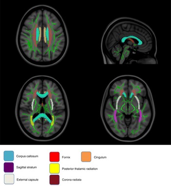
ROIs of the corpus callosum (divided into genu, body and splenium ROIs), fornix, internal capsule (divided into anterior limb, posterior limb and retrolenticular ROIs), external capsule, corona radiata (divided into anterior, posterior and superior ROIs), anterior thalamic radiation, posterior thalamic radiation, superior longitudinal fasciculus, superior fronto-occipital fasciculus, cingulum, the hippocampal end of the cingulum, the uncinate fasciculus and sagittal stratum, including the ILF and the inferior fronto-occipital fasciculus (IFOF), were created with binary masks based on the JHU ICBM-DTI-81 WM labels atlas (Mori et al. Reference Mori, Oishi, Jiang, Jiang, Li, Akhter, Hua, Faria, Mahmood, Woods, Toga, Pike, Neto, Evans, Zhang, Huang, Miller, van Zijl and Mazziotta2008) (Fig. 1). Intersections between anatomical masks and the skeleton (previously created with TBSS) were used in order to extract mean FA values in these regions. After confirming normal distribution of the data by a one-sample Kolmogorov–Smirnov test, one-way analysis of variance with group as the independent variable and diffusion indices as the dependent variables was performed. A statistical significance level of p < 0.05 (Bonferroni correction for multiple comparisons) was used.
Example of regions of interest as determined by the John Hopkins University white matter labels atlases.

Ethical standards
All procedures contributing to this work comply with the ethical standards of the relevant national and institutional committees on human experimentation and with the Helsinki Declaration of 1975, as revised in 2008.
Results
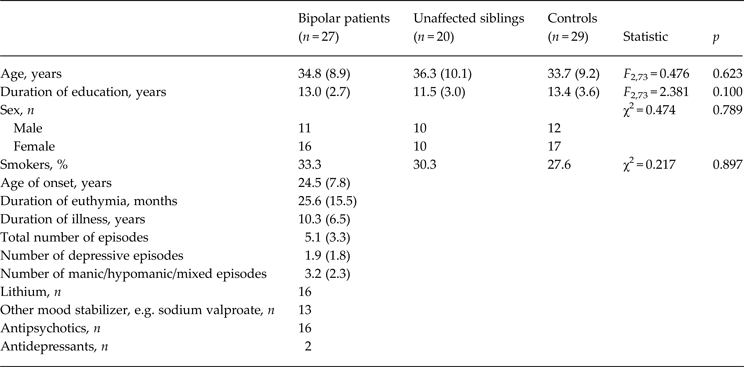
Participants' clinical and sociodemographic information is outlined in Table 1. The three groups did not differ significantly in distributions of age, sex, smoking status or years of education. All patients were taking at least one psychotropic medication whereas no siblings or controls were on any psychotropic medications at the time of the scan.
Participant sociodemographic and clinical characteristics

Data are given as mean (standard deviation) unless otherwise indicated.
TBSS analyses
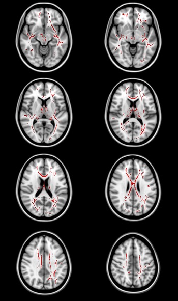
FA was significantly lower in BD patients than in the control group in one large cluster (K = 26 335 voxels, p FWE < 0.05), including the corpus callosum, fornix, bilateral superior longitudinal fasciculus, ILF, IFOF, anterior thalamic radiation, posterior thalamic radiation, cingulum, uncinate fasciculus, superior corona radiata, anterior corona radiata and left external capsule (Fig. 2). RD values in the significant cluster were significantly higher in patients with BD than in controls (U = 158.500, p = 0.0001). AD values did not differ between BD patients and controls (U = 373.000, p = 0.761). We did not detect increased FA values in any voxels in patients relative to healthy controls.
Regions showing reduced fractional anisotropy in bipolar patients relative to healthy comparison subjects (p < 0.05, family-wise error corrected). The right side of the images represents the right side of the brain.

FA was significantly lower in BD patients than in unaffected siblings in one cluster (K = 10 771 voxels, p FWE < 0.05), including the corpus callosum, bilateral superior corona radiata, anterior corona radiata, anterior limb of internal capsule, anterior thalamic radiation and left external capsule. We did not detect increased FA in any voxels in patients relative to unaffected siblings.
Using TBSS no significant clusters of either increased or decreased FA were detected between unaffected siblings of bipolar patients and controls.
ROI analyses
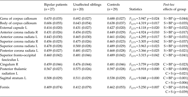
ROI analyses revealed that bipolar patients had significantly reduced FA in the left superior fronto-occipital fasciculus and right cingulum compared with healthy controls. In addition, bipolar patients showed reduced FA in the genu and body of the corpus callosum, bilateral anterior corona radiata, bilateral superior corona radiata, the left posterior corona radiata and the left external capsule compared with unaffected siblings (Table 2).
Between-group differences on mean fractional anisotropy within atlas-based regions of interest

Data are given as mean (standard deviation).
S, Unaffected siblings; BP, bipolar patients; L, left; R, right; C, controls.
Both siblings and bipolar patients had significantly reduced FA in the left posterior thalamic radiation, and left sagittal stratum (ILF + IFOF), and fornix compared with healthy controls. Average FA for siblings was intermediate between the comparison group and bipolar patients within these three ROIs (Table 2).
Correlation analyses
We found a significant negative correlation between mean FA extracted from the significant cluster in BD patients and total number of episodes (r = −0.438, p = 0.025). Mean FA was also significantly negatively correlated with the number of depressive episodes (r = −0.415, p = 0.035), but not with the number of manic/hypomanic/mixed episodes. There was no significant correlation between mean FA and the duration of illness, the age of onset and the duration of euthymia in the patient group.
Discussion
In this DTI study, bipolar patients, unaffected siblings of bipolar patients and healthy controls were examined in order to identify microstructural WM alterations that may be associated with the familial risk for BD. Mean FA values were significantly lower in BD patients and in unaffected siblings in several major WM tracts compared with the controls, while differences between unaffected siblings and controls did not reach statistical significance in the whole-brain DTI analyses. In ROI analyses, we found that both unaffected siblings and bipolar patients had significantly reduced FA in the left posterior thalamic radiation, the left sagittal stratum (ILF + IFOF) and the fornix compared with healthy controls. Average FA for unaffected siblings was intermediate between the healthy controls and bipolar patients within these ROIs. We also found that bipolar patients had greater RD values than controls in the significant cluster and ROIs indicate that demyelination may be a prominent microstructural change, but the interpretation of RD remains controversial (Wheeler-Kingshott & Cercignani, Reference Wheeler-Kingshott and Cercignani2009).
The finding of widespread impaired WM integrity in bipolar patients has been reported in a number of previous DTI studies compared with healthy controls (Kafantaris et al. Reference Kafantaris, Kingsley, Ardekani, Saito, Lencz, Lim and Szeszko2009; Sussmann et al. Reference Sussmann, Lymer, McKirdy, Moorhead, Muñoz Maniega, Job, Hall, Bastin, Johnstone, Lawrie and McIntosh2009; Zanetti et al. Reference Zanetti, Jackowski, Versace, Almeida, Hassel, Duran, Busatto, Kupfer and Phillips2009; Haller et al. Reference Haller, Xekardaki, Delaloye, Canuto, Lövblad, Gold and Giannakopoulos2011; Emsell et al. Reference Emsell, Leemans, Langan, Van Hecke, Barker, McCarthy, Jeurissen, Sijbers, Sunaert, Cannon and McDonald2013). In addition, we found greater RD values in bipolar patients than in controls in the significant cluster. Consistent with our findings, studies in bipolar patients (Chan et al. Reference Chan, Yang, Chia, Woon, Lee, Keefe, Sitoh, Nowinski and Sim2010; Benedetti et al. Reference Benedetti, Absinta, Rocca, Radaelli, Poletti, Bernasconi, Dallaspezia, Pagani, Falini, Copetti, Colombo, Comi, Smeraldi and Filippi2011; Lu et al. Reference Lu, Zhou, Keedy, Reilly and Sweeney2011, Reference Lu, Zhou, Fitzgerald, Keedy, Reilly, Passarotti, Sweeney and Pavuluri2012; Barysheva et al. Reference Barysheva, Jahanshad, Foland-Ross, Altshuler and Thompson2013; Emsell et al. Reference Emsell, Leemans, Langan, Van Hecke, Barker, McCarthy, Jeurissen, Sijbers, Sunaert, Cannon and McDonald2013; Linke et al. Reference Linke, King, Poupon, Hennerici, Gass and Wessa2013) have found that FA differences were mainly due to changes in RD values, suggesting myelin abnormalities. An altered myelination during development, resulting in abnormal MRI findings early in the paediatric stage, has been proposed to play a role in the pathophysiology of BD (Caetano et al. Reference Caetano, Silveira, Kaur, Nicoletti, Hatch, Brambilla, Sassi, Axelson, Keshavan, Ryan, Birmaher and Soares2008). There are several possible explanations for the observed myelin abnormalities in bipolar patients. Oligodendrocytes are the glial cells that form myelin sheaths in the central nervous system. A number of neuropathological studies have reported a decrease in the number of oligodendrocytes in BD; most of these studies focus on the prefrontal cortex (Ongür et al. Reference Ongür, Drevets and Price1998; Uranova et al. Reference Uranova, Vostrikov, Orlovskaya and Rachmanova2004; Vostrikov et al. Reference Vostrikov, Uranova and Orlovskaya2007; Bellani et al. Reference Bellani, Perlini, Ferro, Cerruti, Rambaldelli, Isola, Cerini, Dusi, Andreone, Balestrieri, Pozzi Mucelli, Tansella and Brambilla2012). In addition, oligodendrocyte and oligodendrocyte-related gene expression is modified in BD. Some genetic studies suggest that neuregulin 1-erbB expression is important for the regulation of axonal migration, oligodendrocyte growth and differentiation in BD (Tkachev et al. Reference Tkachev, Mimmack, Ryan, Wayland, Freeman, Jones, Starkey, Webster, Yolken and Bahn2003; Georgieva et al. Reference Georgieva, Dimitrova, Ivanov, Nikolov, Williams, Grozeva, Zaharieva, Toncheva, Owen, Kirov and O'Donovan2008). To our knowledge this is the first study that found a significant negative correlation between total number of episodes and FA values in the bipolar patient group. Moreover, FA values were negatively correlated with the number of depressive episodes, but not with the number of manic and mixed episodes. This finding is consistent with another study by Zanetti et al. (Reference Zanetti, Jackowski, Versace, Almeida, Hassel, Duran, Busatto, Kupfer and Phillips2009), describing inverse correlations between illness duration and DTI scores in ventromedial prefronto-striatal-thalamic and fronto-temporo-parietal WM. Together, these findings could help to explain the cognitive impairment, involving deficits in executive functioning, attention, memory and information processing speed, which may be detected in chronic, multiple-episode BD individuals (Robinson & Ferrier, Reference Robinson and Ferrier2006).
We found that both unaffected siblings and bipolar patients had significantly reduced FA in the fornix, left posterior thalamic radiation and left sagittal stratum in comparison with healthy controls. The fornix is central to the limbic system and it is the major WM bundle projection from the hippocampus to other brain structures, including septal nuclei, the hypothalamus (especially the mammillary bodies), anterior nucleus of the thalamus, cingulate cortex and nucleus accumbens, regions important for memory, emotional regulation and reward processing, which have been implicated in BD (Ahn et al. Reference Ahn, Breeze, Makris, Kennedy, Hodge, Herbert, Seidman, Biederman, Caviness and Frazier2007; Frey et al. Reference Frey, Andreazza, Nery, Martins, Quevedo, Soares and Kapczinski2007). One case study has proposed compression of the fornix as the cause of bipolarity (Xu et al. Reference Xu, Rasmussen, Berntsen, Moss, Shnier, Lagopoulos and Malhi2007). Barnea-Goraly et al. (Reference Barnea-Goraly, Chang, Karchemskiy, Howe and Reiss2009) found that adolescents with BD, who also were offspring of at least one parent with BD, had lower FA values than control subjects in the fornix, suggesting that alterations in the fornix are present early in the course of disease in familial BD. In line with these, a number of DTI studies reported reductions of FA in the fornix of bipolar patients in comparison with healthy controls (Barnea-Goraly et al. Reference Barnea-Goraly, Chang, Karchemskiy, Howe and Reiss2009; Barysheva et al. Reference Barysheva, Jahanshad, Foland-Ross, Altshuler and Thompson2013; Emsell et al. Reference Emsell, Leemans, Langan, Van Hecke, Barker, McCarthy, Jeurissen, Sijbers, Sunaert, Cannon and McDonald2013; Sprooten et al. Reference Sprooten, Brumbaugh, Knowles, McKay, Lewis, Barrett, Landau, Cyr, Kochunov, Winkler, Pearlson and Glahn2013; Oertel-Knöchel et al. Reference Oertel-Knöchel, Reinke, Alves, Jurcoane, Wenzler, Prvulovic, Linden and Knöchel2014). However, previous studies in unaffected siblings (Mahon et al. Reference Mahon, Burdick, Ikuta, Braga, Gruner, Malhotra and Szeszko2013; Sprooten et al. Reference Sprooten, Brumbaugh, Knowles, McKay, Lewis, Barrett, Landau, Cyr, Kochunov, Winkler, Pearlson and Glahn2013) or unaffected relatives (Frazier et al. Reference Frazier, Breeze, Papadimitriou, Kennedy, Hodge, Moore, Howard, Rohan, Caviness and Makris2007; Chaddock et al. Reference Chaddock, Barker, Marshall, Schulze, Hall, Fern, Walshe, Bramon, Chitnis, Murray and McDonald2009; Sprooten et al. Reference Sprooten, Sussmann, Clugston, Peel, McKirdy, Moorhead, Anderson, Shand, Giles, Bastin, Hall, Johnstone, Lawrie and McIntosh2011; Skudlarski et al. Reference Skudlarski, Schretlen, Thaker, Stevens, Keshavan, Sweeney, Tamminga, Clementz, O'Neil and Pearlson2013) did not find reduced FA in the fornix compared with healthy controls which may be due to anatomical characteristics of the fornix. The fornix is a very thin structure, challenging the spatial resolution of current MRI methods.
The posterior thalamic radiation includes axons connecting the cerebral cortex, basal ganglia and caudal parts of the thalamus. WM alterations in the posterior thalamic radiation have been found in bipolar patients compared with controls, especially in studies which used TBSS analyses (Chan et al. Reference Chan, Yang, Chia, Woon, Lee, Keefe, Sitoh, Nowinski and Sim2010; Nortje et al. Reference Nortje, Stein, Radua, Mataix-Cols and Horn2013). Reductions in FA values in the posterior thalamic radiation in siblings of bipolar patients compared with controls have been reported in one study (Sprooten et al. Reference Sprooten, Brumbaugh, Knowles, McKay, Lewis, Barrett, Landau, Cyr, Kochunov, Winkler, Pearlson and Glahn2013) but not in another study (Mahon et al. Reference Mahon, Burdick, Ikuta, Braga, Gruner, Malhotra and Szeszko2013). As the posterior thalamic radiation is also relevant for the connection between the thalamus and the visual cortex, our finding fits well into the findings by MacQueen et al. (Reference MacQueen, Grof, Alda, Marriott, Young and Duffy2004) who showed visual processing impairments in bipolar patients using a visual backward masking task pointing to deficits in neural circuitry involving the occipital lobe. Consistently, Ozerdem et al. (Reference Ozerdem, Güntekin, Tunca and Başar2008, Reference Ozerdem, Güntekin, Saatçi, Tunca and Başar2010) reported significant oscillatory differences in the occipital cortex in response to visual stimuli both in euthymic and manic states. A recent study examining the relationship between WM microstructure and cognitive control in 61 children found that anisotropy of the posterior thalamic radiation is associated with a child's ability to suppress interfering information and exhibit increased attentional control (Chaddock-Heyman et al. Reference Chaddock-Heyman, Erickson, Voss, Powers, Knecht, Pontifex, Drollette, Moore, Raine, Scudder, Hillman and Kramer2013). In particular, another recent study showed that patients with infarctions involving the posterior thalamus showed significant emotional disturbances and elevated anxiety levels compared with patients with more anterior lesions (Liebermann et al. Reference Liebermann, Ostendorf, Kopp, Kraft, Bohner, Nabavi, Kathmann and Ploner2013). It is therefore possible that reduced FA in the posterior thalamic radiation that we found in both bipolar patients and unaffected siblings compared with healthy controls could lead to emotional and cognitive symptoms, especially impaired response inhibition and visual processing, which have been implicated in BD (Clark et al. Reference Clark, Kempton, Scarnà, Grasby and Goodwin2005; Swann et al. Reference Swann, Lijffijt, Lane, Steinberg and Moeller2009a ).
The sagittal stratum is a complex fibre bundle connecting the occipital cortex to the rest of the brain, and includes fibres from many major tracts including the ILF and IFOF. The ILF connects occipital and temporal lobe structures including the amygdala, hippocampus and parahippocampus. Anteriorly, it joins the uncinate fasciculus to relay information to the orbitofrontal region of the brain (Ashtari, Reference Ashtari2012). The IFOF is unique in that it connects all four major lobes of the brain (Martino et al. Reference Martino, Brogna, Robles, Vergani and Duffau2010). WM alterations in both the ILF (Bruno et al. Reference Bruno, Cercignani and Ron2008; Chaddock et al. Reference Chaddock, Barker, Marshall, Schulze, Hall, Fern, Walshe, Bramon, Chitnis, Murray and McDonald2009; Ha et al. Reference Ha, Her, Kim, Chang, Cho and Ha2011; Lu et al. Reference Lu, Zhou, Keedy, Reilly and Sweeney2011; Barysheva et al. Reference Barysheva, Jahanshad, Foland-Ross, Altshuler and Thompson2013; Torgerson et al. Reference Torgerson, Irimia, Leow, Bartzokis, Moody, Jennings, Alger, Van Horn and Altshuler2013) and IFOF (Ha et al. Reference Ha, Her, Kim, Chang, Cho and Ha2011; Lu et al. Reference Lu, Zhou, Keedy, Reilly and Sweeney2011; Barysheva et al. Reference Barysheva, Jahanshad, Foland-Ross, Altshuler and Thompson2013) have been found in bipolar patients compared with healthy controls. In addition, reduced FA in the ILF and IFOF were found in unaffected relatives of bipolar patients compared with controls which was negatively correlated with cyclothymic temperament (Sprooten et al. Reference Sprooten, Sussmann, Clugston, Peel, McKirdy, Moorhead, Anderson, Shand, Giles, Bastin, Hall, Johnstone, Lawrie and McIntosh2011). Consistent with our findings, Mahon et al. (Reference Mahon, Burdick, Ikuta, Braga, Gruner, Malhotra and Szeszko2013) reported that FA values in the IFOF of unaffected siblings were intermediate to and significantly different from those of controls and patients with BD. Furthermore, they also found that FA values in the IFOF were negatively and significantly correlated with trait impulsivity in unaffected siblings. In line with this, correlation between WM integrity in the ILF and IFOF and impulsive traits has also been shown in alcoholics (Zorlu et al. Reference Zorlu, Gelal, Kuserli, Cenik, Durmaz, Saricicek and Gulseren2013), obsessive–compulsive disorder patients (Garibotto et al. Reference Garibotto, Scifo, Gorini, Alonso, Brambati, Bellodi and Perani2010) and healthy subjects (Xu et al. Reference Xu, Kober, Carroll, Rounsaville, Pearlson and Potenza2012). Together, these findings may suggest that WM integrity and anatomical connections among frontal, occipital and temporal lobes are associated with impulsivity, which has been implicated in both unaffected relatives of bipolar patients (Quintin et al. Reference Quintin, Benkelfat, Launay, Arnulf, Pointereau-Bellenger, Barbault, Alvarez, Varoquaux, Perez-Diaz, Jouvent and Leboyer2001) and bipolar patients (Swann et al. Reference Swann, Lijffijt, Lane, Steinberg and Moeller2009b ).
These findings are in conjunction with a meta-analysis of whole-brain DTI studies in BD that reported two significant clusters of decreased FA in the right hemisphere including one cluster that was located close to the parahippocampal gyrus. The arcuate fasciculus, IFOF, ILF and the posterior thalamic radiations were identified as the most likely pathways crossing this cluster (Vederine et al. Reference Vederine, Wessa, Leboyer and Houenou2011). Moreover, there is evidence indicating that neurogulin-1 gene variations are associated with WM abnormalities in the fornix, cingulum and parahippocampal gyrus circuit in bipolar patients (Cannon et al. Reference Cannon, Walshe, Dempster, Collier, Marshall, Bramon, Murray and McDonald2012). Taken together, decreased FA values in the fornix, left posterior thalamic radiation and left sagittal stratum observed both in bipolar patients and unaffected siblings, who share similar genetic and environmental risk factors, but independent of illness effects, might constitute a potential structural endophenotype for BD. These findings are consistent with a proposed neuroanatomical model of BD suggesting that BD is associated with structural abnormalities of the anterior limbic network, which includes the ventrolateral prefrontal cortex, amygdala and hippocampal complex, as well as subcortical structures such as the striatum and thalamus (Strakowski et al. Reference Strakowski, Delbello and Adler2005; Adler et al. Reference Adler, DelBello and Strakowski2006). However, it is noteworthy that FA values in healthy siblings were similar to healthy controls for the rest of the brain including prefrontal cortex-related WM tracts.
The current study has several potential limitations. The most obvious is the cross-sectional nature of the study, so that potential differential changes in WM measurements over the course of illness in BD remain to be directly established. Second, our sample was relatively small, which might reduce the power of the group comparison especially for the unaffected sibling group as not all individuals in this group are likely to be carrying the susceptibility gene to BD. Third, while the TBSS approach strives to avoid the problems of voxel-based morphology related to partial voluming, some of these issues may still remain. TBSS still has limitations in analysing small fibre tracts, data with within-scan head motion, and regions of crossing fibres or tract junctions (Smith et al. Reference Smith, Jenkinson, Johansen-Berg, Rueckert, Nichols, Mackay, Watkins, Ciccarelli, Cader, Matthews and Behrens2006). In addition, the field strength of 1.5 Tesla should be noted. Another methodological limitation was the lack of drug-naive patients. All patients with BD were being treated with psychotropic medications at the time of MRI examination, and the potential effect of these medications on WM integrity cannot yet be determined, as with the majority of other studies of this type. Lack of intelligence quotient assessment should also be noted.
In conclusion, decreased FA in the fornix, left posterior thalamic radiation and left sagittal stratum in both bipolar patients and unaffected siblings may represent a trait-based marker or endophenotype for BD.
Acknowledgements
This work was supported by the Dokuz Eylul University Department of Scientific Research Projects Funding (project no. 2012.KB.SAG.062).
Declaration of Interest
None.