Introduction
Bipolar disorder (BD) and schizophrenia (SZ) are currently classified as separate diagnostic entities (APA, 1994; WHO, 2004). Although this distinction may be justifiable and clinically useful (Lawrie et al. Reference Lawrie, Hall, McIntosh, Owens and Johnstone2010), it has been questioned on the basis of significant overlap between the two disorders in terms of phenomenology (Fischer & Carpenter, Reference Fischer and Carpenter2009), genetic risk factors (International Schizophrenia Consortium, Reference International Schizophrenia, Purcell, Wray, Stone, Visscher, O'Donovan, Sullivan and Sklar2009; Lichtenstein et al. Reference Lichtenstein, Yip, Bjork, Pawitan, Cannon, Sullivan and Hultman2009) and brain morphological changes (Ellison-Wright & Bullmore, Reference Ellison-Wright and Bullmore2010; Yu et al. Reference Yu, Cheung, Leung, Li, Chua and McAlonan2010).
In this study we focus specifically on facial affect processing and examine whether diagnosis-related differences in the engagement of the corresponding neural network support the distinction between SZ and BD. The reasons for this are threefold. First, differences in emotional processing have been highlighted as the most distinctive features differentiating SZ from BD. Both early descriptions and later investigations of the two syndromes (Bleuler, Reference Bleuler1950; Kraepelin, Reference Kraepelin1971; Carpenter et al. Reference Carpenter, Strauss and Bartko1973) emphasize avolition and restricted affect in SZ and excess emotional reactivity in BD. Second, facial affect is the most common probe of the neural circuitry involved in emotional processing because of its biological and behavioral salience (LeDoux, Reference LeDoux1995; Adolphs, Reference Adolphs2002). Facial affect processing engages numerous neural systems with key nodes spatially distributed within the primary and extrastriate visual cortices (particularly within the fusiform gyrus) in the occipital and ventral temporal lobes, the superior temporal sulcus, the medial and ventrolateral prefrontal cortex (PFC), limbic (amygdala and insula, among others) and thalamic areas (Adolphs et al. Reference Adolphs, Damasio, Tranel and Damasio1996; Haxby et al. Reference Haxby, Hoffman and Gobbini2002; Phan et al. Reference Phan, Wager, Taylor and Liberzon2002; Murphy et al. Reference Murphy, Nimmo-Smith and Lawrence2003; Fairhall & Ishai, Reference Fairhall and Ishai2007; Fusar-Poli et al. Reference Fusar-Poli, Placentino, Carletti, Landi, Allen, Surguladze, Benedetti, Abbamonte, Gasparotti, Barale, Perez, McGuire and Politi2009; Vytal & Hammann, Reference Vytal and Hammann2010; Dima et al. Reference Dima, Stephan, Roiser, Friston and Frangou2011). Most of these regions are also implicated in current models of BD and SZ that respectively highlight abnormal interaction within prefrontal–subcortical–limbic (Strakowski et al. Reference Strakowski, Delbello and Adler2005) and prefrontal–temporal–subcortical–limbic networks (Gur et al. Reference Gur, Keshavan and Lawrie2007a). Third, available findings in BD and SZ are suggestive of diagnosis-specific differences, particularly within limbic and posterior parieto-occipital regions. For example, viewing emotional faces compared to neutral faces is associated with increased engagement of the parahippocampus/amygdala in BD (Altshuler et al. Reference Altshuler, Bookheimer, Proenza, Townsend, Sabb, Firestine, Bartzokis, Mintz, Mazziotta and Cohen2005; Strakowski et al. Reference Strakowski, Delbello and Adler2005; Chen et al. Reference Chen, Lennox, Jacob, Calder, Lupson, Bisbrown-Chippendale, Suckling and Bullmore2006; Foland et al. Reference Foland, Altshuler, Bookheimer, Eisenberger, Townsend and Thompson2008; Delvecchio et al. Reference Delvecchio, Fossati, Boyer, Brambilla, Falkai, Gruber, Hietala, Lawrie, Martinot, McIntosh, Meisenzahl and Frangou2012) but not in SZ (Aleman & Kahn, Reference Aleman and Kahn2005; Holt et al. Reference Holt, Kunkel, Weiss, Goff, Wright, Shin, Rauch, Hootnick and Heckers2006; Surguladze et al. Reference Surguladze, Russell, Kucharska-Pietura, Travis, Giampietro, David and Phillips2006; Hall et al. Reference Hall, Whalley, McKirdy, Romaniuk, McGonigle, McIntosh, Baig, Gountouna, Job, Donaldson, Sprengelmeyer, Young, Johnstone and Lawrie2008; Seiferth et al. Reference Seiferth, Pauly, Kellermann, Shah and Ott2009; Li et al. Reference Li, Chan, McAlonan and Gong2010; Anticevic et al. Reference Anticevic, Van Snellenberg, Cohen, Repovs, Dowd and Barch2012). Additionally, recruitment in medial occipital and parietal regions involved in visual processing has been reported to be enhanced in SZ (Farkas et al. Reference Farkas, Wolf, Jaeger, Brodie, Christman and Fowler1984; Taylor et al. Reference Taylor, Kang, Brege, Tso, Hosanagar and Johnson2012) and reduced in BD (Pavuluri & Passarotti, Reference Pavuluri and Passarotti2008). However, inferring potential differences or similarities between the two disorders from existing studies is difficult because direct comparisons between the diagnostic groups are limited. Therefore, meta-analytic techniques are currently the best option to investigate brain regions differentially engaged in SZ and BD, and thus address questions of diagnostic specificity.
We conducted a quantitative voxel-based meta-analysis of available functional magnetic resonance imaging (fMRI) data to test for diagnosis-specific dysfunction in the neural correlates of facial affect processing in SZ and BD. Our key prediction was that functional disruption in limbic and associated areas would differ between the two disorders. We also hypothesized that SZ, but not BD, would be associated with increased engagement of posterior midline cortical regions involved in higher-order visual processing.
Method
Data sources and inclusion criteria
Studies investigating facial affect processing in patients with SZ or BD were identified through computerized literature searches using Medline and PubMed supplemented by inspection of reference lists. We reviewed all papers published in the English language up to 30 April 2012. The search keywords used were ‘bipolar disorder’, ‘schizophrenia’, ‘facial affect’, ‘facial emotion’, ‘emotional processing’, ‘facial affect labeling’, ‘facial affect matching’ ‘mania’ and ‘fMRI’ and their combinations and differing terminations, in addition to terms specifying individual facial affects (e.g. fear, happiness). Studies were included if they (a) reported comparisons between SZ or BD patients and healthy individuals, (b) used fMRI in conjunction with subtraction methodology to identify foci of task-related neural changes contrasting an active (emotional faces) and a control condition (neutral faces or shapes or fixation cross), and (c) reported their results in standard stereotactic coordinates [either Talairach or Montreal Neurological Institute (MNI) space]. Studies were excluded if they used facial stimuli to investigate processes not directly involved in emotional processing (e.g. working memory, attention) or if they focused on pediatric or geriatric patients.
Activation likelihood estimation (ALE) procedure
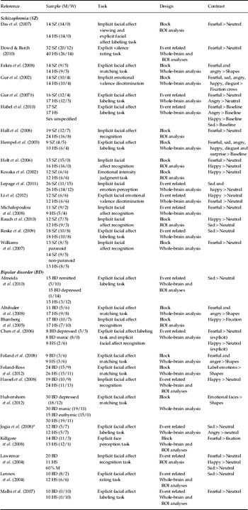
Meta-analyses were performed using the ALE software implemented in GingerALE 2.0.4 (http://brainmap.org). This version uses a random effect model and weighting for sample size of the original studies (Laird et al. Reference Laird, Fox, Price, Glahn, Uecker, Lancaster, Turkeltaub, Kochunov and Fox2005). The contrasts included are shown in Table 1. We accepted results as significant based on the threshold used in the original studies. When both were available, coordinates used derived from whole-brain and not region of interest (ROI) analyses. Coordinates of the foci of activation reported in the original studies were transformed into Talairach space using the Lancaster transform (icbm2tal) in GingerALE. Activation coordinates derived from each study were weighted to yield estimates of activation likelihood at each voxel that were then combined to compute a modeled activation (MA) map. ALE scores from the convergent MA maps were then calculated on a voxel-by-voxel basis to test for convergent (random effects) rather than study-specific foci (fixed effects). All ALE data processing was performed using the BrainMap Search and View software (http://brainmap.org). The threshold of statistical significance was set at p < 0.05, with false discovery rate (FDR) correction for multiple comparisons and a minimum cluster size of 200 mm3. Each ALE map was imported into Mango (http://ric.uthscsa.edu/mango) and overlaid on an anatomical template (www.brainmap.org/ale/colin1.1.nii) for representation purposes. For each suprathreshold cluster, corresponding Brodmann areas (BAs) were identified using the Talairach and Tournoux stereotactic anatomic brain atlas (Talairach & Tournoux, Reference Talairach and Tournoux1988). Finally, to compare diagnostic groups, the ALE maps for SZ and BD were contrasted directly using the subtraction meta-analysis procedure implemented in GingerALE. We report only results where there was convergence of activation between two or more studies.
Details of studies included in the meta-analysis (in alphabetical order)

HS, Healthy subjects; M, men; W, women; ROI, region of interest
a Only baseline data used.
Moderator variables
Demographic information about age and sex and clinical information about level of psychopathology and medication type and dosage were extracted from each primary study. Mean antipsychotic dose was converted to chlorpromazine equivalents (Bezchlibnyk-Butler & Jeffries, 2010). The effect of other medications (e.g. lithium) was not examined because dosage was not mentioned in a sufficient number of studies. We extracted information about participants' scores on the Hamilton Depression Rating Scale (HAMD; Hamilton, Reference Hamilton1960), the Young Mania Rating Scale (YMRS; Young et al. Reference Young, Biggs, Ziegler and Meyer1978) and the positive and negative subscales of the Positive and Negative Syndrome Scale (PANSS; Kay et al. Reference Kay, Fiszbein and Opler1987). We tested for potential group differences in these moderator variables and examined their influence on the ALE results using meta-regression analyses. The impact of symptom severity was examined in each diagnostic group separately. Based on procedures implemented in previous studies (Van Snellenberg et al. Reference Van Snellenberg, Torres and Thornton2006; Anticevic et al. Reference Anticevic, Van Snellenberg, Cohen, Repovs, Dowd and Barch2012), we rescaled each psychopathology scale from 0 to 1, where 0 and 1 indicated respectively the minimum and maximum possible scores on each scale.
Results
Included studies
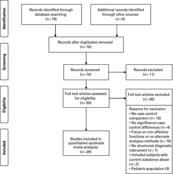
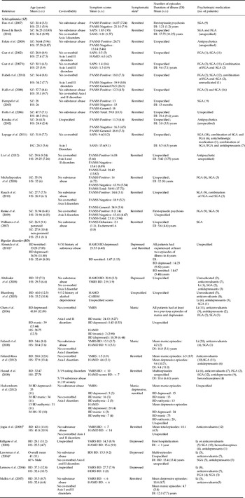
The study selection flowchart is shown in the PRISMA diagram provided in Fig. 1 and Table 1 shows the details of the studies. The total sample comprised 268 patients with SZ, 267 BD patients and 483 healthy individuals. Demographic details for all participants and clinical information of SZ and BD patients are shown in Table 2. There was a significant difference between the two patient groups in age (SZ: mean = 31.69 years, s.d. = 6.1 years; BD: mean = 35.9 years, s.d. = 4.1; t = − 2.33 p = 0.026) but not sex (SZ: 64.3% men, 35.7% women: BD: 56.2% men, 43.8% women, χ2 = 3.55, p = 0.06).
Flowchart of study selection.

Description of participants included in the meta-analysis (in alphabetical order)

BD, Bipolar disorder; SZ, schizophrenia; HS, healthy subjects; Li, lithium; BDI, Beck Depression Inventory; CARSM, Clinician-Administered Rating Scale for Mania; HAMD, Hamilton Depression Rating Scale; FGA, first-generation antipsychotic; PANSS, Positive and Negative Syndrome Scale; SAPS, Scale for the Assessment of Positive Symptoms; SANS, Scale for the Assessment of Negative Symptoms; SGA, second-generation antipsychotic; YMRS, Young Mania Rating Scale; s.d., standard deviation.
a Study also included patients with major depressive disorder (MDD).
b Only baseline data used.
c We used the description of symptomatic state as in the original articles.
Comparison of patients with SZ to healthy individuals
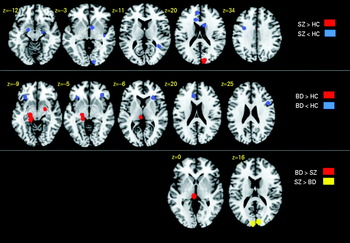
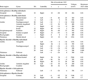
Compared to healthy individuals, patients with SZ showed an increased likelihood of activation in the right cuneus and a decreased likelihood of activation in frontal regions [left precentral gyrus (BA 6) and left medial frontal gyrus (BA 9)], in limbic and paralimbic regions [right amygdala, right insula and left parahippocampal gyrus (BA 28) and anterior cingulate cortex (ACC; BA 32)], in occipital and occipitotemporal regions [right inferior occipital gyrus (BA 17) and right fusiform gyrus (BA 37)], in the basal ganglia (right caudate nucleus) and in the right medial dorsal thalamus (19 studies; 103 foci and 645 subjects). Details are shown in Fig. 2 and Table 3.
Activation likelihood estimation results. Top row: patients with schizophrenia (SZ) compared to healthy controls (HC). Middle row: patients with bipolar disorder (BD) compared to HC. Bottom row: patients with SZ compared to patients with BD. p < 0.05 false discovery rate corrected.

Activation likelihood estimation (ALE) results for facial emotion processing in schizophrenia and bipolar disorder.

BA, Brodmann area.
Talairach and Tournoux coordinates: x = sagittal, y = coronal, z = axial.
No effect of gender was observed but age was positively correlated with the likelihood of activation in the left parahippocampal gyrus (BA 28) (x = −20, y = −26, z = −8; r 2 = 0.66, p = 0.001, voxels = 227). A higher PANSS positive symptoms subscale score correlated with a reduced likelihood of activation in the hippocampus/parahippocampal gyrus (x = 38, y = −18, z = −14; r 2 = 0.56; p = 0.001, voxels = 619). A higher PANSS negative symptoms subscale score correlated with a reduced likelihood of activation in the precentral gyrus (BA 6) (x = −38, y = 2, z = 40; r 2 = 0.56, p = 0.001, voxels = 33).
Comparison of patients with BD to healthy individuals
Compared to healthy individuals, BD patients showed increased activation in the parahippocampal gyrus extending to the amygdala bilaterally and left pulvinar thalamus (15 studies; 51 foci and 473 subjects). Additionally, BD patients showed decreased activation in the inferior frontal gyrus bilaterally (BA 47, BA 44), the left ACC (BA 32) and the right caudate (13 studies; 47 foci and 366 subjects). Details are shown in Fig. 2 and Table 3. Age and sex did not contribute to the results. Higher HAMD scores were associated with a greater likelihood of activation in the right amygdala (x = 22, y = −6, z = −12; r 2 = 0. 53, p = 0.001, voxels = 79) whereas higher YMRS scores were positively correlated with the likelihood of activation in the right thalamus (x = 6, y = −18, z = 10; r 2 = 0.2, p = 0.002, voxels = 70).
Comparison of SZ to BD
Compared to SZ patients, patients with BD were more likely to activate the left pulvinar thalamus (Table 3, Fig. 2). Conversely, patients with SZ were more likely to activate the cuneus bilaterally (BA 18) (Table 3, Fig. 2). Age and sex did not contribute to differences between diagnostic groups. Differences between the two disorders in amygdala activation were negatively correlated with antipsychotic dose (x = 22, y = −6, z = −12; r 2 = 0.98, p = 0.001, voxels = 832).
Discussion
The results of this quantitative meta-analysis demonstrate that SZ and BD are associated with largely distinct patterns of neural engagement during facial affect processing. During facial affect processing: (a) patients with BD showed increased likelihood of activation within subcortical regions of the facial affect processing network (parahippocampus/amygdala, thalamus and putamen) and reduced likelihood of activation within cortical regions, specifically the ventrolateral PFC compared to healthy individuals, (b) patients with SZ showed reduced likelihood of activation throughout the entire facial affect processing network and increased likelihood of activation in the cuneus (and adjacent posterior cortical regions) compared to healthy individuals. Additional differences were found when the two patient groups were compared to each other. Patients with BD showed greater likelihood of activation in thalamic regions compared to patients with SZ; in the reverse comparison, SZ patients showed increased likelihood of activation in the posterior visual association cortices.
Our findings when comparing patients with either SZ or BD to healthy individuals are in line with several recent meta-analyses on this topic (Li et al. Reference Li, Chan, McAlonan and Gong2010; Chen et al. Reference Chen, Suckling, Lennox, Ooi and Bullmore2011; Sugranyes et al. Reference Sugranyes, Kyriakopoulos, Corrigall, Taylor and Frangou2011; Anticevic et al. Reference Anticevic, Van Snellenberg, Cohen, Repovs, Dowd and Barch2012; Delvecchio et al. Reference Delvecchio, Fossati, Boyer, Brambilla, Falkai, Gruber, Hietala, Lawrie, Martinot, McIntosh, Meisenzahl and Frangou2012; Taylor et al. Reference Taylor, Kang, Brege, Tso, Hosanagar and Johnson2012). Therefore, here we focus mostly on the comparison between SZ and BD so as to draw inferences about their pathophysiological boundaries. We begin by describing the functional properties of the neural network involved in facial affect processing as these are relevant to the interpretation of the differences between patients with SZ and patients with BD.
Functional properties of the facial affect processing network
In fMRI studies, facial emotional expressions engage anatomically distributed cortical and subcortical regions with evidence of relative specialization (Adolphs, Reference Adolphs2002; Haxby et al. Reference Haxby, Hoffman and Gobbini2002; Phan et al. Reference Phan, Wager, Taylor and Liberzon2002; Murphy et al. Reference Murphy, Nimmo-Smith and Lawrence2003; Fairhall & Ishai, Reference Fairhall and Ishai2007; Fusar-Poli et al. Reference Fusar-Poli, Placentino, Carletti, Landi, Allen, Surguladze, Benedetti, Abbamonte, Gasparotti, Barale, Perez, McGuire and Politi2009; Pessoa & Adolphs, Reference Pessoa and Adolphs2010; Vytal & Hammann, Reference Vytal and Hammann2010; Dima et al. Reference Dima, Stephan, Roiser, Friston and Frangou2011). Within cortical regions, visual stimuli are first processed in the primary (BA 17) and extrastriate visual cortices (BA 18, 19), where they are rapidly categorized according to their physical characteristics (Lachaux et al. Reference Lachaux, George, Tallon-Baudry, Martinerie, Hugueville, Minottic, Kahanec and Renault2005). Information is then transmitted to temporal regions, notably the fusiform gyrus. The fusiform gyrus is robustly associated with encoding facial features and identity (Haxby et al. Reference Haxby, Hoffman and Gobbini2002) but is also crucially involved in facial affect processing (Pizzagalli et al. Reference Pizzagalli, Regard and Lehmann1999; Vuilleumier & Pourtois, Reference Vuilleumier and Pourtois2007), particularly in the early categorization of facial expressions (Pizzagalli et al. Reference Pizzagalli, Lehmann, Hendrick, Regard, Pascual-Marqui and Davidson2002; Tsuchiya et al. Reference Tsuchiya, Kawasaki, Oya, Howard and Adolphs2008). Involvement of the medial PFC is elicited by multiple tasks that require conscious attribution or judgment of mental states, dispositional traits or intentions of one's self or of others (Phillips et al. Reference Phillips, Drevets, Rauch and Lane2003; Amodio & Frith, Reference Amodio and Frith2006; Etkin et al. Reference Etkin, Egner and Kalisch2011). Engagement of the ventrolateral PFC relates primarily to the contextual or social appropriateness of responses to emotional cues (Berthoz et al. Reference Berthoz, Armony, Blair and Dolan2002; Quirk & Beer, Reference Quirk and Beer2006; Spreng et al. Reference Spreng, Mar and Kim2009). Among the subcortical regions, the amygdala is thought to reflect arousal (Critchley et al. Reference Critchley, Rotshtein, Nagai, O'Doherty, Mathias and Dolan2005) or biological salience (Santos et al. Reference Santos, Mier, Kirsch and Meyer-Lindenberg2010) associated with stimuli. The insula is engaged during observation and imitation of facial emotional expressions and is thought to contribute to the modulation of action representation (mirroring) (Carr et al. Reference Carr, Iacoboni, Dubeau, Mazziotta and Lenzi2003) and agency (self or other) (Farrer et al. Reference Farrer, Franck, Georgieff, Frith, Decety and Jeannerod2003) on emotional processing. Finally, higher-order thalamic nuclei also support facial affect processing. The pulvinar thalamus is directly involved in visual perception (Pessoa & Adolphs, Reference Pessoa and Adolphs2010), particularly in directing and maintaining attention towards salient stimuli (Desimone et al. Reference Desimone, Wessinger, Thomas and Schneider1990). A similar role is ascribed to the medial dorsal thalamus, often referred to as the ‘limbic thalamus’ (Armstrong, Reference Armstrong and Paxinos1990), in addition to its function in emotional responsiveness and memory retrieval (Taber et al. Reference Taber, Wen, Khan and Hurley2004).
Emotional modulation of the face processing network is reduced in SZ and increased in BD
In the contrast between emotional and neutral facial expressions, patients with SZ show reduced activation within the amygdala/parahippocampus compared to both BD patients and healthy individuals. This has been attributed to abnormally elevated responses to neutral facial stimuli (Aleman & Kahn, Reference Aleman and Kahn2005; Holt et al. Reference Holt, Kunkel, Weiss, Goff, Wright, Shin, Rauch, Hootnick and Heckers2006; Surguladze et al. Reference Surguladze, Russell, Kucharska-Pietura, Travis, Giampietro, David and Phillips2006; Hall et al. Reference Hall, Whalley, McKirdy, Romaniuk, McGonigle, McIntosh, Baig, Gountouna, Job, Donaldson, Sprengelmeyer, Young, Johnstone and Lawrie2008; Anticevic et al. Reference Anticevic, Van Snellenberg, Cohen, Repovs, Dowd and Barch2012) and is thought to reflect abnormal salience attribution (Kapur, Reference Kapur2003). However, Gur et al. (Reference Gur, Loughead, Kohler, Elliott, Lesko, Ruparel, Wolf, Bilker and Gur2007b) reported that limbic activation was abnormally elevated in patients with SZ when they failed to identify facial expressions. This information, combined with behavioral evidence that SZ is associated with deficits in correct facial affect identification, suggests that patients find these stimuli ambiguous regardless of valence (Pinkham et al. Reference Pinkham, Gur and Gur2007). It could therefore be argued that it is ambiguity (Santos et al. Reference Santos, Mier, Kirsch and Meyer-Lindenberg2010), rather than inappropriate attribution, that drives limbic recruitment in SZ. Additionally, visual processing of facial stimuli (regardless of emotional expression) in SZ may be impaired from the early stages of sensory perception (Chen et al. Reference Chen, Norton, McBain, Ongur and Heckers2009). Consistent with this observation we found reduced likelihood of recruitment in SZ within the visual and fusiform cortices involved in early stimulus categorization (Pizzagalli et al. Reference Pizzagalli, Lehmann, Hendrick, Regard, Pascual-Marqui and Davidson2002; Lachaux et al. Reference Lachaux, George, Tallon-Baudry, Martinerie, Hugueville, Minottic, Kahanec and Renault2005; Tsuchiya et al. Reference Tsuchiya, Kawasaki, Oya, Howard and Adolphs2008). Difficulties in stimulus categorization are also likely to increase stimulus ambiguity. It is therefore possible that a more general dysfunction in visual perception contributes to abnormal facial affect processing in SZ (Kumar et al. Reference Kumar, Christodoulou, Vyas, Kyriakopoulos, Corrigall, Reichenberg and Frangou2010; Green et al. Reference Green, Lee, Wynn and Mathis2011).
In BD, emotion seems to be associated with increased modulation of subcortical regions of the face processing network compared to healthy individuals and to patients with SZ. BD was associated with increased engagement of the pulvinar, which indicates enhanced attention towards emotionally salient stimuli from the early stages of visual processing (Pessoa & Adolphs, Reference Pessoa and Adolphs2010). Neural engagement in BD patients was also amplified in the parahippocampal gyrus/amygdala. Greater amygdala engagement in BD is congruent with the notion of increased salience of or arousal to emotional stimuli (Critchley et al. Reference Critchley, Rotshtein, Nagai, O'Doherty, Mathias and Dolan2005; Santos et al. Reference Santos, Mier, Kirsch and Meyer-Lindenberg2010) whereas increased parahippocampal activation may index greater contextual appraisal (Gerdes et al. Reference Gerdes, Wieser, Muhlberger, Weyers, Alpers, Plichta, Breuer and Pauli2010). Consistent with the primary literature, our analysis points to a positive association between level of affective symptoms and degree of signal enhancement in subcortical (Altshuler et al. Reference Altshuler, Bookheimer, Proenza, Townsend, Sabb, Firestine, Bartzokis, Mintz, Mazziotta and Cohen2005; Foland et al. Reference Foland, Altshuler, Bookheimer, Eisenberger, Townsend and Thompson2008; Bermpohl et al. Reference Bermpohl, Dalanay, Kahnt, Sajonz, Heimann, Ricken, Stoy, Hägele, Schlagenhauf, Adli, Wrase, Ströhle, Heinz and Bauer2009) but not cortical regions (Foland-Ross et al. Reference Foland-Ross, Bookheimer, Lieberman, Sugar, Townsend, Fischer, Torrisi, Penfold, Madsen, Thompson and Altshuler2012).
The ACC and the ventral PFC are known to play a crucial role in evaluative and regulatory functions during emotional processing, which may map on different subdivisions of these regions (Phillips et al. Reference Phillips, Drevets, Rauch and Lane2003; Etkin et al. Reference Etkin, Egner and Kalisch2011). It has been proposed that dorsal subdivisions are primarily involved in appraisal and generation of affective states and their corresponding somatic markers (Critchley, Reference Critchley2005; Walton et al. Reference Walton, Behrens, Noonan and Rushworth2011). By contrast, ventral subdivisions are thought to exert a regulatory (mostly inhibitory) influence on the amygdala and other subcortical and posterior cortical regions (Ochsner & Gross, Reference Ochsner and Gross2005; Quirk & Beer, Reference Quirk and Beer2006). Therefore, the decreased engagement of the ventral ACC and PFC identified here (Table 3) is indicative of a greater impact of BD on regions relating to emotional regulation.
Increased engagement in posterior midline cortices may typify SZ but not BD
SZ was associated with increased likelihood of engagement within the cuneus compared to healthy individuals and patients with BD (Table 3). This was coupled to reduced engagement within the primary visual cortex in the inferior occipital gyrus compared to healthy individuals. The cluster of overactivation identified corresponds to the medial cuneus, which is part of the extrastriate visual cortex (Iaria et al. Reference Iaria, Robbins and Petrides2008) and thus involved in higher-order processing of visual information. The function most consistently ascribed to the cuneus relates to early stimulus categorization. In this respect the cuneus may enlist mnemonic and/or attentional mechanisms towards features that distinguish categories and thus modulate the quality or quantity of visual information reaching later processing stages (Sergent et al. Reference Sergent, Ohta and MacDonald1992; Vanni et al. Reference Vanni, Tanskanen, Seppä, Uutela and Hari2001). This pattern of overactivation within higher-order visual cortices in SZ was also observed by Taylor et al. (Reference Taylor, Kang, Brege, Tso, Hosanagar and Johnson2012) in a meta-analysis of fMRI studies of emotional perception and by Seiferth et al. (Reference Seiferth, Pauly, Kellermann, Shah and Ott2009) in a study of facial affect processing in adolescents with SZ. Taken together, these findings suggest a significant role for the visual cortices in SZ that is probably already present at the earliest stages of the illness. The nature of this overactivation within higher-order visual regions in SZ is unclear but the prevailing view is that it represents a ‘compensatory’ response to deficits in integrating visual information (Seiferth et al. Reference Seiferth, Pauly, Kellermann, Shah and Ott2009; Taylor et al. Reference Taylor, Kang, Brege, Tso, Hosanagar and Johnson2012). Visual abnormalities in SZ are not limited to the perception of facial expressions but have been reported in multiple paradigms including motion and form perception, spatial frequency and location discrimination, perceptual organization and backward masking (Butler et al. Reference Butler, Silverstein and Dakin2008; Kumar et al. Reference Kumar, Christodoulou, Vyas, Kyriakopoulos, Corrigall, Reichenberg and Frangou2010; Green et al. Reference Green, Lee, Wynn and Mathis2011).
Methodological considerations
Meta-analyses allow the synthesis of data from the primary literature but some limitations are evident. First, we accepted the results of individual studies as reported and not weighted based on the threshold of significance used in each original study. Second, although most patients were remitted when scanned (Table 2), the level of symptomatology impacted on brain activity. The effect of symptoms seems to exaggerate rather than confound diagnostic differences between the two disorders. Third, the impact of medication on our results cannot be easily evaluated. Medication regimes for SZ and BD are inherently different, with the exception of antipsychotic medication. Current evidence suggests predominantly ameliorative effects of antipsychotic medication on facial affect processing in SZ and in BD (Phillips et al. Reference Phillips, Travis, Fagiolini and Kupfer2008; Cabral-Calderin et al. Reference Cabral-Calderin, Mendoza-Quiñones, Garcia, Caballero, Domínguez and Reyes2010). In this study antipsychotic dose seemed to reduce group differences in amygdala/parahippocampal engagement. Antipsychotic medication is known to influence the function of the amygdala (Aleman & Kahn, Reference Aleman and Kahn2005) but the effect observed here suggests that it minimizes diagnostic differences. Fourth, a further limitation is the variability in the types of tasks used in individual source studies, which precludes separate analyses per task type because of small sets sizes. As the primary literature expands, more task-specific meta-analyses should be possible in the future. Fifth, it would be desirable to evaluate a range of study-wise factors that may influence both the nature and the topography of the results. These factors may include clinical information about age of onset or illness severity, task performance and details of data acquisition and analysis. The considerable variation in study design within the primary literature (Tables 1 and 2) unfortunately precludes a quantitative assessment of all of these factors and we therefore focused on those for which we had sufficient information. However, our results achieved a degree of robustness that suggests that we captured the most replicable differences between patients with BD or SZ. Finally, our meta-analysis provides an estimate of the probability of differential activation in brain regions when comparing diagnostic groups and not the mean activation difference in these regions. Therefore, traditional measures of heterogeneity and publication bias that are based on the effect size of group differences are not applicable.
Conclusions
Affective dysfunction is a prominent feature of SZ and BD. This meta-analysis focused on a particular component of affective processing relating to the identification of facial affect, where we demonstrate significant diagnostic differences. We confirmed that, in SZ, affective modulation was reduced in the amygdala and additionally we show the same effect throughout the traditional face processing network. This reduction in affective modulation was previously designated to inappropriate attribution of salience to neutral stimuli (Kapur, Reference Kapur2003). The overactivation in higher-order visual cortices observed here supports an alternative explanation; facial affect identification deficits in SZ may arise from abnormalities in visual perception and represent another dimension of poor stimulus categorization. Our results also indicate that, in BD, there is increased affective modulation within the subcortical regions of the face processing network linked with underengagement of ventral PFC regions considered important in cognitive control. In our previous study on BD we were able to show the differential impact of positive and negatively valenced facial expressions on the face processing network (Delvecchio et al. Reference Delvecchio, Fossati, Boyer, Brambilla, Falkai, Gruber, Hietala, Lawrie, Martinot, McIntosh, Meisenzahl and Frangou2012). The effect of valence on the neurobiology of affective facial processing in SZ remains unresolved because of the limited availability of relevant original studies but it would be worth pursuing in future research. Further work would also be required to assess the effect of diagnosis of SZ or BD on emotional expression and subjective experience of emotion, two aspects of affective processing not addressed here.
Declaration of Interest
None.