Introduction
The Blue Lizard, Green Lizard, Giveaway–Simplot and Markey mines in the Red Canyon portion of the White Canyon district in south-eastern Utah have yielded many new mineral species in recent years (e.g. Kampf et al., Reference Kampf, Olds, Plášil, Nash and Marty2021a). Most of the new species are uranyl sulfates and most, especially from the Blue Lizard mine, contain Na. Of the ten new species that the Markey mine has yielded, all contain uranyl and six contain carbonate; of the six new uranyl carbonates, five also contain essential Ca. The new mineral paramarkeyite, which is included in the aforementioned enumerations, is very closely related to three of the other new minerals from the Markey mine: markeyite (Kampf et al., Reference Kampf, Plášil, Kasatkin, Marty and Čejka2018), natromarkeyite and pseudomarkeyite (Kampf et al., Reference Kampf, Olds, Plášil, Burns and Marty2020a).
A preponderance of uranyl carbonate minerals form structures based on finite clusters of polyhedra that have relatively weak bonding interactions (~0.1–0.4 vu) between bridging and non-bridging cations and carbonate polyhedra (Lussier et al., Reference Lussier, Lopez and Burns2016). The uranyl tricarbonate (UTC) unit, [(UO2)(CO3)3]4-, present in most of the currently known uranyl carbonate minerals is highly stable and only slight differences in pH and cation:U:CO3 content affects uranyl carbonate speciation in solution, leading to crystallisation of a wide variety of finite cluster-based topologies. Likewise, numerous uranyl sulfate minerals based on finite cluster topologies (especially those found in Red Canyon) crystallise relatively rapidly as efflorescent crusts from high ionic-strength solutions, suggesting that the aqueous conformation of polyhedral clusters may be retained from solution to crystal.
Paramarkeyite (IMA2021-024, Kampf et al., Reference Kampf, Olds, Plášil, Burns, Škoda and Marty2021b) is named based on the Greek παρα for ‘near’ and the mineral markeyite to which it is similar. It is also similar to natromarkeyite and pseudomarkeyite. The four minerals are similar in appearance, composition, Raman spectrum and structure. Note that markeyite (/ma:r ’ki: ait/) is named for the locality, the Markey mine. The new mineral and name were approved by the Commission on New Minerals, Nomenclature and Classification of the International Mineralogical Association. The description is based on two cotype specimens deposited in the collections of the Natural History Museum of Los Angeles County, 900 Exposition Boulevard, Los Angeles, CA 90007, USA, catalogue numbers 67487 and 67488; 67487 is also the holotype for natromarkeyite and 67488 is also the cotype for natromarkeyite.
Occurrence
Paramarkeyite was found underground in the Markey mine, Red Canyon, White Canyon District, San Juan County, Utah, USA (37°32′57″N, 110°18′08″W). The Markey mine is located ~1 km southwest of the Blue Lizard mine, on the east-facing side of Red Canyon, ~72 km west of the town of Blanding, Utah, and ~22 km southeast of Good Hope Bay on Lake Powell. The geology of the Markey mine is quite similar to that of the Blue Lizard mine (Chenoweth, Reference Chenoweth1993; Kampf et al., Reference Kampf, Plášil, Kasatkin, Marty and Čejka2017), although the secondary mineralogy of the Markey mine is notably richer in carbonate phases. Underground gas measurements collected in 2016 using a hand-held Crowcon Gasman CO2 monitor showed consistently elevated CO2 levels at the Markey mine, averaging ~1000 ppm CO2 with a maximum recorded value of 1600 ppm CO2, levels considerably higher than at the nearby Blue Lizard mine where carbonate mineral species are less abundant. Higher CO2 concentration at the Markey mine may be connected to an abundance of calcite present in the ores, released by the action of acidic waters derived from decaying sulfides.
The following information regarding the history and geology is taken largely from Chenoweth (Reference Chenoweth1993). Jim Rigg of Grand Junction, Colorado began staking claims in Red Canyon in March of 1949. The Markey group of claims, staked by Rigg and others, was purchased by the Anaconda Copper Mining Company on June 1, 1951. After limited exploration and production, the mine closed in 1955. The mine was subsequently acquired from Anaconda by Calvin Black of Blanding, Utah under whose ownership the mine operated from 1960 to 1982 and was a leading producer in the district for nearly that entire period.
The uranium deposits in Red Canyon occur within the Shinarump member of the Upper Triassic Chinle Formation, in channels incised into the reddish-brown siltstones of the underlying Lower Triassic Moenkopi Formation. The Shinarump member consists of medium- to coarse-grained sandstone, conglomeratic sandstone beds and thick siltstone lenses. Ore minerals (uraninite, montroseite, coffinite, etc.) were deposited as replacements of wood and other organic material and as disseminations in the enclosing sandstone. Since the mine closed in 1982, oxidation of primary ores in the humid underground environment has produced a variety of secondary minerals, mainly carbonates and sulfates, as efflorescent crusts on the surfaces of mine walls.
Paramarkeyite is a very rare mineral in the secondary mineral assemblage at the Markey mine. It occurs on gypsum-coated asphaltum in association with andersonite, calcite, gypsum and natromarkeyite (Kampf et al., Reference Kampf, Olds, Plášil, Burns and Marty2020a). Other new minerals recently described from the Markey mine are feynmanite (Kampf et al., Reference Kampf, Olds, Plášil, Marty and Perry2019a), leószilárdite (Olds et al., Reference Olds, Sadergaski, Plášil, Kampf, Burns, Steele, Marty, Carlson and Mills2017), magnesioleydetite (Kampf et al., Reference Kampf, Plášil, Kasatkin, Nash and Marty2019b), markeyite (Kampf et al., Reference Kampf, Plášil, Kasatkin, Marty and Čejka2018), meyrowitzite (Kampf et al., Reference Kampf, Plášil, Olds, Nash, Marty and Belkin2019c), pseudomarkeyite (Kampf et al., Reference Kampf, Olds, Plášil, Burns and Marty2020a), straβmannite (Kampf et al., Reference Kampf, Plášil, Kasatkin, Nash and Marty2019b) and uroxite (Kampf et al., Reference Kampf, Plášil, Nash, Němec and Marty2020b).
Physical and optical properties
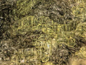
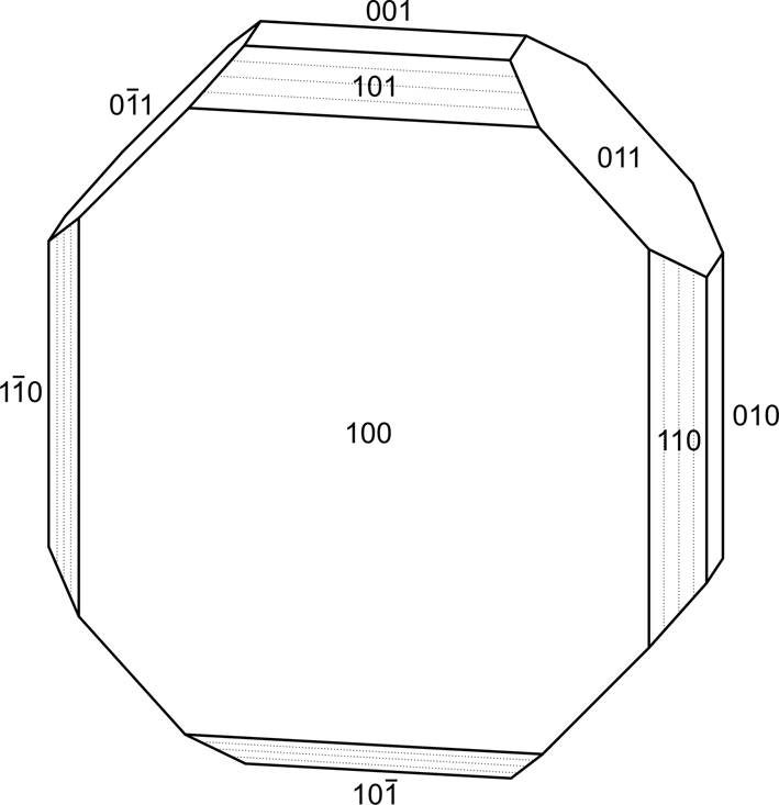
Paramarkeyite crystals are striated tablets (Fig. 1) up to ~0.11 mm across, commonly in parallel intergrowths. Tablets are probably flattened on {100} and striated parallel to [010] and [001]; however, because crystals occur in parallel intergrowths and easily break into irregular fragments, it was not possible to extract complete crystals for measurements. The morphological description is based, in part, on the structure of paramarkeyite by analogy with relation of morphology to structure in the closely related minerals markeyite, natromarkeyite and pseudomarkeyite. The forms {100}, {010}, {001}, {110}, {101}, {10 $\bar{1}$} and {011} (Fig. 2) are based upon observed morphology, but were not measured. No twinning was observed.
Paramarkeyite on gypsum; field of view 0.84 mm across (catalogue number 67487).

Crystal drawing of paramarkeyite; clinographic projection.

Crystals are pale green yellow and transparent with vitreous lustre. The streak is white. The mineral fluoresces moderate bluish white under a 405 nm laser. The Mohs hardness is 2½, based upon scratch tests. Crystals are very brittle with irregular, curved fracture. There is excellent cleavage on {100}, and probably two good cleavages on {010} and {001}. In room-temperature H2O, the mineral slowly loses birefringence, but does not dissolve; it dissolves immediately with effervescence in dilute HCl. The density measured by flotation in a mixture of methylene iodide and toluene is 2.91(2) g cm–3. The calculated density based on the empirical formula and unit–cell parameters obtained from single-crystal X-ray diffraction data is 2.905 g cm–3.
Optically, paramarkeyite is biaxial (–), with α = 1.550(2), β = 1.556(2), γ = 1.558(2) (measured in white light). The 2V measured directly on a spindle stage is 60(2)°; the calculated 2V is 59.8°. Dispersion is r > v, strong. The mineral is nonpleochroic. The partially determined optical orientation is Y = b. The Gladstone–Dale compatibility index 1 – (K P/K C) for the empirical formula is –0.025, in the excellent range (Mandarino, Reference Mandarino2007), using k(UO3) = 0.118, as provided by Mandarino (Reference Mandarino1976).
Raman spectroscopy
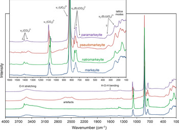
A Raman spectrum was collected from a loose crystal fragment of paramarkeyite using a Horiba LabRAM HR Evolution spectrometer equipped with a 532 nm diode laser. The Raman spectrum of paramarkeyite is very similar to those of markeyite (Kampf et al., Reference Kampf, Plášil, Kasatkin, Marty and Čejka2017), natromarkeyite (Kampf et al., Reference Kampf, Olds, Plášil, Burns and Marty2020a) and pseudomarkeyite (Kampf et al., Reference Kampf, Olds, Plášil, Burns and Marty2020a). All four spectra are compared in Fig. 3.
Comparison of the Raman spectra of paramarkeyite, pseudomarkeyite, natromarkeyite and markeyite.

Multiple broad bands in the 3700–3100 cm–1 range are attributed to ν O–H stretching vibrations of symmetrically distinct hydrogen-bonded H2O groups. According to the correlation given by Libowitzky (Reference Libowitzky1999), this corresponds to approximate O–H⋅⋅⋅O hydrogen bond-lengths between 3.2 and 2.7 Å, which is consistent with what is reported in the structures of markeyite and pseudomarkeyite. The broad bands in the 2800–2300 cm–1 range in the markeyite and pseudomarkeyite spectra were originally interpreted as corresponding to strong (short) hydrogen bonds (Kampf et al., Reference Kampf, Plášil, Kasatkin, Marty and Čejka2018); however, no such bonds appear to exist in these structures and no such bands are seen in the paramarkeyite spectrum. We now think that these may be spectral artefacts because such bands have also been observed in spectra of anhydrous minerals, and we have recorded other spectra for markeyite that do not exhibit this feature.
The weak bands between ~1450 and 1300 cm–1 can be attributed to the ν3 (CO3)2– antisymmetric stretching vibrations of the (CO3)2– units. Medium to strong multiple bands between 1100 and 1050 cm–1 are connected with the ν1 (CO3)2– symmetric stretching vibrations of several structurally non-equivalent carbonate units (Koglin et al., Reference Koglin, Schenk and Schwochau1979; Anderson et al., Reference Anderson, Ch, Irish and Tong1980; Čejka, Reference Čejka, Burns and Finch1999 and Reference Čejka2005).
The very strong bands centred at 826, 827, 829 and 830 cm–1 for markeyite, pseudomarkeyite, natromarkeyite and paramarkeyite, respectively, are due to the ν1 (UO2)2+ symmetric stretching vibrations and provide inferred U–O bond lengths of ~1.78–1.79 Å (Bartlett and Cooney, Reference Bartlett and Cooney1989). The ν2 (δ) (CO3)2– bending vibration may coincide with this band.
Several weak to strong bands in the range 780 to 680 cm–1 are assigned to the doubly degenerate ν4 (δ) (CO3)2– bending vibrations. The medium-strength band in the spectra centred near 240 cm–1 is assigned to the split doubly degenerate ν2 (δ) (UO2)2+ bending vibrations. Bands between 200 and 120 cm–1 are due to lattice modes (Koglin et al., Reference Koglin, Schenk and Schwochau1979; Anderson et al., Reference Anderson, Ch, Irish and Tong1980; Čejka, Reference Čejka, Burns and Finch1999 and Reference Čejka2005).
Chemical composition
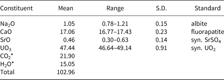
Chemical analyses (6) were performed using a Cameca SX100 electron microprobe at Masaryk University, Brno, operating in wavelength dispersive spectroscopy mode using an accelerating voltage of 15 kV, beam current of 1 nA and a 15 μm beam diameter. Such mild conditions were used in order to minimise the decomposition of the analysed area by the electron beam. The analysed area showed no visible changes in back-scattered electron spectroscopy or reflected light after the analysis. Concentrations of elements other than those reported in Table 1 were below detection limits. Matrix correction by the X-PHI algorithm (Merlet, Reference Merlet1994) was applied to the data. Insufficient material was available for CHN analysis; however, the fully ordered structure and detailed bond-valence analysis unambiguously established the identities of O, H2O and CO2 and the corresponding quantitative contents of H2O and CO2, which were calculated on the basis of 16 O and 1 U atoms per formula unit (apfu). The high analytical total is probably attributable to H2O loss in the vacuum during the deposition of the conductive carbon coat and in the microprobe chamber, which resulted in higher concentrations for the remaining constituents. Analytical data are given in Table 1.
Chemical composition (in wt.%) for paramarkeyite.

* Based on the structure. S.D. – standard deviation.
The empirical formula (calculated on the basis of 16 O apfu) is Ca1.83Na0.20Sr0.03U1.00C3.00O16.00H10.07, or (Ca1.83Na0.20Sr0.03)Σ2.05(UO2)(CO3)3⋅5H2O (+0.07 H). The simplified formula is (Ca,Na,Sr)2(UO2)(CO3)3⋅5H2O and the ideal formula is Ca2(UO2)(CO3)3⋅5H2O, which requires CaO 18.08, UO3 46.11, CO2 21.29 and H2O 14.52, total 100 wt.%.
X-ray crystallography and structure refinement
Powder X-ray diffraction was done using a Rigaku R-Axis Rapid II curved imaging plate microdiffractometer, with monochromatised MoKα radiation. A Gandolfi-like motion on the φ and ω axes was used to randomise the sample and observed d values and intensities were derived by profile fitting using JADE Pro software (Materials Data, Inc.). The powder data are presented in Supplementary Table S1. This table is deposited with the Principal Editors of Mineralogical Magazine and is available as Supplementary material (see below).
The single-crystal structure data were collected at room temperature using the same diffractometer and radiation noted above. The Rigaku CrystalClear software package was used for processing structure data, including the application of an empirical multi-scan absorption correction using ABSCOR (Higashi, Reference Higashi2001). The structure was solved using the intrinsic phasing algorithm of the SHELXT program (Sheldrick, Reference Sheldrick2015a). SHELXL-2016 (Sheldrick, Reference Sheldrick2015b) was used for the refinement of the structure. Most atoms were located and subsequently refined at full occupancies; however, several Ca and O sites exhibited disorder manifest as split sites, partial occupancies and/or prolate anisotropic displacement ellipsoids. This prompted us to perform several tests. Despite a strong indication that the data are centrosymmetric (|E 2–1| = 1.01), seventy reflections (I > 3σI) were found to violate systematic absence expectations for the n glide plane, and we attempted to solve the structure in the non-centrosymmetric space group P21 using a racemic twin matrix. The resulting structure, while reasonable, provides a generally poor refinement with numerous non-positive definite anisotropic displacement ellipsoids not encountered in the P21/n refinement.
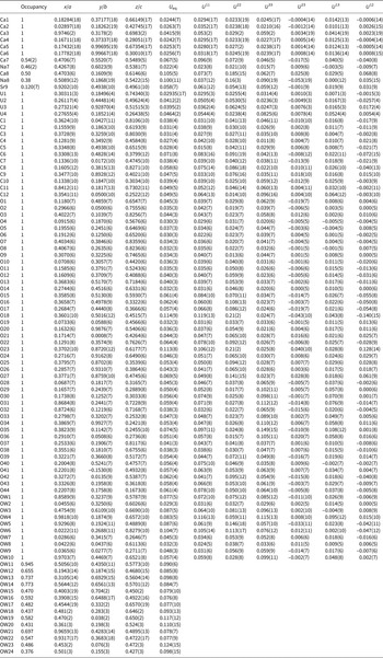
The occupancies of the O sites in the disordered interlayer portion of the structure were refined but still exhibited unrealistically large displacement parameters (U eq/U iso = 0.12–0.33 Å) indicating that their refined occupancies were too high. To account for this, the occupancies for sites OW11 to OW24 were refined with U iso set to 0.08. The occupancies obtained were then set in subsequent refinement cycles with U iso refined. The total of the OW site occupancies provide 4.55 H2O pfu; however, because there is additional residual electron density in the interlayer region and because the interlayer OW sites could accommodate additional occupancy, 5 H2O pfu is used for the ideal formula.
Three pairs of split cation sites were noted in the interlayer region. Based on bond-valence sums, these sites were assigned as Ca7 and Na7 (separated by 1.18 Å), Ca8 and Na8 (separated by 1.74 Å) and Sr9 (separated by 2.83 Å from another Sr9 site). The occupancies of the Ca7 and Na7 sites were refined jointly with Ca + Na = 1. Because of the large displacement parameters of the Ca8 and Na8 sites, joint occupancy refinement was not deemed appropriate; instead, we set the Ca8 occupancy to 0.50 Ca and the Na8 site occupancy to 0.38, which provided U iso values close to 0.1 for each site. The occupancy of the Sr9 site was refined. It is worth noting that Ca is greater than Na in both the Ca7/Na7 and Ca8/Na8 paired sites and the occupancy of the Sr9 site is only 0.120(7); hence, only Ca is provided in the ideal formula. Furthermore, the site occupancies for all of the Ca, Na and Sr sites provides (Ca1.76Na0.21Sr0.03)Σ2.00 pfu, which compares very well with the analysed composition (Ca1.83Na0.20Sr0.03)Σ2.05.
The refined anisotropic displacement parameters of several of the equatorial O atoms bonded to U and C are strongly prolate. It seems likely that this is in response to the Ca disorder, i.e. the planarity of the uranyl tricarbonate (UTC) units is disordered to accommodate the disordered Ca positions. Most of the impacted O atoms are in equatorial sites and are shared by Ca7/Na7 or Ca8/Na8.
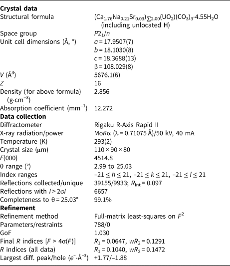
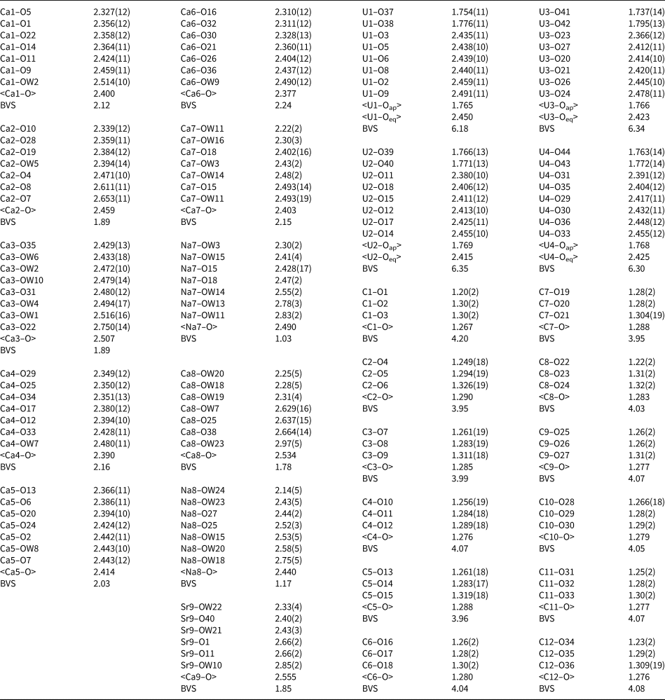
Data collection and refinement details are given in Table 2, atom coordinates and displacement parameters in Table 3 and selected bond distances and cation bond-valence sums (based on bond-valence parameters from Gagné and Hawthorne, Reference Gagné and Hawthorne2015) in Table 4. The crystallographic information files have been deposited with the Principal Editor of Mineralogical Magazine and are available as Supplementary material (see below).
Data collection and structure refinement details for paramarkeyite.*

* R int = Σ|F o2–F o2(mean)|/Σ[F o2]. GoF = S = {Σ[w(F o2–F c2)2]/(n–p)}½. R 1 = Σ||F o|–|F c||/Σ|F o|. wR 2 = {Σ[w(F o2–F c2)2]/Σ[w(F o2)2]}½; w = 1/[σ2(F o2)+(aP)2+bP] where a is 0.0337, b is 176.6 and P is [2F c2+Max(F o2,0)]/3.
Atom coordinates and displacement parameters (Å2) for paramarkeyite.

Selected bond distances (Å) and cation bond-valence sums (BVS in valence units) for paramarkeyite.

Description and discussion of the structure
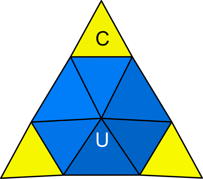
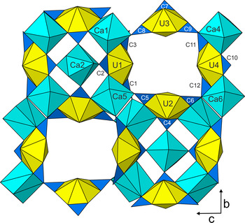
The four U sites (U1, U2, U3 and U4) in the structure of paramarkeyite are each surrounded by eight O atoms forming a squat UO8 hexagonal bipyramid. These bipyramids are each chelated by three CO3 groups, forming uranyl tricarbonate (UTC) clusters of formula [(UO2)(CO3)3]4- (Burns Reference Burns2005; Fig. 4). Six Ca sites (Ca1, Ca2, Ca3, Ca4, Ca5 and Ca6) are not split and have well defined coordinations; all are seven coordinated except Ca2, which is eight coordinated. The Ca7, Na7, Ca8 and Na8 sites have seven-fold coordinations and the Sr9 site has six-fold coordination. It is worth noting that in the closely related mineral natromarkeyite, Na cations fill pore space between multiple UTC units, sharing oxygen atoms of outstretched CO3 groups and apical uranyl oxygen atoms; these spaces are occupied only by O atoms of disordered H2O groups in paramarkeyite.
The uranyl tricarbonate cluster (UTC) of formula [(UO2)(CO3)3]4-.

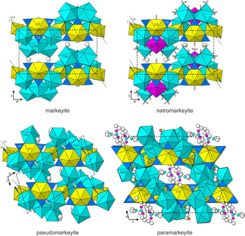
The Ca–O polyhedra share edges and corners with the UTCs. The UTCs and ordered polyhedra (except the Ca3–O polyhedron) form heteropolyhedral layers parallel to {100} (Fig. 5). The heteropolyhedral layer in the structure of paramarkeyite is essentially identical to the layers in the structures of markeyite, natromarkeyite and pseudomarkeyite. Viewed down the b axis (Fig. 6), the structures are seen to differ in the arrangement of additional Ca–O polyhedra (and a disordered CO3 group in the markeyite and natromarkeyite structures). We conjecture that the disorder in the interlayer region of the paramarkeyite structure and the different arrangement of non-layer polyhedra (including the Ca3–O polyhedron) is driven by the presence of minor Na and Sr, which can only be accommodated by splitting the cation sites.
The portion of the uranyl–carbonate layer that is common to the structures of markeyite, natromarkeyite, pseudomarkeyite and paramarkeyite. The cell directions and polyhedral labels apply specifically to paramarkeyite.

The structures of paramarkeyite, pseudomarkeyite and markeyite viewed down [010]. The atoms with disordered coordinations in paramarkeyite are shown as balls: Ca7 and Ca8 are turquoise, Na7 and Na8 are pink, Sr9 is orange and all OW are white.

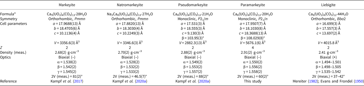
The structure of liebigite, Ca2(UO2)(CO3)3⋅11H2O (Mereiter, Reference Mereiter1982), contains the same structural components as those in the structures of markeyite, natromarkeyite, pseudomarkeyite and paramarkeyite. The same types of polyhedral linkages occur in all five structures, where the Ca–O polyhedra link UTCs forming heteropolyhedral layers. In the structure of liebigite, as in that of markeyite, these layers link to one another and to interlayer H2O groups only via hydrogen bonds. However, the topology of the layer in liebigite is quite different from those in markeyite, natromarkeyite, pseudomarkeyite and paramarkeyite. Selected data for markeyite, natromarkeyite, pseudomarkeyite, paramarkeyite and liebigite are compared in Table 5.
Comparison of markeyite, natromarkeyite, pseudomarkeyite, paramarkeyite and liebigite.

* To facilitate comparison, the ideal formulas are all based on 4 U apfu.
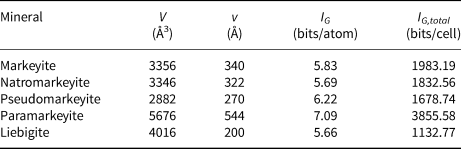
The structures of the aforementioned minerals were further evaluated with regard to their structural complexities (after Krivovichev Reference Krivovichev2012, Reference Krivovichev2013, Reference Krivovichev2014, Reference Krivovichev2018) (Table 6). Paramarkeyite, with a complexity of 3855.58 bits/cell, is classified as very complex. It is not only the most complex within this group of related minerals, but it is the fourth most complex uranyl carbonate mineral known (see Gurzhiy et al., Reference Gurzhiy, Kalashnikova, Kuporev and Plášil2021). It is noteworthy that paramarkeyite also has the greatest density of the five related minerals listed in Tables 5 and 6; of the five, it has the lowest H2O content and thus the structure is the most densely packed regardless of having the largest unit-cell volume in the group.
Information measures (IG) for markeyite, natromarkeyite, pseudomarkeyite, paramarkeyite and liebigite including H atoms.

Acknowledgements
Aaron Lussier and an anonymous reviewer are thanked for their comments on the manuscript. Comments by Structures Editor Peter Leverett were particular helpful in improving the structure refinement. A portion of this study was funded by the John Jago Trelawney Endowment to the Mineral Sciences Department of the Natural History Museum of Los Angeles County. This research was also financially supported by the Czech Science Foundation (project 20-11949S to JP) and by the Operational Program – Research, Development and Education (OP VVV) (project Geobarr CZ.02.1.01/0.0/0.0/16_026/0008459 to RS).
Supplementary material
To view supplementary material for this article, please visit https://doi.org/10.1180/mgm.2021.100