With an incidence of nearly 1% of all live births, congenital heart disease significantly contributes to the rate of birth defects in the population.Reference Hoffman 1 The aetiology of congenital heart disease is linked to genetic causes, as well as non-genetic causes such as environmental teratogens, maternal exposures, and infections.Reference Faheed, Gelb, Seidman and Seidman 2 Known genetic causes include large chromosome abnormalities, single gene disorders, and submicroscopic chromosome abnormalities, otherwise termed copy number variants. In patients with congenital heart disease with a genetic basis, efficient diagnosis of the underlying aetiology is essential to providing optimal clinical care, prognostic information, and an accurate recurrence risk.
Classical cytogenetic analysis by G-band karyotyping has been the gold standard for the detection of chromosome abnormalities, but its resolution is generally limited to genomic imbalances of 5 Mb or larger.Reference Thienpont, Mertens and de Ravel 3 With the evolution of array comparative genomic hybridisation, it has been recognised that novel submicroscopic copy number variants also contribute significantly to the aetiology of congenital heart disease. Copy number variants are defined as any DNA segment >1 kb in length with variable copy number in comparison to a reference genome.Reference Breckpot, Thienpont and Peeters 4
22q11.2 deletion or DiGeorge syndrome is one well-studied copy number variant that is estimated to account for 5% of all congenital heart disease.Reference Wilson, Burn, Scambler and Goodship 5 In a previous study at our institution, the detection rate of chromosome abnormalities by classical cytogenetic analysis was 14% among infants with congenital heart disease, and fluorescence in situ hybridisation for 22q11.2 increased the diagnosis rate by 5%.Reference Baker, Sanchez-de-Toledo and Munoz 6 Array comparative genomic hybridisation is able to detect well-known regions of genomic imbalance associated with congenital heart disease, including a 22q11.2 deletion, as well as other potentially causal copy number variants across the entire genome with a resolution at least 10-fold greater than classical cytogenetic analysis.Reference Miller, Adam and Aradhya 7
The American College of Medical Genetics recommends array comparative genomic hybridisation as a first-tier test to be used in concert with classical cytogenetic analysis for evaluation of individuals with developmental delay/intellectual disability, autism spectrum disorders, and multiple congenital anomalies,Reference Manning and Hudgens 8 but data to support this recommendation for patients with congenital heart disease are lacking. Wapner et al and Reddy et al published evidence of the efficacy of array comparative genomic hybridisation compared with karyotype for prenatal diagnosis and genetic diagnosis after stillbirth. In both settings, array comparative genomic hybridisation identified additional copy number variants, which were clinically significant but not detected by karyotype alone.Reference Wapner, Martin and Levy 9 , Reference Reddy, Page and Saade 10 Notably, no studies to date have prospectively studied array comparative genomic hybridisation as a first-tier test for neonates with congenital heart disease in the critical care setting.
Lu et al reported a detection rate of 21.8% for array comparative genomic hybridisation among neonates with congenital heart disease, including those with apparently isolated and apparently syndromic congenital heart disease.Reference Lu, Phung and Shaw 11 Breckpot et al analysed the causality of copy number variants detected by array comparative genomic hybridisation among patients with congenital heart disease from previous studies; most categorised patients into syndromic and isolated congenital heart disease, and did not limit inclusion by age. The detection rate of copy number variants was 19% among patients with apparently syndromic congenital heart disease, and 3.6% among patients with isolated congenital heart disease, suggesting a higher utility only in apparently syndromic cases.Reference Breckpot, Thienpont and Arens 12 However, Erdogan et alReference Erdogan, Larsen and Zhang 13 showed that copy number variants detected by array comparative genomic hybridisation had a similar frequency among patients with isolated congenital heart disease (17%), and that array comparative genomic hybridisation can identify a child with a syndrome even before additional symptoms manifest.
The use of array comparative genomic hybridisation among neonates with congenital heart disease has the potential to increase the efficiency of genetic diagnoses and accuracy of recurrence risks provided to families, as well as to potentially identify novel candidate genes for congenital heart disease. Array comparative genomic hybridisation has been added as a first-tier diagnostic test along with classical cytogenetic analysis for all neonates with congenital heart disease admitted to the Cardiac Intensive Care Unit at Children’s Hospital of Pittsburgh of UPMC. The primary aim of our study was to determine whether array comparative genomic hybridisation significantly increased the detection of chromosome abnormalities over classical cytogenetic analysis alone. The secondary aim was to determine the prevalence of additional syndromic features in this population and whether the identification of dysmorphic features varied between providers.
Methods
This study was approved by the University of Pittsburgh Institutional Review Board before initiation. Neonates diagnosed with congenital heart disease and admitted to the Cardiac Intensive Care Unit at Children’s Hospital of Pittsburgh of UPMC from April, 2011 to March, 2012 were included. Patients received classical cytogenetic analysis, array comparative genomic hybridisation, and a dysmorphology examination by a geneticist. After obtaining written informed consent from a parent or guardian, 58 patients were enrolled and data were compiled into a secure, password-protected database.
Data collection included patient demographics, growth percentiles, type of congenital heart disease, results of any genetic testing, results of imaging studies – for example, renal ultrasound, head ultrasound, and brain magnetic resonance imaging – and physical examination findings reported by Medical Genetics and Cardiology providers. For the purposes of this study, dysmorphic features were defined as any physical features noted on physical examination that were deemed “abnormal” rather than a variant of normal. Extracardiac abnormalities included extracardiac diagnoses, additional congenital anomalies, and abnormalities on imaging studies unless noted to be normal variation. Clinical features attributed to an environmental cause – that is, hyperbilirubinaemia attributed to prematurity – were not categorised as extracardiac features. Patients were considered to have “apparently isolated” congenital heart disease only if they exhibited no dysmorphic features or extracardiac abnormalities.
Genetic testing was performed at the CLIA-approved Pittsburgh Cytogenetics Laboratory. Classical cytogenetic analysis was performed by high-resolution G-banding to visualise prometaphase chromosomes at a 675 band-level resolution. A total of 20 cells were counted and six metaphase cells were analysed and karyotyped in each analysis. Array comparative genomic hybridisation was performed using the Roche NimbleGen 135 K (Madison, WI, United States of America) oligonucleotide array platform by SignatureChipOS™, version 2.0 (based on UCSC 2006 hg 18 assembly). This platform analyses 3397 loci at a 135 Kb resolution, and includes clones for subtelomeric regions, pericentromeric regions, and known genetic syndromes. Copy number variants <300 Kb in size were not reported, with the exception of those containing genes with known clinical significance. Variants of unknown clinical significance were evaluated and reported per standard laboratory protocol.Reference Manning and Hudgens 8 , Reference Kearney, Thorland, Brown, Quintero-Rivera and South 14
For the assessment of pathogenicity, Genoglyphix Genome Browser® and DECIPHER, v5.1 were utilised to identify any genomic imbalance syndromes that overlap with the region of interest, as well as any genes that are deleted or duplicated. 15 , 16 Online Mendelian Inheritance in Man was used to determine dosage sensitivity of genes in the region and any association with previously defined genetic syndromes. The Database of Genomic Variants was used to determine whether the copy number variant has been reported in healthy individuals. 17 The inherited versus de novo nature of the copy number variant was reported in cases in which parental studies were performed.
McNemar’s test was used to compare detection rates of array comparative genomic hybridisation and classical cytogenetic analysis at a significance level of α=0.05. The null hypothesis was equal detection rates of both tests. Only paired data – patients tested by both array comparative genomic hybridisation and classical cytogenetic analysis – were captured in this analysis. The sensitivity, specificity, and positive predictive value of a physical examination for dysmorphic features were also calculated, as performed by Genetics providers in comparison with non-genetics providers.
Results
Of all patients enrolled in the study (n=58), 45 had both array comparative genomic hybridisation and classical cytogenetic analysis. Neonates diagnosed with an aneuploidy receiving palliative care were not likely to be admitted to the Cardiac Intensive Care Unit; thus, no cases of Trisomy 18 and 13 were identified in our study population.
The total detection rate by array comparative genomic hybridisation was 26.1% (12/46). In comparison, the total detection rate by classical cytogenetic analysis among all patients tested was 9.4% (5/53), which included two patients with Trisomy 21 who were not tested by array comparative genomic hybridisation. The three other chromosome abnormalities detected by classical cytogenetic analysis were a 46,XX,t(6;10)(p21.2;15) balanced translocation, a 7.7 Mb deletion of 3p22.2–3p23, and mosaic Trisomy 21. Array comparative genomic hybridisation detected 10 copy number variants that were not detected by classical cytogenetic analysis, increasing detection by 22.2% above standard chromosome analysis alone (p=0.008) (Table 1). The 6;10 balanced translocation was not detected by array comparative genomic hybridisation, but was inherited from a healthy parent and thought unlikely to be causal for the patient’s congenital heart disease. The 7.7 Mb 3p deletion and mosaic Trisomy 21 were detected by both array comparative genomic hybridisation and classical cytogenetic analysis and were therefore not included in the reported 22.2% (n=10) increase in detection by array comparative genomic hybridisation.
Quantity of chromosome abnormalities detected based on the method of chromosome analysis.

CGH=comparative genomic hybridisation
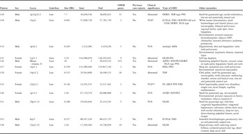
The chromosome aberrations detected by array comparative genomic hybridisation are listed in Table 2. The type of heart defect is also listed, following the classification system used by Botto et al,Reference Botto, Lin, Riehle-Colarusso, Malik and Correa 18 as well as the patients’ other clinical features. Four of the abnormalities were associated with a known syndrome and were a likely cause for the patient’s congenital heart disease, yielding at least a 10.9% (5/46) detection rate of pathogenic copy number variants among all patients tested by array comparative genomic hybridisation. Diagnoses included a interstitial deletion of 3p22.2–p23 (Patient 110), 1q21.1 duplication syndrome (Patient 121), mosaic Trisomy 21 (Patient 126), 16p11.2 microdeletion syndrome (Patient 128), and 22q11.2 deletion syndrome/DiGeorge syndrome (Patient 158). Only the 3p deletion and mosaic Trisomy 21 were also detected by classical cytogenetic analysis. In all, seven additional copy number variants were classified as variants of uncertain significance that require clinical follow-up and reassessment.
Clinical features of patients with copy number variants detected by array CGH.

AS=aortic stenosis; ASD=atrial septal defect; AVSD=atrioventricular septal defect; CGH=comparative genomic hybridisation; CHD=congenital heart disease; COA=coarctation of the aorta; DORV=double-outlet right ventricle; D-TGA=d-transposition of the great arteries; HLHS-IVS=hypoplastic left heart syndrome with intact ventricular septum; HSV=herpes simplex virus; OMIM=Online Mendelian Inheritance in Man; PDA=patent ductus arteriosus; PFO=Patent foramen ovale; PVS=pulmonary valve stenosis; RVOTO=right ventricular outflow tract obstruction; SV, DILV=single ventricle, double-inlet left ventricle; TGA=transposition of the great arteries; TOF=tetralogy of Fallot; VSD=ventricular septal defect; VUS=variant of uncertain significance
* Maternally inherited
** Paternally inherited
Of all patients enrolled, 72.2% had syndromic congenital heart disease by our criteria, including all patients with a copy number variant detected by array comparative genomic hybridisation. However, of these patients, only 25% (3/12) were noted to have dysmorphic features by a cardiologist, whereas 75% (9/12) were noted to have dysmorphic features by a geneticist. The majority of patients with apparently syndromic congenital heart disease presented with both dysmorphic features and extracardiac abnormalities.
Discussion
Our results confirm a significant increase in detection of chromosome aberrations by array comparative genomic hybridisation compared with classical cytogenetic analysis in patients with congenital heart disease [22.2% (p=0.008)]. Considering only copy number variants of known pathogenicity, the diagnostic yield of array comparative genomic hybridisation was 10.9%. In comparison, the diagnostic yield of array comparative genomic hybridisation among patients with developmental delay/intellectual disability, autism spectrum disorders, and multiple congenital anomalies was 12.2%.Reference Miller, Adam and Aradhya 7 On the basis of these data, we recommend that array comparative genomic hybridisation be utilised as a first-tier test among patients with congenital heart disease. Other copy number variants of uncertain significance detected in our population may yet be considered causal, but follow-up and additional information about these copy number variants in healthy and affected individuals is needed in order to reclassify them. Therefore, the diagnostic yield of 10.9% is likely an underestimate of the true diagnostic yield of array comparative genomic hybridisation in patients with congenital heart disease.
The diagnostic yield of classical cytogenetic analysis for chromosome abnormalities with known pathogenicity was 7.5% (4/53), which is similar to array comparative genomic hybridisation. However, all likely causal chromosome abnormalities in our study would have also been detected by array comparative genomic hybridisation. Array comparative genomic hybridisation can miss some cases of low-level mosaicism, but the case of mosaic Trisomy 21 in this population (Patient 126) was detected. This patient had ~10% trisomic cells by classical cytogenetic analysis. Array comparative genomic hybridisation has significant clinical utility as a first-tier diagnostic test in this population.
Multiple known syndromes were diagnosed by array comparative genomic hybridisation in this study, and clinical features of each patient are listed in Table 2. Patient 121 was diagnosed with chromosome 1q21.1 duplication syndrome, which has variable expression and is associated with developmental delay, neuropsychiatric abnormalities, macrocephaly, mild dysmorphic features, and congenital anomalies. There is at least one reported case of complex congenital heart disease and another patient with a bicuspid aortic valve.Reference Brunetti-Pierri, Berg and Scaglia 19 Patient 128 was diagnosed with 16p11.2 microdeletion syndrome, which also has variable expression and is associated with neurologic manifestations, primarily autism spectrum disorder. Ghebranious et al reported monozygotic twins with a 16p11.2 microdeletion and aortic valve abnormalities.Reference Ghebranious, Giampietro, Wesbrook and Rezkalla 20 Patient 158 was diagnosed with DiGeorge syndrome; classic features include conotruncal heart defects, immune deficiency, hypocalcaemia, cleft lip/palate, dysmorphic facial features, and developmental delay.Reference Kobrynski and Sullivan 21 In three of these copy number variant conditions, parents may be affected and previously undiagnosed at the time of a child’s diagnosis because of a missed diagnosis or significantly milder features in the parent. Although most copy number variants have high phenotypic variability, this would lead to a recurrence risk of 50%, in contrast to the typical empiric recurrence risk of 3%.Reference Erdogan, Larsen and Zhang 13 This highlights the importance of a timely diagnosis in order to provide the family with an accurate estimate of recurrence risk.
Although array comparative genomic hybridisation increased detection of pathogenic abnormalities in our study population, a proportion of the copy number variants detected (58.3%) were classified as variants of uncertain significance because of a lack of previous reports of similar imbalances or dosage sensitivity of genes in the region. Standard laboratory protocol involves a vigorous assessment of copy number variants before assigning clinical significance.Reference Kearney, Thorland, Brown, Quintero-Rivera and South 14 Those that remain a variant of uncertain significance are rare and not seen in databases of healthy controls, but still lack the definitive support to be called pathogenic or benign. Clinicians who can compare the patient’s features with the copy number variant, in the context of family history, can gain some additional understanding. Therefore, copy number variants that are classified as variants of uncertain significance still warrant clinical follow-up, including family studies, as information about genetic imbalances is constantly improving, and the copy number variant may be reclassified in the future.
Incidental findings
Patient 110 highlights one important consideration of genetic testing by array comparative genomic hybridisation, which is the potential for incidental findings. His clinical features are summarised in Table 2. This patient had a 7.7 Mb deletion from 3p22.2–p23, which was detected by both classical cytogenetics and array comparative genomic hybridisation and is a likely cause for the patient’s congenital heart disease. A previously reported patient with a similar deletion had global developmental delay, mild dysmorphic features, and short stature.Reference Liu, Oei, Mitchell and McGaughran 22 A total of 35 genes listed in the Online Mendelian Inheritance in Man database are located in this region, including seven genes with known disease associations: MLH1, ACVR2B, GPD1L, SCN5A, GLB1, CRTAP, and MYD88. The deletion of the mismatch repair gene MLH1 is consistent with an incidental diagnosis of Lynch syndrome, or Hereditary Non-Polyposis Colorectal Cancer.Reference Hemminki, Peltomaki, Mecklin, Jarvinen, Salovaara and Nystrom-Lahti 23 The effect of whole gene deletions of MLH1 has not been reported in the literature, but a mutation in intron 14 of MLH1, which results in silencing the gene, has been reported in Lynch syndrome patientsReference Vasen, Watson, Mecklin and Lynch 24 who appeared to have a slightly lower frequency of extracolonic tumours.Reference Jager, Bisgaard, Myrjoj, Bernstein, Rehfeld and Nielsen 25 It is likely that a whole gene deletion, which will have a similar silencing effect of one allele, would cause a similar phenotype.
In addition, other genes located within Patient 110’s deletion have been associated with cardiac channelopathies with incomplete penetrance. Mutations in GPD1L are one known cause of Brugada syndromeReference London, Michalec and Mehdi 26 and mutations in SCN5A may cause familial atrial fibrillation, Brugada syndrome, dilated cardiomyopathy, progressive familial heart block type 1A, nonprogressive heart block, long QT syndrome, and familial ventricular fibrillation. 27 To our knowledge, the effect of whole gene deletions of GPD1L and SCN5A has not been reported, but this patient may have an increased risk for a channelopathy. Finally, GLB1, CRTAP, and MYD88 are associated with autosomal recessive disorders, including GM1-gangliosidosis, osteogenesis imperfect a type VII, and recurrent pyogenic bacterial infections, respectively. Patient 110 is likely only a carrier of these recessive conditions and would only be affected in the case of a mutation in the remaining allele. Children are not typically tested for adult-onset disorders or cancer predisposition genes because of ethical considerations; therefore, it is important for the physician and parents to be aware of the potential for incidental findings when ordering array comparative genomic hybridisation and to have a plan for follow-up.
The possibility of finding an incidental result is not unique to array comparative genomic hybridisation, but is a risk faced by practitioners whenever he or she orders a diagnostic test. Potential results should be discussed at the time of parental consent for the testing. Although incidental results are often thought of as something to be dreaded, they can at times be life saving. In a child with a symptom suggestive of a genetic condition, such as a congenital heart disease, the most efficient diagnostic testing that can clarify prognosis, recurrence risks, and medical management should be pursued despite the risk of incidental findings.Reference Ross, Saal, David and Anderson 28 Consultation with a genetic counsellor, or other professional with a strong understanding of genetic test results, to understand and explain the result may be warranted.
Clinical application of array comparative genomic hybridisation
The combined prevalence of extracardiac abnormalities and dysmorphic features within this study population is high (72%). This finding may be a result of all patients being admitted to a critical care unit, which could increase the prevalence of complex and syndromic congenital heart disease. All patients with a copy number variant had additional syndromic features, but 25% (3/12) of these patients would likely have been categorised with isolated congenital heart disease had they not received a head and renal ultrasound and physical examination by a geneticist. Identification of extracardiac features is important not only to identify patients with potentially syndromic congenital heart disease, but also to assist in interpreting the clinical significance of array comparative genomic hybridisation results.
Some providers have suggested limiting array comparative genomic hybridisation testing to patients with apparently syndromic congenital heart disease, under the assumption that a child with an apparently isolated heart defect is unlikely to have a chromosome abnormality. However, our data highlight that this approach can result in missed diagnoses. Physical examination in an inpatient setting is often limited by tubes, appliances, tape, and patient instability, all of which impair identification of subtle dysmorphic features. Baker et al suggested that all neonates and infants with critical congenital heart disease receive a baseline head and renal ultrasound,Reference Baker, Sanchez-de-Toledo and Munoz 6 and if these studies are not routinely done, then occult abnormalities may be undetected. Finally, some features such as neurologic abnormalities may present later in patients who were initially thought to have isolated congenital heart disease.
There is some debate on whether array comparative genomic hybridisation should replace classical cytogenetic analysis as a single first-tier test for various clinical indications. The advantage of this strategy is that it would streamline the testing process and reduce the cost of testing. In addition, the turn-around time for array comparative genomic hybridisation is typically shorter than classical cytogenetic analysis. All causal chromosomal aberrations in this study would have been detected by array comparative genomic hybridisation. Although array comparative genomic hybridisation is limited in its detection of mosaicism and balanced rearrangements, our study suggests that classical cytogenetic analysis may be moved to the second-tier diagnostic work-up if clinical suspicion of a chromosome abnormality remains high after a normal array comparative genomic hybridisation.
In conclusion, array comparative genomic hybridisation shows great promise as a first-tier test in the evaluation of patients with congenital heart disease, even those with apparently isolated malformations, though careful follow-up is always warranted. Such testing requires close collaboration between cardiologists and medical geneticists to optimise clinical care.
Acknowledgements
The authors thank Dana Shiderly, R.N., and Jacqueline Hoover, M.S., for assistance with recruitment. Statistical assistance was provided by John Wilson, PhD, and Richard Day, PhD, as well as the University of Pittsburgh Clinical and Translational Science Institute, which was supported by the National Institutes of Health through Grant Numbers UL1 RR024153 and UL1TR000005. This study makes use of data generated by the DECIPHER Consortium. A full list of centres who contributed to the generation of the data is available from http://decipher.sanger.ac.uk and via email from decipher@sanger.ac.uk. Ms Bachman wrote the first draft of the manuscript and received no compensation.
Financial Support
Funding for the project was provided by the Wellcome Trust. Partial funding in the form of a grant was provided by the Cardiovascular Special Interest Group of the National Society of Genetic Counselors. This group had no involvement in study design, collection, analysis, and interpretation of data, writing of the report, and the decision to submit the paper for publication.
Conflicts of Interest
None.