The definition of foetal congestive heart failure is similar to that after birth – inadequate tissue perfusion. Inadequate cardiac output results in a series of complex reflexes and adaptations to improve forward flow or to direct flow towards vital organs. This state can be described as a deficiency of flow of blood to the tissues such that certain reflexes are triggered for the survival of the foetus.
Secretion of excess circulating catacholamines, which are produced in response to peripheral vascular detection of abnormal perfusion, is one of these reflexes. Powerful hormonal reflexes are triggered, including those that control salt and water retention in an attempt to increase myocardial preload and those that control adrenocorticoid excess, which mobilise additional calories for the increased metabolic demand that is present.
The diagnosis of foetal congestive heart failure must be addressed in a clinical manner similar to that after birth. The classical clinical tetrad of cardiomegaly, tachycardia, tachypnea, and hepatomegaly has been used in neonates and children. This clinical state in the foetus can be characterised by findings in at least five categories that are obtained during the ultrasonographic examination. The following categories are each worth 2 points in a 10-point scoring system used quantitatively to assess the cardiovascular system: hydrops, umbilical venous Doppler, heart size, abnormal myocardial function, and arterial Doppler. Abnormalities in the cardiovascular profile score may occur before the clinical state of hydrops foetalis.
Faced with a foetus with non-immune hydrops foetalis, one must first determine whether the hydrops is cardiac, inflammatory, or metabolic. Many cases of hydrops are attributed to foetal systemic infection. New markers are identifying aetiological agents such as parvovirus or adenovirus. The associated hepatitis with these infections can compromise the protein-producing capability of the foetus, thereby decreasing the foetal oncotic pressure. Immune hydrops must always be considered in the differential diagnosis, but other causes of anaemia can also cause hydrops, such as haemoglobinopathies. Infections can cause haemolytic anaemia, which can be treated by foetal transfusion.
Anaemia may cause hydrops; however, ventricular shortening fraction did not significantly change among 111 foetuses with haemoglobin Bart’s disease with and without hydrops.Reference Tongsong, Wanapirak and Piyamongkol 1 On the other hand, left and right ventricular shortening fraction were significantly decreased (mean Z-scores 5SD and 8SD below the mean, respectively) in 21 hydropic foetuses as a result of CHDs (p<0.001). These authors concluded that foetuses with hydrops foetalis secondary to cardiac defects and anaemia have different patterns of shortening fraction. Defects present with cardiac decompensation have major re-distribution of cardiac output, whereas in foetal anaemia failure is probably caused by hypervolaemia with cardiac decompensation occurring when the cardiac compensatory mechanism is exhausted. Foetal cardiomyopathy described as a case of isolated left ventricular non-compaction, a severe congenital cardiomyopathy, which presents in the neonatal period as foetal hydrops has been described.Reference Richards, Mao and Dobson 2
Maternal complications
The mother may develop oedema, hypertension, and proteinuria during conservative management of hydrops – a condition known as Mirror syndrome and also known as pseudotoxaemia or Ballantyne syndrome. Symptoms may persist even after delivery.
Prognosis
In 2007, a retrospective review of a large national data set showed a total of 253,651 discharges from 162 neonatal intensive care units, and 598 patients were identified with a report of hydrops foetalis. The most common associated diagnoses were congenital heart problems (13.7%), abnormalities in heart rate (10.4%), twin-to-twin transfusion (9%), congenital anomalies (8.7%), chromosomal abnormalities (7.5%), congenital viral infections (6.7%), congenital anaemia (5%), and congenital chylothorax (3.2%). Of those 598 neonates, 115 were transferred either to another hospital or to another service, 215 died before discharge, and 267 were discharged from the hospital. Mortality was highest among neonates with congenital anomalies (57.7%) and lowest among neonates with congenital chylothorax (5.9%). Factors that were associated independently with death in the logistic regression analyses were younger gestational age, low 5-minute Apgar score, and need for high levels of support during the 1st day after birth – that is, higher levels of inspired oxygen support and more often treated with high-frequency ventilation.Reference Abrams, Meredith, Kinnard and Clark 3
Huang studied 28 live-born neonates with hydrops and most of them presented with pleural effusions (21 of 28) and ascites (22 of 28). The majority of patients had hydrops due to cardiovascular diseases (7 of 28), haematological disorders (6 of 28), lymphatic malformations (6 of 28), and idiopathic origins (6 of 28). The overall survival rate was 50% and was highest (83%) in infants with lymphatic malformations. By univariate analysis, risk factors for mortality are earlier ages at diagnosis and at birth, low Apgar scores, need for resuscitation in the delivery room, low serum albumin levels, and severe acidaemia. After using stepwise multiple logistic regression analysis, the most significant factors associated with fatality were younger gestational age at birth and lower serum albumin levels. They concluded that hydrops foetalis remains a complex condition with a high mortality rate. Hydrops resulting from lymphatic malformations has a favourable outcome. Preterm birth at <34 weeks and serum albumin concentration <2 g/dl are two poor prognostic factors for survival.Reference Huang, Tsay, Chiang, Lien and Chou 4
Multiple mechanisms of hydrops may coexist and the primary cause may not be immediately obvious. Of more importance is the determination of the prognosis of hydrops. This task would be aided by a semi-quantitative measure of foetal heart failure. This article presents such an assessment tool called the cardiovascular profile score.
Foetal hydrops for the perinatal cardiologist
The challenge of hydrops assessment can be summarised by several questions for the perinatal cardiologist: Is hydrops from the heart? Is foetal heart failure a result of CHD? Is it becoming more severe? Is there foetal myocardial dysfunction?
After birth, the prognosis will depend on the answers to these and many other questions. The long-term outcome is dependent on whether the insult is reversible and whether there are periods of ischaemia and/or brain injury. There are several possible causes of heart failure in the foetus after ruling out foetal infection. The most useful predictor of perinatal death in foetal hydrops is the presence of umbilical venous pulsations,Reference Gudmundsson, Huhta, Wood, Tulzer, Cohen and Wein 5 because the most common pathway of perinatal demise is compromised foetal cardiac output – foetal congestive heart failure. What follows is a method to detect this entity and to determine which foetuses should be referred to a foetal centre. Initially, the following data are collected during foetal echocardiography:
-
∙ Cardiac size:thoracic size (C:T): cardiac divided by thoracic area ratio (normal, 0.25–0.35) or C:T circumference ratio (normal, <0.5).
-
∙ Venous Doppler: inferior caval – or hepatic venous; increased atrial reversal – and umbilical cord vein (pulsations).
-
∙ Four-valve Doppler: any leak of the valve should be evaluated further. If there are abnormalities in any of these measurements, then a cardiac cause or associated physiological problem may be present and a detailed study is indicated to rule out serious cardiovascular involvement.
Within specific disease entities, more emphasis is placed on certain areas to predict the prognosis. This information can only comprise a portion of the total picture and must be integrated by the attending physician into the diagnostic and treatment plan for the patient.
Counselling
Long-term prognosis depends on the underlying cause and severity of the heart failure. If the cause of non-immune hydrops fetalis cannot be determined, the perinatal mortality is ~50%. Prognosis is much poorer if diagnosed at <24 weeks and if pleural effusion or structural abnormalities are present. Pulmonary hypoplasia is a common cause of death in neonates with pleural effusions. Foetal hydrops associated with a structural heart defect is associated with an almost 100% mortality rate. If hydrops is found early in pregnancy – <24 weeks – with no treatable cause, the option of termination may be considered. Recurrence is uncommon unless related to blood group incompatibility – isoimmunisation – or inheritable disorders. A cardiovascular profile score <7 is associated with mortality.
The cardiovascular profile score in hydrops and heart failure
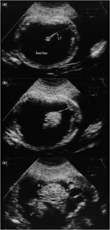
In early stages, hydrops foetalis may present with ascites, pleural effusion, pericardial effusion (Fig 1), or a combination of these findings. In advanced hydrops, there is generalised skin oedema seen easily over the scalp and abdominal wall. In scoring hydrops for the cardiovascular profile score, 1 point is deducted for early hydrops and 2 points for skin oedema.
( a – c ) Ascites fluid in a foetus with hydrops foetalis showing the LT and the R and L lobes of the liver. L=left; LT=ligament teres; R=right.

A cardiovascular profile score is calculated by assigning 2 points for each of the five categories and using the score in serial studies to provide a method of uniform physiological assessment. By taking a multivariate approach, this type of multifactorial score can combine the assessments of direct and indirect markers of cardiovascular function.
Initial validation of the cardiovascular profile score in hydrops was shown by Falkensammer et al;Reference Falkensammer, Paul and Huhta 6 seven foetuses with hydrops, including three with CHD, had correlation of the cardiovascular profile score with the myocardial performance index (Tei index). Right ventricle and left ventricle Tei indices were assessed in controls and showed no change with gestational age. Hofstaetter et alReference Hofstaetter, Hansmann, Eik-Nes, Huhta and Luther 7 measured the cardiovascular profile score in 59 hydropic foetuses. Mortality was 21/59. The median score in those who died prenatally or postnatally was 5, whereas the median score in the survivors was 7. The use of the score appears to aid in the assessment of intrauterine growth retardation.Reference Makikallio, Rasanen, Makikallio, Vuolteenaho and Huhta 8 Recently, the American Heart Association review of foetal echocardiography included the cardiovascular profile score as an evidence-based technique.Reference Donofrio, Moon-Grady and Hornberger 9
Rationale for the five cardiovascular profile score categories
Umbilical and ductus venous Doppler
Foetal venous blood velocities have been examined and investigations have been clinically promising. Significant increase in flow reversal with atrial contraction is evident in altered haemodynamic states in the foetus after 12 weeks of gestation. Studies in the foetal lamb have shown that this increase in the percentage of reversed flow seen in normal pregnancy is related to the pressure gradient between the right atrium and the right ventricle during end diastole. It appears to be related to both ventricular compliance and ventricular end diastolic pressure, and therefore is a reflection of central venous pressure. Recording venous blood velocity might, thus, give important information on foetal cardiac pump function. Transmission of the venous pulsations into the portal and umbilical circulation correlates with increasing degrees of cardiac compromise. Tulzer et alReference Tulzer, Gudmundsson, Wood, Cohen, Weiner and Huhta 10 studied the cardiac factors related to prognosis in hydrops and noted that umbilical venous pulsations could stand in for a number of cardiac variables in predicting prognosis, including ventricular shortening fraction, ejection velocities, and percentage inferior caval vein atrial reversal. Abnormal venous Doppler develops with increasing heart failure in the following order:
-
∙ Increased atrial reversal in the inferior caval vein.
-
∙ Ductus venosus atrial reversal.
-
∙ Portal venous atrial pulsations.
-
∙ Umbilical venous atrial pulsations.
The cardiovascular profile score has deductions for abnormal venous Doppler as follows: ductus venosus atrial reversal, deducts 1 point, umbilical venous atrial pulsations, deduct 2 points. The maximum deduction in any category is 2 points.
Cardiomegaly
Enlargement of the cardiac chambers is a universal sign of heart failure. This is true in the foetus as well, but few of the mechanisms are understood. It is likely that neurohumoral reflexes are triggered, resulting in retention of extracellular volume leading to increased end diastolic volume of the ventricles. At some point, this increased ventricular size indicates increased end diastolic pressure. On the other hand, unlike in the postnatal human, it is uncommon to encounter persistent tachycardia with signs of catacholamine excess.
Small heart size with external compression has been correlated with hydrops and poor outcome in foetuses with cystic adenomatoid malformation. When the heart size was <20% of the chest area, foetal outcome was affected. In foetuses with cystic adenomatoid malformation, a small C:T ratio (<0.2) is associated with a poor prognosis. In utero, the heart area can be easily compared with the area of the thorax, and the ratio should be less than one-third and greater than one-fourth in the presence of normal chest development. Cardiac size calculations are as follows: C:T area ratio=cardiac area/chest area (normal, 0.2–0.35) and C:T circumference ratio=cardiac circumference/chest circumference (normal, <0.5).Reference Mahle, Rychik and Tian 11
Cardiovascular profile scoring for cardiac size is as follows: normal heart:chest area ratio is <0.35 and >0.20; mild cardiomegaly: area ratio >0.35, deduct 1 point; severe cardiomegaly: area ratio >0.50, deduct 2 points; small heart ratio (<0.2), deduct 2 points. Maximum deduction is −2 points.
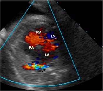
An example of cardiomegaly is shown in Figure 2. This foetus at 28 weeks of gestation had cardiomegaly caused by a pulmonary arteriovenous fistula in the right lung. A positive response to digoxin administered transplacentally was possible in this case and the pregnancy proceeded to term and a coil occlusion of the fistula was performed by catheterisation.
Foetal PAVF in the right lung. LA=left atrium, LV=left ventricle, PAVF=pulmonary ateriovenous fistula; RA=right atrium, RV=right ventricle. Note the mild cardiac enlargement.

Abnormal myocardial function
The cardiac function is assessed indirectly by the global shortening – and thickening – of the walls of the ventricles and by the function of the atrioventricular and semi-lunar valves. The diameters of both the right and the left ventricles should shorten >28% in systole compared with diastole. Measurements of cardiac dimensions with time are performed using M-mode echocardiography. The shortening fraction (FS) of a ventricle is calculated by taking the difference between the diastolic (DD) and systolic dimensions (SD) and dividing it by the diastolic dimension:
Fractional shortening⩾
 $${\rm FS}={\rm DD}{\minus}{\rm SD}/{\rm DD}\quad {\rm Normal}\,⩾ \,0\!\!:\!\!{\rm 28}$$
An abnormal shortening fraction could reflect myocardial compromise or an increase in the foetal ventricular workload. Regardless, an increase in diastolic dimension is often related to a decrease in shortening fraction and should be regarded as an indication for more intensive monitoring.
The atrioventricular and semi-lunar valves are competent in the normal foetus, and if regurgitation is detected it is usually a sign of altered cardiovascular physiology. Respondek et alReference Mahle, Rychik and Tian 11 showed that 7% of foetuses with a foetal echocardiogram displayed trivial (holosystolic) or significant tricuspid valve regurgitation. Most of these cases had some reason for this, such as constriction of the ductus arteriosus from indomethacin treatment of preterm labour, but 93% had no trace of regurgitation with state-of-the-art equipment and careful examination. As tricuspid valve regurgitation is common after birth, we may speculate that the foetal right ventricle adapted well to the systemic pressure work. Therefore, valvular competence is normal, and only in disturbances of cardiovascular physiology in which there is increased ventricular wall stress is holosystolic tricuspid valve regurgitation present. Trace tricuspid regurgitation, defined as non-holosystolic regurgitation lasting at least 70 ms, is not normal. This may be the first sign of a problem but has little prognostic importance. Holosystolic tricuspid regurgitation is abnormal and indicates the need for further investigation.Reference Respondek, Kammermeier, Ludomirsky, Weil and Huhta 12 When regurgitation is detected by color Doppler, it must be confirmed and graded by pulsed Doppler. With congenital diseases of the tricuspid valve, hydrops and foetal death can occur.Reference Neves, Mathias and Wilhm 13
Regurgitation of the tricuspid, mitral, aortic, or pulmonary valves is usually a sign of more advanced congestive heart failure and may occur in the moribund foetus with acidosis and severe heart failure as a sign of myocardial compromise. With severe myocardial failure, the support for the semi-lunar valves is compromised and pulmonary or aortic valve regurgitation can occur. Tricuspid valve regurgitation can be a reversible sign of heart failure, because successful therapy for anaemia or tachycardia in foetuses in utero has been performed. Progression to mitral valve regurgitation is a sign of foetal congestive heart failure and usually means that a significant increase in left ventricular wall stress is present.
The nature of the velocity waveform of atrioventricular valvular regurgitation has prognostic value in the calculation of foetal ventricular dP/dt. With holosystolic tricuspid valve regurgitation, the time interval from one right ventricle–right atrium pressure difference to another can be used to calculate the change in pressure over time or the dP/dt. A value of <800 mmHg/second is abnormal, and a value <400 mmHg/second predicts poor foetal outcome.Reference Tulzer, Gudmundsson, Huhta, Wood and Tews 14 This measurement requires continuous-wave Doppler, and the peak velocity may be 2.5–4.5 m/second. We have found that the most useful range for dP/dt measurement in foetal tricuspid regurgitation is 0.5–2.5 m/second – that is, a right ventricle–right atrium gradient of 1–25 mmHg or a 24-mmHg difference. The foetal ventricles are at equal systemic pressure throughout gestation, and therefore the blood pressure of the foetus is estimated using this technique. The filling pattern of the ventricles in diastole is an indicator of the diastolic function of the heart. Normal values show that the proportion of atrial filling during the atrial contraction is constant from 14 to 40 weeks of gestation. Monophasic filling of the ventricles is a sign of compromised diastolic function and foetal heart failure.
Abnormalities of diastolic function can be present in the foetus and should be excluded by comparing the filling patterns of the ventricles using pulsed Doppler to standardised normal values. A rule of thumb is that the A wave of the ventricular filling is always greater than the E wave. Monophasic filling of the ventricles occurs in severe diastolic dysfunction and with severe external cardiac compression. Tissue Doppler may be useful to assess the early filling rate of the early ventricular filling.
Arterial Doppler re-distribution of foetal cardiac output
It is well-established that the blood velocities measured by Doppler echocardiography in the umbilical artery and in other peripheral vascular beds can be used as indirect indicators of the relative vascular impedances. Findings of an increased pulsatility index in the umbilical artery and descending aorta and a decreased index in the middle cerebral artery are non-invasive signs of re-distribution of flow. The most common cause of elevated vascular resistance in the foetus is placental dysfunction secondary to vasculopathy leading to asymmetrical growth retardation. This complex pathophysiololgical state is poorly understood, but there is evidence that there is hypoxaemia resulting from placental dysfunction and additional compromise of nutrition severe enough to impair growth. Once the normal pattern of growth is disturbed, usually asymmetrical such that the brain continues growing but the body does not, the foetus is at risk of organ damage from hypoxaemic/ischaemic injury. The umbilical artery manifests this problem with a loss or reversal of diastolic blood flow. There is re-distribution of flow to the brain (brain sparing) due to reflex vasodilatation of the cerebral vessels. This is manifested by a decrease in the pulsatility index (PI) in the middle cerebral artery such that the diastolic flow is relatively increased (PI<2SD below the mean (25,26)). In the foetus with hypoxaemia, the peripheral foetal vessels are vasoconstricted and the larger arteries are suspected to be non-compliant compared with controls with increased blood pressure. In other words, this is a physiological state characterised by increased vascular resistances and, at end-stage, decreased vascular compliance and cardiac output. Right ventricular enlargement occurs in some cases. Foetal brain sparing is a marker of cardiac output re-distribution that is sensitive for the detection of significant hypoxaemia and placental dysfunction; however, there is evidence that reversal of diastolic flow in the umbilical artery or in the aortic isthmus may be significant risk factors for abnormal outcomes. Growth restriction prognosis has been described by the use of the cardiovascular profile score.Reference Makikallio, Rasanen, Makikallio, Vuolteenaho and Huhta 8
As a sign of foetal heart failure, the vasoconstriction resulting from decreased cardiac output and the compensatory sign of vasodilatation in the brain can be included in the cardiovascular profile score: absent end diastolic flow in the umbilical artery+brain sparing, increased middle cerebral artery diastolic velocity, deduct 1 point, and reversed end diastolic flow in the umbilical artery, deduct 2 points.
Hydrops
The cardiovascular profile score (see below) gives a semi-quantitative score of foetal cardiac well-being and uses known markers by ultrasound that have been correlated with poor foetal outcome. This profile is normal if the score is 10 and signs of cardiac abnormalities result in a decrease of the score from normal – for example, if there is ascites but no other abnormalities, there would be a deduction of 1 point for hydrops – ascites but no skin oedema – and no deductions for the other categories, for a score of 9 out of 10.
Treatment
(Table 1) Treatment of foetal cardiovascular problems can be classified into five groups based on the aetiology of congestive heart failure: abnormal peripheral impedances causing re-distribution of flow and growth failure, high output due to anaemia or arteriovenous fistula, primary or secondary valvular regurgitation, heart failure due to myocardial dysfunction, and tachycardia/bradycardia. Interventions aimed at improving the effective cardiac output are also aimed at prolonging the pregnancy and preventing prematurity and prenatal asphyxia.
Treatment for foetal hydrops by cause.

AV=arteriovenous
Treatment with digoxin for evidence of decreased ventricular shortening is controversial but it appears that digoxin can reduce the afterload in the foetus by altering favourably the neurohumoral state. Digoxin is known to decrease the catecholamine response to congestive heart failure and decrease sympathetic tone, and if there is diastolic dysfunction in the foetus it may lower filling pressures. Terbutaline appears to have promise as an inotropic and chronotropic agent and is effective in complete heart block, but studies of the possible negative effects on the foetal myocardium are needed. At present, we use digoxin for foetal cardiac failure due to arrhythmias and high-output states such as fistula and anaemia. In a case of acardiac twinning in which the normal foetus was supporting two circulations, digoxin appeared to improve cardiac function and result in a prolonged and successful gestation for the normal twin. Laser treatment of the twin–twin communications or cord ligation with acardiac twins can be performed to improve cardiac failure.
Conclusion
Foetal cardiac findings must be integrated into the clinical management of the foetus with hydrops foetalis or foetal heart failure by the perinatologist. The cardiovascular profile score can be used by the perinatal cardiology team to assess the urgency of abnormalities and the prognosis. Serial studies using the cardiovascular profile score are necessary to obtain the value from this test. Using it, uniform treatment strategies can be planned.